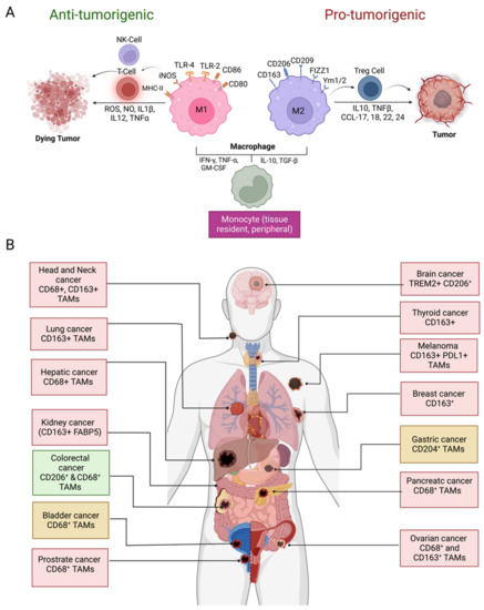
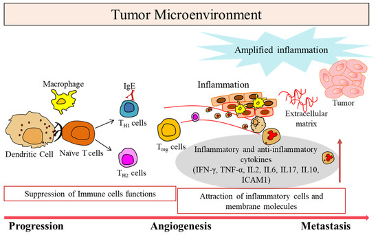
In addition to intrinsic growth-promoting changes within neoplastic cells, fertile ground is essential for tumor progression and metastasis. TAMs play a crucial role in the process of inducing EMT, as well as providing metastatic ground. During the EMT process, the interaction between the tumor cell and the TAMs plays a pivotal role. TAMs-derived TGF-β, TNF-α, CCL18, IL-6, IL-8, and IL-10 have been linked to EMT induction and metastasis by regulating various intracellular pathways such as TGF-β-SMAD signaling, MAPK signaling, WNT-β-Catenin pathway, NF-KB signaling, and PI3K-AKT signaling pathway. By inducing theses signaling pathways, TAMs promote the expression of mesenchymal cell markers while inhibiting the expression of epithelial cell markers, resulting in EMT in tumor cells. TAMs-secreted TGF-β binds to its receptors TGF-βR1 and TGF-βR2 and phosphorylates SMAD2 and SMAD3 proteins, which then combines with SMAD4 and forms the trimeric SMAD complex. The subsequent translocation of this trimeric SMAD complex into nucleus regulates the expression of EMT-associated genes through transcriptional mechanisms [
56]. Similarly, TNF-α induces EMT by inhibiting the expression of epithelial marker E-cadherin, upregulating the expression of mesenchymal markers, such as vimentin, N-cadherin, and fibronectin, and activating matrix metalloproteinase-9 (MMP-9) by interacting with its receptors TNFR1 and TNFR2, thereby inducing different signaling pathways [
57]. Moreover, TGF-β and TNF-α released from macrophages work collaboratively to induce EMT. A synergistic role of TGF-β and TNF-α has been found to induce EMT-mediated breast cancer cell migration and metastasis [
58,
59]. CCL18 produced by TAMs promotes breast cancer metastasis by downregulating miR98 and miR27b expression via the N-Ras/ERK/PI3K/NF-κB/Lin28b signaling pathway by inducing EMT [
60]. IL-8 promotes the migration and EMT of triple-negative breast cancer (TNBC) and ovarian cancer cells via PI3K-Akt signaling and the WNT/β-catenin pathway, respectively [
61,
62]. The inflammatory cytokine IL-6 secreted by TAMs induces EMT and promotes tumor cell invasion in lung cancer via the COX-2/PGE2/β-catenin signaling pathway [
63]. IL-10 is another cytokine that is abundantly produced by TAMs. TAMs release IL-10 upon activation of TLR4 signaling. The TLR4/IL-10 signaling pathway has been found to be involved in the promotion of EMT in pancreatic cancer cells [
64]. Moreover, M2-polarized macrophages promote the migration and EMT of HCC cells via the TLR4/STAT3 signaling pathway [
65]. In addition, tumor hypoxia induced an HIF-1α/IL-1β signaling loop between cancer cells and TAMs that leads to EMT in HCC [
66]. EGF derived from TAMs induces EMT in head and neck squamous cell carcinoma by activating the EGFR/ERK1/2 signaling pathway [
15,
67]. Moreover, M2-like TAMs secrete CCL20 to activate CCR6 in cancer cells, thereby enhancing the metastasis of primary cutaneous melanoma tumors [
68]. In addition, a long noncoding RNA (LncRNA) AFAP1-AS1 in the exosome derived from TAMs induces EMT-associated gene expression and metastasis in esophageal cancer by downregulating miRNA-26a and thereby upregulating its target transcription factor ATF2 [
15,
69].

留言 (0)