1. IntroductionHepatocellular carcinoma (HCC) is one of the major causes of death in patients with end-stage liver disease [
1]. Liver transplantation (LT) can be the best therapy for patients who are within the Milan criteria, which is a single nodule less than 5 cm, or up to 3 nodules less than 3 cm each, without macrovascular invasion and extrahepatic spreading [
2]. Tumor recurrence rates may be up to 20% (or more frequently) for patients beyond the Milan criteria [
3]. Adequate patient selection, pre-transplantation locoregional therapies and prioritization within the waiting list can be done in order to prevent HCC recurrences [
4]. “Incidental hepatocellular carcinoma” (iHCC) is a clinical condition, which is unexpectedly diagnosed after the histopathological evaluation of explanted livers in patients who have undergone LT for a reason other than tumors. Despite achievements due to radiologic evaluation, the diagnosis of iHCC in the explanted livers can still be a common condition for the patients undergoing LT for end-stage liver disease. Various prevalence rates were reported for iHCC (4.2% to 40%) [
5,
6,
7,
8,
9]. Different prognostic factors of iHCC were reported in the previous studies [
7,
10,
11]. In some studies, there is no prognostic effect either in disease-free survival (DFS) or in overall survival (OS); on the other hand, a worse prognostic effect can be observed in iHCC patients than “preoperatively diagnosed HCC” (pdHCC) groups [
5,
6,
7,
8,
9,
10,
11]. We aimed to compare the clinicopathologic and prognostic features of pdHCC and iHCC patients who received LT at the Inonu University Liver Transplantation Institute. We hypothesized that iHCC patients have better clinicopathologic features and better prognosis. 2. Materials and Methods
The liver transplantation Institute of the Inonu University database, which is recorded prospectively for every LT consecutively, was queried retrospectively. This study was approved by Institutional Ethics Committee of Inonu University (Approval number: 2022/3931). The present clinical investigation has been managed according to the principles contained in the Declaration of Helsinki. Liver transplantations between January 2002 and April 2022 were reviewed (n = 3204). Patients with post-transplant follow up period less than 90 days and who transplanted for non-tumoral reasons were excluded, leaving 406 patients who have pathologically confirmed HCC to be included in the study. Of these, 54 were iHCC. Demographic features, underlying liver disease, Child–Pugh, MELD scores, pretransplant tumor marker alpha-feto protein (AFP), gamma glutamyl transferase (GGT) levels and explanted liver’s histopathological data of these patients were recorded. Number of nodules and the size of the tumors were described. Additionally, the grade and microvascular invasion of the tumors were recorded.
Liver transplantation is not recommended for patients with a MELD score of less than 15 unless there are clinical signs of liver decompensation. Cirrhotic patients were screened by liver ultrasound every 6 months according to international guidelines. If there was a suspected liver nodule detected by ultrasound or a raised AFP level, computed tomography (CT) and/or magnetic resonance imaging (MRI) was ordered pre-operatively. During post-transplant follow-up, serum AFP levels and imaging modalities, such as CT or MRI, were evaluated in order to diagnose recurrence at every four months for the first three years, and then the patients were checked for a recurrence every year by PET-CT scan. Recurrence rates, OS and DFS were assessed both for pdHCC and iHCC. Then, we compared the survival rates of patients within Milan in the pdHCC group versus iHCCs; lastly, we analyzed the survival rates of iHCC patients according to Milan criteria.
Statistical Methods
Normal distribution of quantitative variables was assessed by Shapiro–Wilk test and summarized by median, minimum and maximum values. Comparisons of the two independent groups according to quantitative variables were performed by Mann–Whitney U test. Qualitative variables were expressed as count and percentage. For comparisons, Pearson’s chi-square, continuity-corrected chi-square tests and exact significance of Pearson’s test were used where appropriate. Additionally, for comparisons according to qualitative variables with more than two categories, Bonferroni adjustment was used for comparison of the column proportions of the two independent groups. Kaplan–Meier method and log-rank test were used for survival analysis. In all analyses, significance level was considered as 0.05.
4. DiscussionThe incidence of iHCC in our series was 1.6% (54/3204), which is lower than previously reported series (4.2% to 40%) [
5,
6,
7,
8,
9]. If we observed suspicious nodules in the liver during the pre-transplant evaluation of the transplant candidate we routinely performed liver-specific contrast-enhanced MRI in order to identify tumor presence. We think that this approach contributed to the low incidence rate of iHCC in our series. Only 54 out of 406 HCC patients were diagnosed after the histopathologic examination of the explanted livers. Demographic features, such as age, gender and body mass index, do not have any significant difference between the pdHCC and iHCC patients in our series.The etiology of the underlying liver disease between pdHCC and iHCC groups had some differences in our study population. Viral hepatitis were statistically more frequent in the pdHCC group (83%) than in the iHCC group (59.3%). We think that the interaction between viral replication and tumor growth may be related to this result. Nevertheless, cryptogenic cirrhosis and Budd–Chiari syndrome are more frequent underlying liver diseases in the iHCC group. We think that the appearance of macronodular cirrhosis in the imaging of Budd–Chiari patients may increase the rate of iHCC by superposing small tumors. In the previous reports, HCV hepatitis is higher in iHCC patients [
10,
11]. Although we cannot explain the reason with strong evidence-based data, it is the first study to show that iHCC patients may differ from pdHCC patients in terms of etiological features.MELD/PELD scores of iHCC patients were higher than pdHCC patients in our study. When we compare the Child–Pugh scores of these two groups, Child–Pugh A patients were much higher in the pdHCC group (37.8% vs. 5.6%), and even the Child–Pugh C patients were higher in the iHCC group (44.4% vs. 19.9%). In our study, pdHCC patients had better liver function than iHCC patients. Worsened liver function, inflammation, severe liver fibrosis and regeneration are particular features in the pathogenesis of HCC, and these features were also reported in the previous studies about iHCC [
12,
13,
14,
15,
16]. So, patients with advanced cirrhosis may be included into a specific surveillance protocol that consist of more frequent high resolution imaging and biopsy of suspicious nodules in order to diagnose missed HCC’s.
AFP levels calculated before liver transplantation were higher in pdHCC patients, as expected, because of the multifocal and larger tumors. Additionally, GGT levels were higher in the pdHCC group as a proinflammatory marker.
In our study population, most of the iHCC patients had uninodular lesions (66.7%); on the other hand, pdHCC group had more multinodular tumors (52.8%). Additionally, maximum tumor diameter was statistically higher in pdHCC group. In the pdHCC group, patients had more poor and moderate tumor differentiation, but in the iHCC group patients had more well and moderately differentiated tumors. There was no macrovascular invasion and only 7% microvascular invasion in İHCC patients. In contrast to our study, Perez et al. reported that the predominant part of the iHCC patients had multinodular tumors (55.6%) and they had macrovascular (3.7%) and microvascular invasion (14.8%). They also had more poorly and moderately differentiated iHCC tumors (70.4%) [
11]. On the contrary, Rajakannu et al. reported that majority of iHCC’s have well differentiated tumors without micro/macrovascular invasion within the Milan criteria, similar to our study [
17]. We should also mention that in our study, most of the iHCC tumors were within the Milan criteria as well (88.9%).In our study, iHCC recurrence rate was only 1.85 %; this may be due to our iHCC patients’ tumor characteristics, as we found that iHCC patients had mostly uninodular, relatively small tumors within the Milan criteria and also microvascular invasion rate was only 7.4% versus 38% in the larger diameter pdHCC group. Some previous reports about iHCC found similar tumor features with negligible tumor recurrence [
6,
8]. Perez et al. reported that iHCC tumors often had poor histopathological features beyond the Milan criteria. They mentioned that inadequate selection and prioritization of candidates without bridging locoregional therapies for iHCC patients have similar recurrence rates with pdHCC [
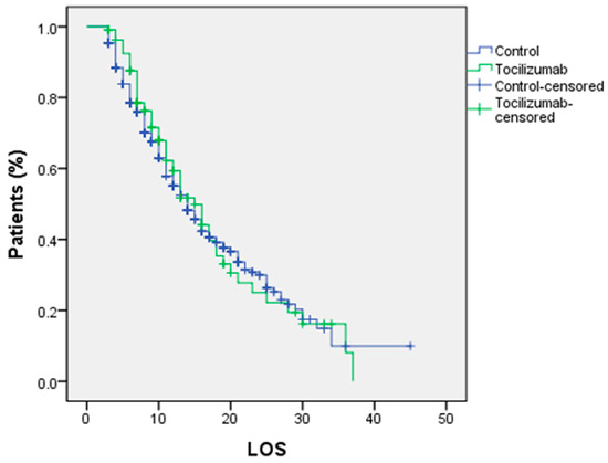
11].OS and DFS rates at 1, 3, 5 and 10 years were better in iHCC patients than pdHCC patients in our series (
Figure 1). It may be due to better histological features and low recurrence rate in our series. Most of the iHCC patients of our study group were within the Milan criteria, so we compared them with pdHCC patients who met the Milan criteria. DFS, OS and also recurrence rates were not statistically different (
Figure 2). Thus, we believe that iHCC tumor characteristics may be similar with pdHCC patients who met the Milan criteria. Finally, we also compared the survival rates of iHCC patients who met the Milan criteria and iHCC patients out of the Milan criteria. There were only six patients who did not meet the Milan criteria in the iHCC group and interestingly, they were still alive and had no recurrence. Statistical evaluation could not reach any significant result because of the small sample size (
Figure 3). In addition, the Milan criteria patients in the iHCC group with multiple tumors had a poor prognostic factor, but these patients had at least 3 years OS and DFS 100% of the time. Additionally, some previous reports about liver transplantation criteria for HCC ignore the number of tumors [
17,
18,
19,
20,
21].
The limitations of this study include its retrospective design and the relatively small number of patients. Meanwhile, the strengths of this study include the use of the largest volume of the European liver transplant center’s databank, which has each transplant patient consecutively recorded in a prospective manner, and being the first study that compares the survivals of iHCC versus pdHCC within the Milan criteria.


留言 (0)