
記住我
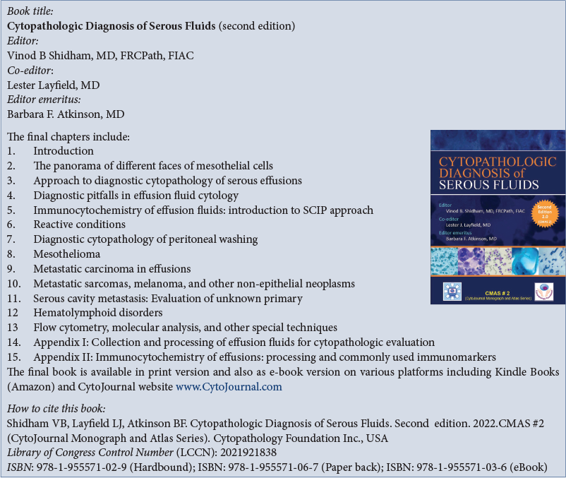
This second edition of Cytopathologic Diagnosis of Serous Fluids as CMAS #2 provides an essential tool to aid in the diagnosis of serous effusions,[1] which can be challenging and fraught with pitfalls. It is lavishly illustrated, with the first 12 chapters devoted to different topics, ranging from benign reactive effusions, common metastatic carcinomas, and mesothelioma to the more unusual and rare non-epithelial neoplasms and hematolymphoid disorders.
There is a chapter describing the cytology of peritoneal fluids and washings which is essential reading, providing an excellent overview of this topic, including the pitfalls associated with the diagnosis of Mullerian inclusions or endosalpingiosis compared to low-grade serous carcinomas of ovarian origin and an update on BRCA-related peritoneal washing before prophylactic oophorectomy.
Each chapter demonstrates classic examples of different neoplasms with their characteristic architectural and cellular features, easily relatable to the diagnostic cytologist. Examples of different entities are clearly displayed using both Giemsa and Papanicolaou stains. In addition, cell block tissue stained with H&E is used frequently, adding a further diagnostic dimension to each entity, as well as providing essential tissue on which to perform batteries of immunocytochemical stains,[2-4] which are critical to perform to refine a malignant diagnosis, so that precise chemotherapy or targeted biological therapy may be instituted.
Throughout the book, there are tables containing an algorithmic approach to the diagnosis of different entities. For example, in chapter 10, an initial table is provided summarizing a diagnostic approach to non-epithelial tumors causing a malignant effusion that includes mesothelioma, melanoma, sarcomas, and other neoplasms. This table is further subdivided into the differential diagnosis of small round blue cell tumors (SRBCTs) and other sarcomas and is supplemented by another comprehensive table characterizing the immunoprofile and important molecular characteristics of SRBCTs.
The chapter covering hematolymphoid disorders is a comprehensive review of both benign and malignant lymphoid proliferations involving the serous cavities, but also includes rare but important conditions such as post-transplant lymphoproliferative disorder, primary cardiac lymphoma, plasma cell myeloma, and primary effusion lymphoma. It also covers the differential diagnosis of nonneoplastic causes of effusions such as tuberculosis, which while not common in the USA, is a frequent cause of pleural effusion in other geographic locations. Useful tables are provided describing the salient features of predominantly mature B cell neoplasms in fluid, comprising cytomorphology, and immunophenotype by both immunocytochemistry (ICC) and flow cytometry with useful comments detailing differences from what one would expect to see in a tissue section or direct FNA smear, from a lymphomatous mass versus what is possible to see in an effusion. For example, in SLL/CLL, the classic prolymphocytes and paraimmunoblasts seen in tissue are usually not present in fluids.
The chapter on mesothelioma is beautifully illustrated showing classic mesothelioma and a few examples of carcinomas that may mimic mesothelioma. However, throughout the book and in different chapters, there are examples of reactive mesothelial proliferations and adenocarcinomas that could also be confused with the cytologic appearance of certain subtypes of mesothelioma. A useful algorithm is provided that includes updated molecular pathology information to be used in making a definitive diagnosis of mesothelioma, including the use of BAP1 and MTAP by ICC, as well as deletion of CDKNA by FISH.
The last three chapters are more technical and provide guidelines for the use of flow cytometry to diagnose lymphoproliferative infiltrates in effusions, including tables describing major molecular genetic abnormalities found in lymphoma, acute leukemia, and soft-tissue tumors. Although there is a paucity in the literature regarding the use of FCM for lymphocyte-rich effusions as opposed to fine needle aspirates of lymphoid lesions, very similar principles apply. The use of FISH for genetic abnormalities in fluids, however, is questionable, considering that neoplastic cells may be sparse, and the specimen may not be representative.
Useful protocols for the different commonly used stains such as Romanowsky and Diff-Quik are presented in tabular form. In addition, a comprehensive catalog of cell block techniques is provided including use of Histogel, gelatin, agar, plasma-thrombin, collodion bag embedding, and Nano NextGen CelBloking ™ kits.
With the advent of the application of the International System for reporting serous fluid cytopathology, together with the assessment of the risk of malignancy, the spotlight has fallen on the ability of the international community of practicing cytologists and cytopathologists to accurately diagnose malignancy in serous fluids. Thus, the penultimate chapter, covering optimal specimen handling of direct smears, liquid-based cytology preparations, cytospins, and filter preparations, is welcomed. This chapter also outlines a 5-tier reporting system recently described for serous fluids. It is noteworthy that for fluids diagnosed as non-diagnostic, negative for malignancy, or atypia of uncertain significance, there is a significant risk of missing a malignancy, thus emphasizing the importance of meticulous specimen preparation and clinical correlation.
The final chapter provides in depth information regarding methods for ICC, and commonly used and less commonly used immunomarkers for diagnosing different malignancies, which are richly detailed regarding their derivation, which clones to use and methods for antigen retrieval.
The different chapters of this monograph have been authored by experienced and expert pathologists who provide detailed cytologic descriptions, as well as presenting updated panels of ICC stains, supplemented as necessary by FISH and molecular studies. The chapters are well organized and contain a comprehensive list of references including updated references. This monograph is both practical and comprehensive and is strongly recommended for cytology professionals from beginning students to the most experienced practitioners.
Overall, this second edition while quite similar to the first edition has been thoroughly updated with addition of extra images and new immunohistochemistry information and molecular pathology especially in relation to hematolymphoid conditions and mesothelioma.
留言 (0)