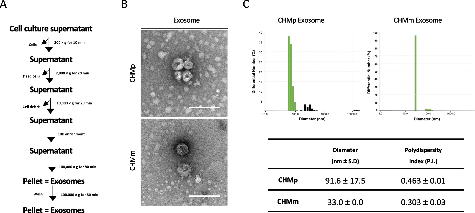
JWDSD preparation
The herbs used in the JWDSD were purchased from the First Affiliated Hospital of Hunan University of Chinese Medicine (Changsha, China) and were composed of Dan Shen (batch number, 20201125), Tan Xiang (batch number, 20201031), Chuan Xiong (batch number, 20201129), Dang Gui (batch number, 20201121), Hong Hua (batch number, 20201110), Chi Shao (batch number, 20201019), and Sheng Di Huang (batch number, 20201127). The botanical raw materials were crushed into pieces and mixed in the ratio of 10:3:3:3:3:5:4(w/w) dissolved in 10×8× total drug weight water, and heated to reflux for 1 h and combined with the drug solution, concentrated to 2 g/ml and stored at 4°C for later use. Furthermore, our laboratory has established JWDSD's strict quality control method [16, 17].
Research animals and experimental design
Male Sprague-Dawley rats weighing 160~180 g were obtained from the Hunan University of Chinese Medicine Animal Experiment Center (Changsha, China License number: SCXK (Hunan) 2019-0004). All rats were fed at 22±3 °C, 50%±5% relative humidity, and 12 h light/dark cycle. Food and drinking were available ad libitum for one week. The animal experiments were performed with the approval of the Animal Ethics Committee of the Hunan University of Chinese Medicine. They were carried out concerning the Chinese Guide for the Care and Use of Laboratory Animals.
All animals were handled with humane care throughout the experiment. The rats were randomly divided into seven groups (n=10): the control group, the model group, the sham-operated group (Sham: normal saline, 1 mL/100g), Diltiazem Hydrochloride Tablets group (Diltiazem-H: 4.32mg/kg), and three groups which fed JWDSD in a low, moderate, or high dosage (JWDSD-D, JWDSD-Z, JWDSD-H: 12.10g, 6.05g and 3.03g/kg, botanical raw materials extracted by distilled water). Each group received continuous gavage twice a day for seven days before modeling. The rats were anesthetized with chloral hydrate (300 mg/kg, intraperitoneal injection) 1 h after the last gavage. Their limbs and heads were immobilized, and a ventilator was inserted (initial frequency 60 times/min, 90 times/min after thoracotomy). To expose the heart, the thoracic cavity is opened, a 6-0 suture needle with a thread about 2 mm is inserted below the left atrial appendage, and the left anterior descending (LAD) coronary artery is ligated [18]. The rats were subjected to LAD coronary artery ligation for 30 min and then to reperfusion for 30 min after the same surgical operation. The sham operation group was threaded but not ligated. At the end of the reperfusion period, blood samples were drawn from the abdominal aorta to separate serum for further analysis by centrifugation at 3000 g for 15 min, and cardiac tissues were collected from each group and stored in a -80 °C refrigerator for later use.
Creatine kinase and lactate dehydrogenase assays
The blood of rats in each group was centrifuged at 3000 R / min for 20 min, and the upper serum was taken. The Chemray 240 automatic biochemical analyzer was used to measure creatine kinase (CK) and lactate dehydrogenase (LDH) levels in serum.
Heart histological examination
The fresh cardiac tissue was fixed with 4% paraformaldehyde for more than 24 h, dehydrated with different ethanol concentrations (75%, 85%, 90%, 95%, and 100%), and then immersed in paraffin for embedding. After the wax solidifies, they were cut into slices with a thickness of 4 μm. After being immersed in xylene and ethanol, the paraffin sections were stained with H&E (hematoxylin-eosin, servicebio, China) to observe any histopathological changes.
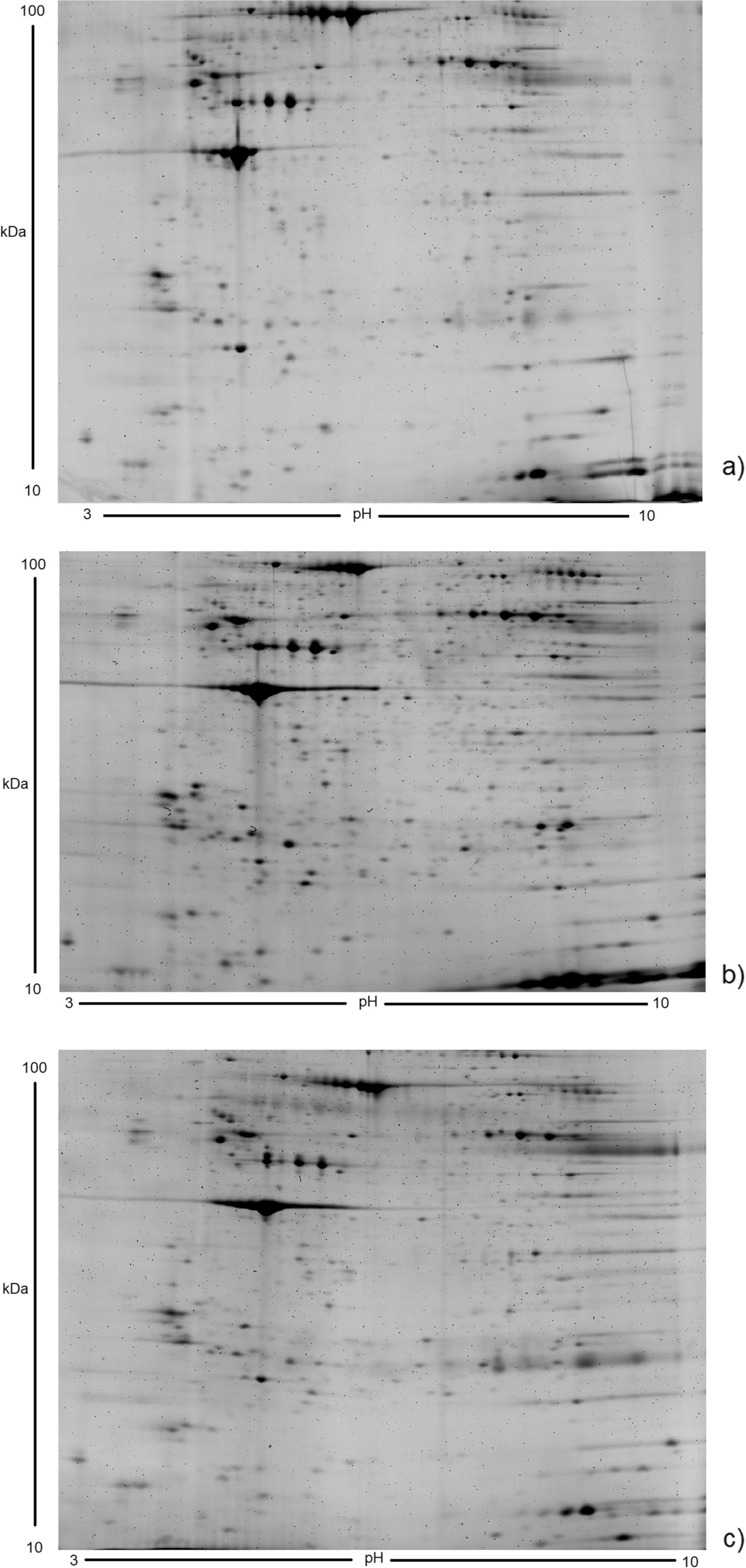
TMT -labeled quantitative proteomicsProtein extraction
The cardiac tissue samples from the Control group, Model group, and JWDSD-H group were ground into a powder with liquid nitrogen and were lysed with SDT lysis buffer (4% (w/v) SDS, 100 mM Tris/HCl pH 7.6, 0.1 M DTT). The lysate was homogenized by MP homogenizer (24×2, 6.0 M/S, 60 s, twice), and the mixture was homogenized on ice for 20 min, then sonicated three times. Later the mixture was centrifuged at 14000 g for 15 min to get the supernatant. All samples were quantified using the BCA method, and aliquots were stored at -80 °C for further use.
Protein digestion, TMT labeling and RP classification
Proteins from each sample were processed by Filter aided proteome preparation (FASP) method [19] was used for trypsin digestion, the filtrate was collected, and the peptide was quantified (OD280). Tandem mass tags TMT10 (Thermo Fisher Scientific, USA) with varying molecular weights (126–131 Da) were used as isobaric tags for relative and absolute quantification. According to manufacturer's protocols, the digested samples were individually labeled with TMT10 reagents for 1 h as follows: 100 μg of aliquots of digested peptides of the Control group, Model group, or JWDSD-H group were each labeled with a different isobaric tag (TMT126, 127, 128, 129, 130 and 131, respectively). The labeling reaction was stopped with 5% hydroxylamine. As directed, the Pierce high pH reversed-phase fractionation kit (Thermo scientific) was then used to fractionate TMT-labeled digest samples into 10 fractions using an increasing acetonitrile step-gradient elution.
LC-MS/MS analysis
Each fraction was injected for nano LC-MS/MS analysis. The peptide mixture was loaded onto a reverse-phase trap column (Thermo Scientific Acclaim PepMap100, 100μm*2cm, nanoViper C18) connected to the C18 reversed-phase analytical column (Thermo Scientific Easy Column, 10 cm long, 75 μm inner diameter, 3 μm resin) in buffer A (0.1% Formic acid) and separated with a linear gradient of buffer B (84% acetonitrile and 0.1% Formic acid) at a flow rate of 300 nl/min controlled by IntelliFlow technology. Subsequently, peptides were eluted over 90 min using the following gradient: 0-55% buffer B for 80 min, 55-100% buffer B for 5 min, and held in 100% buffer B for 5 min.
The peptides were separated using an HPLC system, then injected into a capillary ion source for ionization and analyzed using Q-Exactive (Thermo Scientific) mass spectrometry. In positive ion mode, the mass spectrometer was used. MS data for HCD fragmentation were collected using a data-dependent top 10 method that dynamically selected the most abundant precursor ions from the survey scan (300-1800 m/z). The automatic gain control (AGC) target was set to 3e6, and the maximum injection time to 10 ms. The dynamic exclusion duration was 40.0 s.
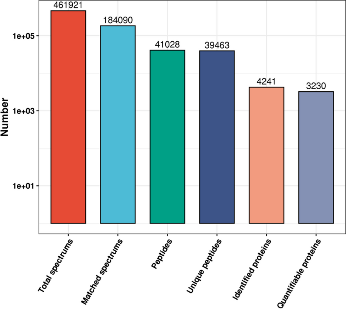
Database search
MS/MS spectra were searched using MASCOT engine (Matrix Science, London, UK; version 2.2) embedded into Proteome Discoverer 1.4. The parameters were set as follows: specific enzyme was trypsin; Peptide Mass Tolerance was ±20 ppm; Fragment Mass Tolerance was 0.1Da; Peptide FDR≤0.01; the protein ratios are calculated as the median of unique peptides of proteins. The other parameters were default. DEPs were satisfied following conditions: unique peptides ≥ 2 with average ratio-fold change > 1.2 (up-regulation) and < 0.83 (down-regulation), as well as p-value < 0.05.
Bioinformatics analysis
The protein sequences of DEPs were in batches retrieved from the UniProtKB database (Release 2016_10) in FASTA format. The term gene ontology (GO) (http: //geneontology. org/) was used to describe cellular components (CC), elucidate biological process (BP), and molecular function (MF). KAAS (http: //www.genome. jp/kaas-bin/kaas_main) was applied to annotate the description of DEPs in KEGG database. KEGG pathway enrichment analysis was performed using Fisher’s exact test.
Western blot analysis
Protein concentrations were determined using a BCA Protein Concentration Determination Kit after extracting proteins from approximately 100 mg of cardiac tissue (Beyotime). A 40 g protein sample was extracted, separated by SDS-PAGE gel, transferred to PVDF membrane, and blocked with 3% BSA-TBST for ten minutes at 28 °C. The membranes were then incubated overnight at 4 °C with corresponding primary antibodies. Santa Cruz Biotechnology provided TIAM1, GAPDH genes (Dallas, TX, USA. After washing three times (5 min each time) using TBST, the membranes were incubated with secondary antibody for 60 min at room temperature. ECL solution was added to regulate the exposure conditions, and the optical density value of the target zone was analyzed using ImageJ software processing system.
Statistical analysis
IBM SPSS Statistics V21.0 was used to analyze data, expressed as mean±standard deviation (SD). Independent t-tests or one-way analyses of variance were used to make comparisons. At P < 0.05, differences were considered statistically significant.


留言 (0)