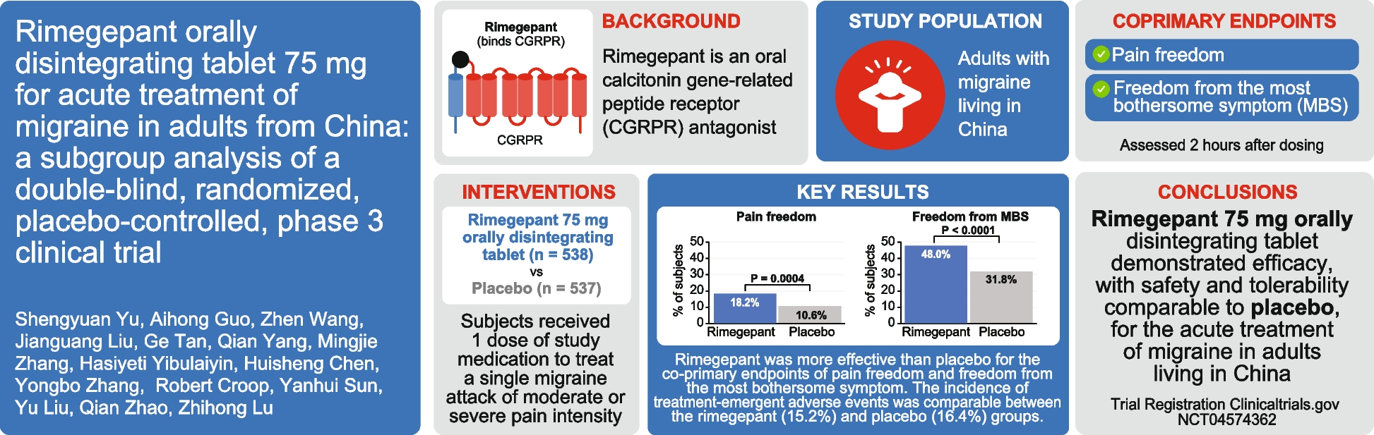
The main finding of this study is that effective migraine treatment with erenumab is associated with changes in brain activations in response to extracranial thermal pain and with changes in resting-state functional connectivity. fMRI differences between erenumab-responders vs non-responders were seen within 8 weeks of initiating erenumab treatment, with some differences starting to emerge at 2 weeks. Brain regions impacted across the different analyses in this study included the hypothalamus, inferior lateral parietal, temporal pole, supramarginal gyrus, amygdala, and periaqueductal gray, amongst others. As discussed further below, these regions are known to have important roles in migraine pain processing including their likely roles in migraine attack generation, pain perception, pain modulation, and multisensory integration.
Pain-induced activations
Our pain-induced activation analyses identified differences between erenumab responders vs. non-responders in activations of regions in the cingulate cortex, putamen, frontal lobe, and periaqueductal gray matter. Brain activations occurring immediately (i.e., within 12.5 seconds) after painful stimuli and delayed activations occurring between 12.5 s to 25 seconds after painful stimuli were investigated. Prior studies have demonstrated a biphasic hemodynamic brain response to painful heat stimuli, consisting of a stimulus-locked response and a second peak delayed by about 12.5 seconds [25]. It has been suggested that the initial BOLD response might be related to pain processing via myelinated A-delta fibers, while the delayed BOLD response could be associated with processing via the slower C fibers [25, 41]. It has also been theorized that the initial BOLD response might represent a fast, non-conscious processing of pain that helps to quickly determine the threat level, while the delayed response might represent a more conscious processing of the painful stimulus [25, 41, 42]. Our study identified differences in immediate and delayed BOLD responses to pain when comparing erenumab-responders vs. non-responders.
For immediate BOLD responses, there were differences in the middle and posterior cingulate at 8 weeks following the first erenumab treatment and in the putamen only at 2 weeks following the first erenumab treatment. Changes in BOLD responses in the middle cingulate and posterior cingulate were already seen at 2 weeks after the first erenumab treatment, but the differences between erenumab-responders and non-responders were not significant until the eight-week MRI. The middle cingulate participates in rapid behavioral adaptive responses to the threat associated with pain, as well as cognitive and affective components of pain processing [43, 44]. A prior fMRI study of migraine demonstrated a strong positive correlation between pain-induced activation in the middle cingulate and headache frequency (r = 0.627, p = .001), providing relatively strong evidence for its role in migraine [24]. In our study, it is possible that the increase in pain-induced activation of the middle cingulate amongst erenumab-responders is due to the pain stimulus as being perceived as more novel and thus as a greater threat compared to the erenumab non-responders who are experiencing more frequent pain, with the painful stimulus thus losing its novelty. The posterior cingulate, a key region of the default mode network, participates in self-referential processing of stimuli, including externally generated pain [45]. Numerous pain and migraine studies have demonstrated atypical functional connectivity of default mode network regions [46,47,48,49]. A prior migraine study demonstrated that migraine improvement (i.e. reduced time with headache) was associated with cortical thickness changes in the left posterior cingulate [50]. The putamen participates in sensory-discriminative aspects of pain processing including the determination of pain sensitivity [51]. Prior migraine studies have demonstrated atypical functional connectivity and structure of the putamen [52, 53]. In our study, differences in putamen activation were detected at two-weeks following the first erenumab treatment, but not at 8 weeks, a pattern that is difficult to interpret.
Delayed BOLD response differences between erenumab-responders and non-responders were identified in the periaqueductal gray at 8 weeks following initiation of erenumab treatment and in the frontal supplementary area prior to starting erenumab (and thus not related to treatment). The periaqueductal gray is a key region of the pain modulating pathway, predominantly involved in pain inhibition. Numerous studies have identified atypical periaqueductal gray structure and function associated with migraine, the extent to which might correlate with migraine disease severity [54,55,56,57,58]. Less effective pain modulation by the periaqueductal gray could be a mechanism by which those with migraine experience more severe pain, allodynia, and increased frequency of migraine attacks [54,55,56,57]. Increased pain-induced activation of the periaqueductal gray amongst erenumab-responders might reflect a stronger pain-inhibitory response.
Overall, our pain activation studies suggest that effective erenumab treatment is associated with changes in cognitive, affective, sensory-discriminative, and modulating aspects of pain processing.
Resting-state functional connectivity
Our resting-state functional connectivity analyses included investigation of graph theory network parameter differences and ROI-to-ROI static functional connectivity differences between erenumab-responders vs. non-responders. Graph theory provides a method for quantitatively describing the topological organization of brain networks [38]. Graph theory measures in our analyses included global and regional efficiency, betweenness, clustering coefficient, node degree, and modularity [39, 40]. Efficiency is the inverse of the minimum path length between an ROI and all other ROIs in a network [38]. Global and local efficiency reflect a network’s ability to transmit information at the global and local levels [40]. Betweenness of a ROI is the number of shortest paths between any two ROIs that run through the ROI [38]. Clustering coefficient describes the local connectedness of a network and is calculated by determining the number of connections that a ROI has with its immediate neighbors divided by all of its possible connections [38]. Node degree is a measure of the number of connections to an ROI [40]. Modularity measures how connected ROIs are to members of their own group, identifying subnetworks within a larger network.
Erenumab responders had an increase in global efficiency from pre-treatment to eight-weeks post-treatment and greater global efficiency at eight-weeks compared to erenumab non-responders. An increase in global efficiency suggests that erenumab-response was associated with an improvement in the ability of the studied regions, those that participate in various aspects of pain processing and modulation, to functionally communicate on a global scale. At eight-weeks post-treatment, compared to erenumab non-responders, responders had ROIs with higher efficiency, cluster coefficient, node degree, and modularity, findings suggesting a greater ‘small world’ quality of the network. Regions most strongly highlighted by these differences included those in the hypothalamus, amygdala, and inferior parietal lobe. The hypothalamus likely plays an important role in the generation of migraine attacks [59, 60]. The amygdala contributes to affective and attentional responses to pain and pain modulation [61]. The inferior parietal region, like the posterior cingulate discussed above, is a key region of the default mode network [62]. Other regions included the temporal pole, supramarginal gyrus, caudate, cuneus, trigeminal nucleus, lingual gyrus, and middle cingulate.
ROI-to-ROI differences in functional connectivity at 8 weeks between erenumab-responders vs. non-responders included regions that were also identified as having differences in graph theory network measures, including the supramarginal gyrus, inferior lateral parietal, hypothalamus, and temporal pole. Additionally, several other regions were involved in these functional connections including regions in the middle temporal, middle occipital, and middle frontal lobes, dorsolateral and ventromedial prefrontal cortices, and the pulvinar.
Study results in context of prior studies
Prior studies have interrogated the impact of migraine treatment on brain functional connectivity and brain activations. Krebs and colleagues demonstrated that treatment with sphenopalatine ganglion blocks was associated with changes in functional connectivity amongst salience network and executive network regions [8]. Russo and colleagues investigated pain-induced brain activations before and after 60 days of treatment with external trigeminal neurostimulation [9]. Neurostimulation treatment was associated with a reduced BOLD response in the anterior cingulate cortex. Acupuncture treatment has been associated with an increase in periaqueductal gray functional connectivity with anterior cingulate cortex among individuals who have migraine without aura [63]. A pre-treatment and 2–3 week post-treatment study of galcanezumab, a CGRP ligand mAB, for migraine demonstrated that galcanezumab decreased hypothalamic activation in response to nociceptive trigeminal stimulation to a greater extent in galcanezumab-responders vs. non-responders [12]. There were also responder-specific decreases in BOLD activation in the inferior parietal lobule, insula and parahippocampal gyrus. Spinal trigeminal nucleus functional connectivity changes from the pre-treatment to post-treatment scans were interrogated for all treated patients and demonstrated weakened connectivity with hypothalamus and superior temporal gyrus and stronger connectivity with the cerebellum, middle temporal gyrus, and insula at the post-treatment timepoint. The study most closely related to ours investigated the impact of erenumab on brain activations in response to nociceptive trigeminal stimulation [10]. In that study, 27 individuals with migraine underwent fMRI prior to and 2 weeks after treatment with 70 mg of erenumab. During the fMRI paradigm, intranasal ammonia was used as a painful stimulus. 63% of participants were considered erenumab treatment responders, which was defined as at least a 30% reduction in headache days during the first month following treatment. Amongst all patients there were post-treatment decreases in pain-induced activations in the thalamus, lingual gyrus, middle temporal gyrus, operculum, and cerebellum. Compared to non-responders, erenumab-responders had a significant reduction of activation in the hypothalamus, insula, and cerebellum. Analysis of hypothalamic functional connectivity amongst all treated patients demonstrated a reduction in connectivity strength with the temporal lobe, hippocampus, parahippocampus, fusiform gyrus, cerebellum, red nucleus, and spinal trigeminal nucleus, and an increase in connectivity strength with the anterior insula. This study and ours complement one another, both demonstrating changes in pain-induced activation and resting functional connectivity associated with erenumab treatment and response. Our study adds to the literature since it determined treatment response during weeks 5–8 after starting treatment, studied 140 mg of erenumab rather than 70 mg, investigated early (i.e. 2 weeks) and later (i.e. 8 weeks) fMRI changes after initiating treatment, utilized two treatments with erenumab rather than one, used an extra-trigeminal painful heat stimulus for the event-related paradigm, and interrogated graph theory network measures of functional connectivity.
Relationship between fMRI changes and Erenumab treatment
How treatment with erenumab is associated with changes in pain-induced brain activations and functional connectivity is a matter of debate. It is perhaps unlikely that the small amount of erenumab that might cross the blood-brain barrier could exert a meaningful central effect and have a direct impact on brain processing of painful stimuli. Alternatively, erenumab might alter brain pain processing indirectly, via its impact on peripheral structures such as the trigeminal ganglia, trigeminal nerves, or dura mater [16,17,18]. Finally, it is possible that the changes in pain processing demonstrated in this study are attributable to the reduction in migraine days associated with erenumab response, but not specifically attributable to the mechanisms by which erenumab exerts therapeutic effects.
Study considerations and limitations
Considerations and limitations of our study include: 1) Although the number of MRIs completed and included in this study is relatively large (n = 86), the number of erenumab-responders (n = 18) and non-responders (n = 14) is relatively small. Larger sample sizes might allow for more stringent statistical corrections for multiplicity. 2) Our study is not able to determine if the pre- to post-erenumab changes in pain-induced brain activations and functional connectivity are directly attributable to erenumab or if there would be similar findings associated with reductions in migraine frequency regardless of the specific reason for such a reduction. Optimally, future studies would include migraine treatments that work via different mechanisms and individuals who have longitudinal reductions in migraine frequency in the absence of treatment. 3) Inclusion of a healthy control group would allow for better interpretation of the pre-to-post treatment changes, whether the changes are consistent with a “normalization” of brain function or adaptive changes, for example. 4) Neuroimaging research studies use different statistical corrections and cluster forming thresholds for determining the significance of results. More stringent methods increase the likelihood for type II error, while less stringent methods increase the likelihood for type I error. The approaches taken in this study should be considered when interpreting the results. Like with all neuroimaging studies, replication of results would further strengthen the assessment of their validity. 5) The sample size of research participants included in this study prevented us from performing additional subgroup analyses, such as differences that might be present based on headache frequency (e.g. episodic migraine vs. chronic migraine), participant sex, use of concurrent migraine preventive medications, and frequency of using migraine as-needed medications. 6) Prior to starting erenumab, a larger proportion of participants who became erenumab responders were concurrently taking other migraine preventive medications. The use of concurrent migraine preventive medications could have an impact on pre-treatment fMRI comparisons between responder and non-responder groups but is unlikely to have impacted differences that were first seen at 2 weeks or 8 weeks following initiation of erenumab. 7) We did not limit the use of medications within 48 hours of the MRI and QST. Thus, some participants had used abortive medications within that time-period. No participants were using opiates, which might directly impact pain sensation during QST and thermal stimulation. 8) QST and fMRI results could be impacted by the participants headache and migraine state, meaning that findings might differ according to whether headache or migraine is present during the test. However, in our study and as presented in the Results, the frequency of testing during headache or migraine was similar between erenumab-responders vs. non-responders and there were few post-treatment QSTs and MRIs collected during headache and none during migraine. Thus, there was not ample justification or sample sizes for analyzing QST and MRI data according to headache and migraine status. 9) Future fMRI studies could use other timepoints for determining treatment response, such as the 9–12-week period after starting erenumab. It is possible that longer durations of response to erenumab could be associated with more substantial changes in fMRI measurements.

留言 (0)