
記住我

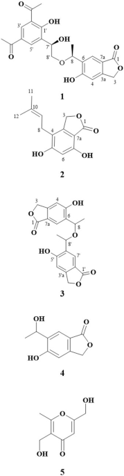
Our previous study demonstrated that the cultivation of a fungus of the genus Talaromyces in potato dextrose broth (PDB) yielded 1, vanitaracin B (2), 3,5-dihydroxy-2-(2-(2-hydroxy-6-methylphenyl)-2-oxoethyl)-4-methylbenzaldehyde, 7-hydroxy-5-methyl-2-(2-oxobutyl)-4H-chromen-4-one, and 2,7-dihydroxy-5-methyl-2-(2-oxobutyl)chroman-4-one [7]. To isolate further novel compounds related to 1, we cultured the fungus under various conditions. First, procaine, a DNA methyltransferase inhibitor, which reportedly enhances secondary metabolite production in fungi [20], was added to the PDB culture medium. The crude extract from this fungal culture broth was subjected to TLC-guided fractionation using silica gel chromatography and HPLC. Under these culture conditions, 1 was obtained together with unknown compound 3. The molecular formula of compound 3 (C24H34O6) was determined using HRMS (FAB). 1H NMR spectroscopy suggested that the structure of compound 3 was similar to that of 2, except for the presence of a methine proton signal (δ 5.36) and the absence of a ketone signal at C-10 (Fig. 1 and Table 1). Based on the correlation between H2-9 and H-10 in the 1H–1H COSY spectrum of 3, the methine proton signal was assigned to H-10, which was supported by HMBC correlations of H3-16 with C-10, C-11, and C-12 (Fig. 2). The consecutive 1H–1H COSY correlations of H-2′–H-8′, and the HMBC correlations of H-3′ and H-7′ with C-1′ (δ 176.5) revealed the presence of a 2,4-dimethylhexanoate unit, which is also present in 1 and 2. The HMBC correlation of H-10 with the ester carbon (δ 176.5) for 3 suggested that the 2,4-dimethyl hexanoate side chain was connected to C-10, whereas this side chain was connected to C-11 in the structures of 1 and 2. The differences in the molecular formulas of 2 and 3 indicated the presence of a hydroxy group at the C-11 position in 3. The remaining proton and carbon signals were assigned using 1H–1H COSY, HMQC, and HMBC experiments (Table 1). Thus, the structure of 3 was determined, as shown in Fig. 1, and this compound was named vanitaracin C. A NOESY correlation between H-2 and H-4 was observed, which indicated a syn relationship between OH-2 and H3-15 (Fig. 2). The NOESY correlation between H3-16 and H-9 (δ 3.34) suggested that these two protons were on the same face of the ring. Cross peaks were observed between H-9 (δ 3.34) and H-10 but not between H-9 (δ 3.09) and H-10, indicating that H-10 was also on the same face. Thus, the relative configuration of the tricyclic moiety was determined, as shown in Fig. 2.
Table 1 1H NMR (400 MHz, CDCl3) and 13C NMR (100 MHz, CDCl3) data for compound 3Fig. 2
Key 1H–1H COSY, HMBC, and NOESY correlations for 3
The relative configurations of the 2,4-dimethyl hexanoate moiety in 2 and 3 were determined using Schmidt’s method, which we previously applied to determine the configuration of the moiety in 1 (refs. 16, 21). The differences in the chemical shifts (Δδ) of the C-3′ geminal protons of compounds 2 and 3 were calculated to be 0.64 and 0.60 ppm, respectively (Figure S17). These Δδ values were all greater than 0.4 ppm, suggesting a syn relationship between C-2′ and C-4′ in 2 and 3, similar to that in 1 (refs. 16).
Because the conformational characteristics between the tricyclic skeleton and the side chain of 3 is still unclear, we employed computational methods to predict the absolute configuration of the stereogenic centers in 3. Based on the data described above, we set four possible diastereomers, (10S,11S,2′S,4′S)-3a, (10S,11S,2′R,4′R)-3b, (10R,11R,2′S,4′S)-3c, and (10R,11R,2′R,4′R)-3d, which were analyzed using the DP4 analysis program [22, 23] (Figure S18). Initially, all the diastereomers were submitted to a conformational search using the MMFF94s conformer research algorithm. Thereafter, all the conformers with relative differences within 5 kcal/mol were optimized via DFT calculations at the B3LYP/6-31G(d,p) level based on the solvent effect for PCM (CHCl3) in the Gaussian 16 package [24]. The optimized conformers were identified using the GIAO method at the mPW1PW91/6-31G(d,p) level, and the NMR spectra of the resultant conformers within 5 kcal/mol were averaged based on the Boltzmann populations to give the estimated chemical shifts. The 1H and 13C chemical shifts of all diastereomers with the statistical DP4+ value resulted in agreement with (10S,11S,2′S,4′S)-3a, which suggested that compound 3 has the structure of 3a or its enantiomer. Furthermore, we calculated CD spectra of each diastereomer using TD-DFT at the cam-B3LYP/6-311++G(2d,p) level. The calculated ECD spectrum of 3a matched the experimental data well as shown in Figure S19. Thus, the absolute and relative stereo configurations were determined as (2S,4R,10S,11S,2′S,4′S)-3a.
Next, we cultured the fungus in a malt extract broth. Purification of the crude extract via silica gel chromatography yielded 1 as well as unknown compounds 4 and 5. The molecular formula of compound 4 (C25H32O7) was determined by HRMS (FAB). The 1H NMR spectrum implied that the scaffold of 4 differed from that of vanitaracin. The 13C NMR and HMQC spectra revealed the presence of 25 carbons, including 3 ketone carbons, 1 ester carbon, 4 quaternary carbons, 8 methine carbons, 4 methylene carbons, and 5 methyl carbons (Table 2). The 1H–1H COSY and HMBC correlations revealed the presence of a 2,4-dimethylhexanoate unit, which is present in all the vanitaracins (Fig. 3). The 1H and 13C NMR data, the HMBC correlations of the methyl protons (H-17) with ketone carbons C-12 (δ 192.9) and C-14 (δ 193.0) as well as oxygenated quaternary carbon C-13 (δ 83.9), and the HMBC correlations of olefinic protons H-9, H-11, and H-16 with C-8, C-10, C-14, and C-15 indicated the presence of an azaphilone skeleton. The presence of a 3-hydroxy-5-methylcyclohexanone moiety was established based on the consecutive 1H–1H COSY correlations for H-2–H-7 and the HMBC correlation of H-2 with C-1 (δ 202.3). The HMBC correlation between H-9 and C-6 indicated that the cyclohexanone moiety was connected to the azaphilone skeleton. Thus, the planer structure of 4 was determined, as shown in Fig. 3, and this compound was named vanitaraphilone A. The relative configuration of the 3-hydroxy-5-methylcyclohexanone moiety in 4 was determined based on NOESY correlations (Fig. 3). The NOESY correlation between H-3 and H-5 suggested a syn relationship between OH-3 and H3-7. The relative configurations of C-5 and C-6 were defined based on the typical trans-diaxial coupling constant (JH-5/H-6 = 12.2 Hz), as supported by the NOESY correlation between H-2α (δ 2.44) and H-6. Compound 4 is structurally related to azaphilones, cohaerin B isolated from the stromata of the xylariaceous ascomycete Hypoxylon cohaerens [25], and penicilone A isolated from the marine-derived fungus Penicillium janthinellum HK1‑6 (ref. 26). The difference between the chemical shifts (Δδ) of the C-3′ geminal protons of 4 was 0.64 ppm (Figure S17), suggesting a syn relationship between C-2′ and C-4′ (ref. 21). Given that the stereochemistry of the C-13 positions of several azaphilones, including cohaerins and penicilones, have been established via ECD spectroscopy [25, 26], we measured ECD spectrum of 4. Compound 4 showed a negative Cotton effect at 354 nm, which clearly indicated (R)-configuration for C-13 (Figure S20). However, due to shortage of materials, the absolute configurations of other stereogenic centers in 4 were not determined.
Table 2 1H NMR (400 MHz, CDCl3) and 13C NMR (100 MHz, CDCl3) data for compound 4Fig. 3
Key 1H–1H COSY, HMBC, and NOESY correlations for 4
The molecular formula of compound 5 (C9H10O3) was determined using HRMS (FAB). The 1H NMR and 13C NMR data suggested the presence of one aldehyde carbon, six aromatic carbons, one oxymethylene carbon, and one methyl carbon. All the proton and carbon signals were assigned based on the HMBC correlations of H-7 with C-1 and C-2, H2-8 with C-3, C-4, and C-5, and H3-9 with C-1, C-5, and C-6 (Figure S21). Thus, compound 5 was determined to be 2-hydroxy-4-(hydroxymethyl)-6-methylbenzaldehyde (Fig. 1).
None of the newly identified compounds (3–5) exhibited anti-HBV activity (Figures S22 and S23). Our previous study demonstrated that 2 has much lower anti-HBV activity than 1 (ref. 7). These observations suggested that the overall structure of 1, especially the methyl and hydroxy groups at the C-9 position, is essential for anti-HBV activity. Additionally, we examined the antiviral spectrum of 1 by evaluating its antiviral activities against zoonotic viruses such as rabies virus, Borna disease virus 1, and BLV. Compound 1 had no significant effect on rabies virus or Borna disease virus 1 (Figure S24), but displayed dose-dependent anti-BLV activity without cytotoxicity (Fig. 4a, b). Compound 1 inhibited syncytia formation, a typical cellular morphology caused by BLV infection. We also evaluated the anti-BLV activities of all the other metabolites obtained from a 1-producing fungus. At a concentration of 10 µM, 2 and 5 exhibited significant anti-BLV activities. However, vanitaracin C (3) and vanitaraphilone A (4) did not show any such behavior (Fig. 5a and b). Compound 4 only exhibited significant anti-BLV activity at a concentration of 30 µM (Fig. 5c). Our results also indicated that at concentrations in the range 30–100 µM, 3 significantly decreased the viability of CC81 cells. Further, 4 showed cytotoxicity at high concentration (50 and 100 µM) (Figure S25).
Fig. 4
Cytotoxicity and anti-BLV activity of 1. a Cell viability of CC81 cells treated with various concentrations of 1. b Anti-BLV activity measured via syncytium assays. CC81 cells were cultivated with FLK-BLV supernatant supplemented with various concentrations of 1. The counts of the infectious virus are shown as relative syncytium-forming units (SFU). Data are presented as mean ± SE (n = 3). *P < 0.05, **P < 0.01, ***P < 0.001: ANOVA with post-hoc Tukey test
Fig. 5
Anti-BLV activities of compounds 2–5. CC81 cells were treated with FLK-BLV supernatant containing BLV and compounds 2–5 (10 μM) (a, b) and compound 4 at 30 μM (c). Cells containing more than five nuclei were defined as syncytia. *P < 0.05: ANOVA with post-hoc Tukey test (a) and Student’s t-test (b, c). Data are presented mean ± SE (n = 3)
In conclusion, the cultivation of 1-producing fungus in PDB containing DNA methyltransferase inhibitor or malt extract broth revealed three novel metabolites. The structures of vanitaracin derivative 3, azaphilone derivative 4, and benzaldehyde derivative 5 were established based on spectroscopic data. Notably, compounds 1, 2, 4, and 5 showed anti-BLV activity. Although BLV infection is prevalent worldwide and causes significant economic losses, no anti-BLV drugs have yet been developed. Thus, compounds 1, 2, 4, and 5 could be useful for controlling BLV infection. In future, we will evaluate the antiviral activities of these compounds against HTLV, a human retrovirus that is closely related to BLV, and other viruses. Zoonotic diseases caused by the emergence of new viral pathogens, which are a serious global public health problem, could potentially be treated using such broad-spectrum antiviral small molecules [5]. Among the antiviral compounds that we previously identified via chemical library screening, only 1 exhibited both anti-HBV and anti-BLV activities. This observation indicated that the vanitaracin scaffold shows potent broad-spectrum antiviral activity. Further studies on the antiviral spectrum of vanitaracins and their mechanisms of action are currently underway.
留言 (0)