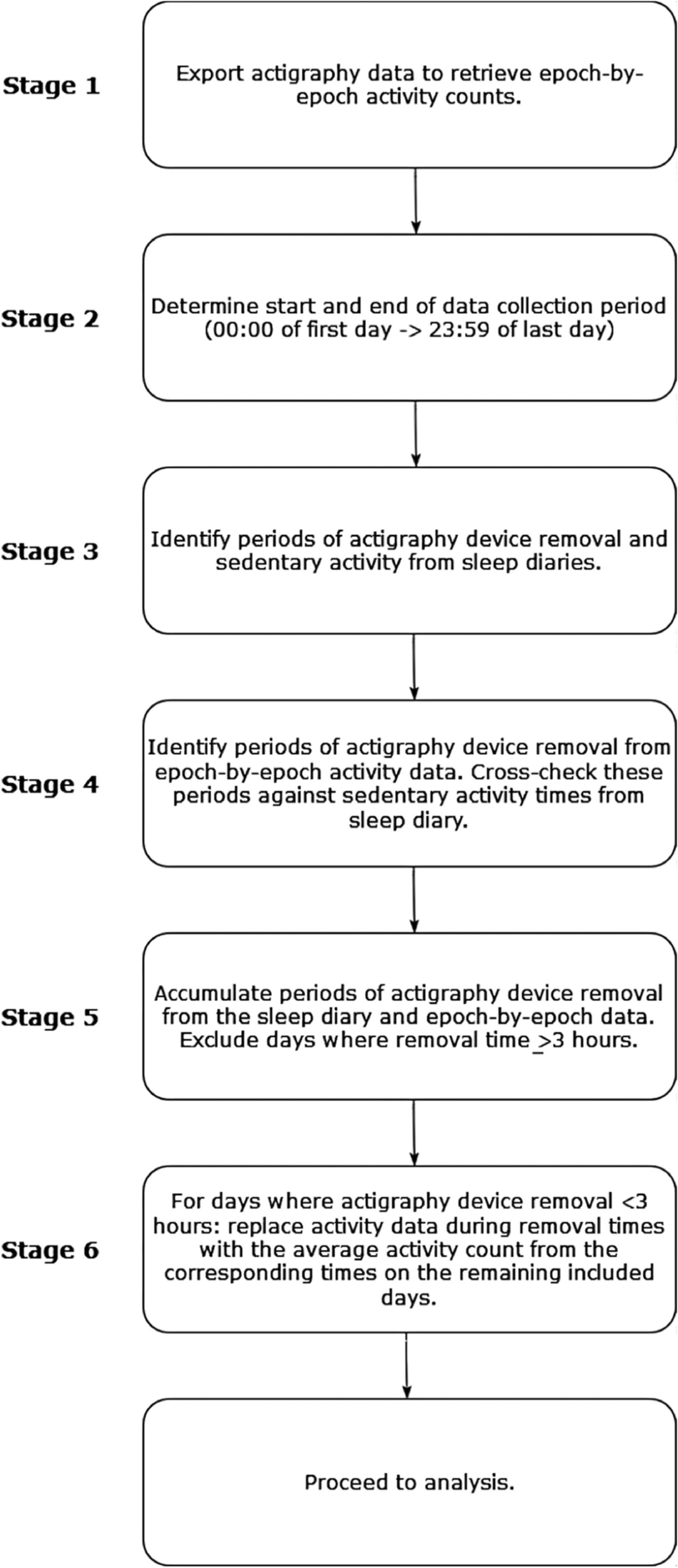
This was a small, single-center (University of Massachusetts Chan Medical School (UMass Chan)), prospective, nondrug pilot feasibility study of the repeated collection of hair follicles by plucking in individuals, 3 to 22 years of age, with FXS and those who were typically developing for the quantitative measurement of FMR1 mRNA and FMRP. The repeated collection of hair follicles was compared with the collection of blood and buccal swabs for detection of FMR1 mRNA and FMRP and related molecules in children and young adults with FXS and age- and sex-matched children and young adults without FXS (healthy controls). For this study, most participants completed three study visits: screening visit, visit 1, and visit 2. An unscheduled visit was allowed for additional sample collection, should any of the samples be inadequate for analysis, or if there was a reason the participant could not provide one of the samples at the regularly scheduled visits. If requested by the family and deemed appropriate by the principal investigator (PI), the screening visit and visit 1 could be combined. There were two sample collection visits, one in the child’s home and one at the clinic. The order of the location of the visits could vary (home visit first, office visit second or office visit first, home visit second), depending on the preference of the family. The feasibility for the repeated collection was determined by the two separate visits, 1–59 days apart. There could be one additional unscheduled visit, either in the office or at home, in case of missed or incomplete scheduled visits.
Measurement of FMRP in hair follicles and peripheral blood lymphocytes (PBLs) was performed by MSD ELISA on whole protein extracts. For PBLs, an independent, flow cytometric method was used to simultaneously measure FMR1 mRNA and FMRP in addition to the MSD ELISA [20].
PrimeFlow™ flow cytometric assay: For a detailed protocol, see Roth et al. (2021) [5]. PrimeFlow™ was carried out per manufacturer instructions with the addition of surface and intracellular protein staining. Surface CD markers identified were CD8a, CD19, CD14, CD3, and CD4. A fixable viability dye was also included. Samples were fixed, washed twice, and permeabilized. Anti-FMRP or mouse IgG1 isotype was added for internal staining. After incubation and washes, samples were fixed and washed again. Probes to detect FMR1 or dapB mRNA were added. Following hybridization of probes, the samples were washed and stored overnight at 4 °C. The next day, the PreAMP mix was added followed by incubation and washing. RNA AMP Mix was added. After incubation and washes, PrimeFlow™ RNA-labeled probes were added. After incubation and washes, the data was acquired on an ATTUNE NXT cytometer. Data was analyzed using FlowJo software.
Absolute quantification of FMRP by electrochemiluminescence ELISA (MSD): For a detailed protocol, see Roth et al. (2021) [5]. A participant’s lysate, 150 μg/mL FMRP-positive control lysate, 150 μg/mL FMRP negative control lysate, or a standard curve of recombinant FMRP were mixed 1:1:1 with custom biotinylated rabbit, polyclonal anti-FMRP, and mouse monoclonal 6B8/FMRP in a V-bottom, polypropylene, 96-well plate. The plate was sealed and placed on a shaker at 4 °C overnight. A total of 5 μL of the mixture was added to each well of a 384-well avidin-coated MSD plate in quadruplicate. Mixtures were incubated followed by three washes. The plate was blocked using 3% MSD Blocker A in the MSD wash buffer. The plate was washed followed by addition of sulfo-tagged anti-mouse. After incubation and washes, 2× MSD read buffer T was added. The plate was immediately acquired using the MESO SECTOR S 600 reader. Data was analyzed using MSD software. A standard curve in fmol was created from the recombinant FMRP. The fmol of FMRP for each lysate was calculated from the standard curve.
Total protein concentration: For a detailed protocol, see Roth et al. (2021) [5]. The sample lysates’ concentrations were below the detection limit of a BCA reaction. Therefore, the highly sensitive ProteinSimple Total Protein Detection Module was modified to determine lysate concentrations. A PBMC or hair follicle lysate standard was created from a large batch of isolated PBMCs or hair follicles, respectively. The protein concentrations of the standards were determined using the BCA assay. A 4-point standard lysate curve and lysates from the participants’ samples were prepared following the protocol provided with the total protein detection module. The prepared lysates were run in the 12–230 kDa separation module on the JESS. Data analysis was performed using Compass software. For each point on the standard cure, the area under the curve (AUC) for peaks at 48, 75, and 190 kDa was calculated. The AUCs for the same peaks in the sample lysates were determined. The values from the standard curve were used to determine the concentrations of the sample lysates.
Hair follicle qPCR protocol: Plucked hair follicles were immediately submerged in RNA later. Samples were processed using the TRI-Reagent protocol with homogenization using the Qiashredder and the Zymo Research Direct-zol MicroPrep kit. Quantitated mRNA was prepared using SuperScript IV for first-strand synthesis and Applied Biosystems PreAmp Master Kit. Commercially available TaqMan probes were used for the FMR1 transcript (Invitrogen cat. no. Hs00924547_m1). The fast advanced master mix (Applied Biosystems 4444557) was employed, and samples were read on the QuantStudio 7 thermocycler.
Participants
Individuals 3 to 22 years of age were recruited for this study. Some participants were invited to participate from the FXS clinic at UMass Chan, while other participants were recruited from the greater New England area via referrals from patient organizations and other methods. A parent focus group was formed as part of recruitment efforts for education on the rationale for the study and to gain parent support. Recruitment materials prepared by UMass Chan were used to facilitate awareness and communication between interested families at the Center for Autism and Neurodevelopmental Disorders (CANDO) clinic at UMass Chan and University of Massachusetts Memorial Health Care. Informed consent was obtained from the parent/legally authorized representative (LAR) for children ages 3–17 years, by self (ages 18–22), or by proxy (parent or LAR of the individual). If they were capable, assent by the child or the adult, in case of a proxy or LAR, of their willingness to have samples of hair, blood, and buccal swabs collected was obtained.
The determination of the FXS phenotype regarding expressive language delay and/or social anxiety was performed by the principal investigator (JF) based on the history and physical examination, review of available medical records, and results of standardized testing during the screening visit. All subjects were assessed at screening with standard and age-appropriate tests of language development, social anxiety, social communication impairment, and other FXS phenotypes. As described above, given that social communication challenges are a key area of difficulty in FXS, it is imperative to include measures that can identify these deficits. The overarching aim of this proof-of-concept study is the generation of data for future clinical trials focused on reactivation of FMR1 mRNA and FMRP. Thus, the primary endpoint for these studies would involve measures of social communication and expressive language, unlike IQ, which is a complex construct that cannot be used as a primary endpoint for pharmacotherapeutic interventions. Hence, the OWLS-II Listening Comprehension (LC) and Oral Expression (OE) portions were used to measure expressive language and LC [29]. The Gilliam Autism Rating Scale 3rd Edition (GARS-3) was used for the determination of the presence and severity of restrictive/repetitive behaviors, social interaction impairment, social communication impairment, altered emotional responses, cognitive style, and maladaptive speech by parent report [30].
ProceduresHair follicle collection and analysis
Plucking refers to the process of removing human hair by mechanically pulling hair follicles from the scalp. In this study, hair follicles were collected via plucking at visit 1, visit 2, and any unscheduled visits, using methodology and tweezers provided by the Epistem Ltd. Epistem and Fulcrum provided training to UMass Chan personnel via Webex with ongoing support as needed, including on the collection and the quality assessment of plucked hairs. Required supplies were provided for hair follicle storage. It was anticipated that about 20 hair follicles would be plucked per visit, of which about 15 would pass quality control. Any plucked hairs missing the follicle were discarded by Epistem or Fulcrum.
Collection and analysis of blood cells, cellular DNA, and plasma
Blood was collected via venipuncture at visit 1, visit 2, and any unscheduled visits. At visit 1, blood was collected for both FMR1 mRNA methylation assessment and FMR1 mRNA/FMRP measurement. The blood collection at visit 2 or any unscheduled Visit was optional for subjects with FXS.
Measurement of FMRP in hair follicles was performed at Fulcrum by MSD ELISA on whole protein extracts from individual hair follicles. Measurement of mRNA in hair follicles was performed by TaqMan® analysis [31]. The blood collection tubes were shipped to Fulcrum where peripheral blood lymphocytes, DNA, and serum isolation were performed. Claritas Genomics also performed the analysis for the FMR1 gene mutation for mosaicism and length of the triple repeat expansion when not available from the clinical records. Blood lymphocytes and plasma were evaluated for the measurement of FMR1 mRNA or FMRP. FACS analysis was performed at Fulcrum. For complete MSD ELISA and FACS methods, refer to Roth et al. (2021).
Measurement of FMRP by IHC was discontinued early due to fragility of youth hair follicles (Epistem Ltd., UK). Measurement of FMRP in serum using MSD ELISA was performed at Fulcrum or by NanoSomiX by isolation and characterization of exosomes. Buccal swabs were obtained using the manufacturer’s instructions (Puritan HydraFlock from Puritan Diagnostics [Guilford, ME]) at visit 1, visit 2, and any unscheduled visits. Measurement of FMRP was performed at Fulcrum by MSD ELISA of protein extracts. Measurement of FMR1 mRNA in buccal swabs was performed at Fulcrum by TaqMan analysis, as described above. Measurement of FMR1 mRNA by qRT-PCR was not done in PBMCs due to lack of sufficient sample to run both protein assays and qRT-PCR. We were able to obtain FMR1 mRNA expression by PrimeFlow™ because the detection of FMR1 mRNA and FMRP was done simultaneously on the same sample.
All hair follicles, blood tubes, and buccal swabs were shipped by the primary study site on the same day as the collection under conditions specified in the study reference manual. All samples were labeled with study identification number and fully anonymized prior to shipping. Fulcrum scientists shipped a portion of the hair follicles (about 5 per subject per visit) to Epistem for analysis by IHC.
Statistical analysis
Data analysis was performed using statistical analysis software (SAS) Version 9.4. Descriptive statistics for continuous variables included number of subjects (n), mean, standard deviation (SD), median, minimum, and maximum. Summaries of change from baseline variables included only subjects who had both a baseline value and corresponding value at the timepoint of interest. Descriptive statistics for categorical data included frequency and percentage. Where appropriate, descriptive statistics were presented with 95% confidence intervals (CIs). Measures of spread (e.g., standard deviation) were reported to 2 degrees of precision more than the recorded data. Pearson’s correlation coefficients were used to assess relationships between FMR1 mRNA and FMRP levels with measures of OWLS-II LC and OE portions and the GARS-3 subscales. For the OWLS-II and GARS-3, standard scores were used in analyses. Higher OWLS-II scores indicate better language comprehension; higher GARS-3 scores indicate more severe autistic behavior.

留言 (0)