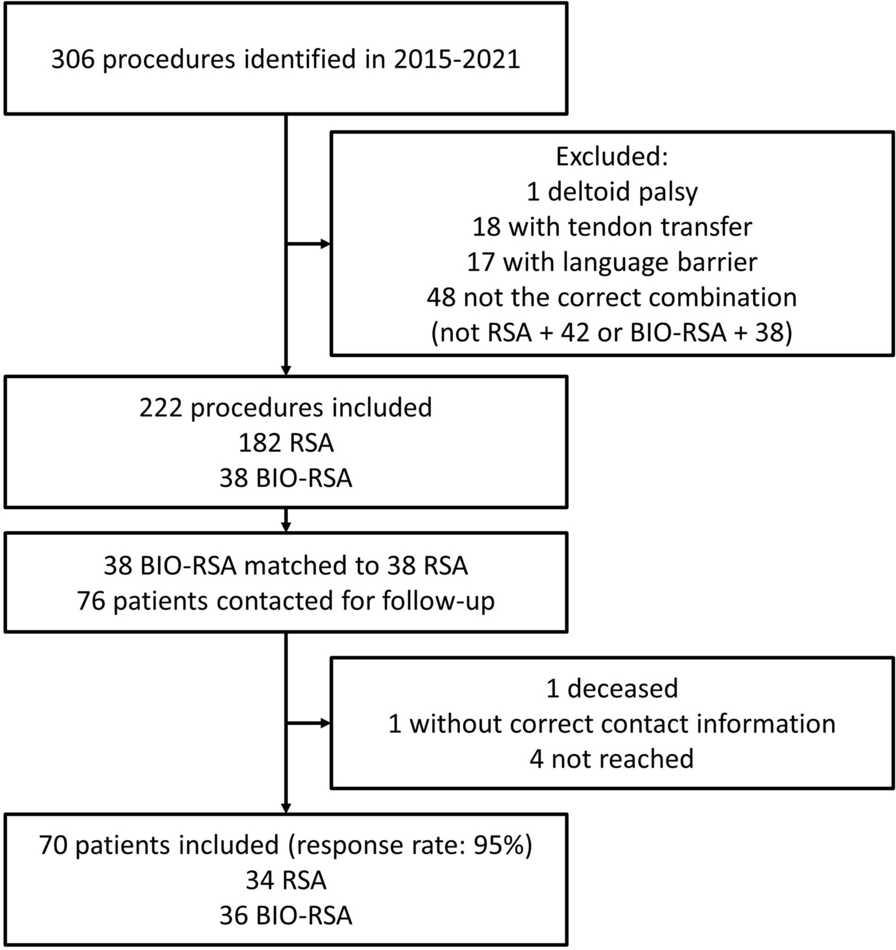
In this study, a coronoid prosthesis was developed based on the morphology of the upper 40% of the coronoid process, and a simulated model with a coronoid process defect that was reconstructed by the coronoid prosthesis was analyzed using a finite element analysis to investigate the fixation strength of the prosthesis when applying different fixation methods at various elbow flexion angles. As hypothesized, prosthesis fixation with a single bolt 4.5 mm in diameter fixed from posterior to anterior showed better strength and fixation stability.
The coronoid process of the ulna is the key element in the stability of the elbow joint [1]. In the case of a comminuted coronoid process fracture, when the reduction and fixation of the fracture fragment could not be achieved, reconstruction using autograft or allograft was regarded as a choice [5, 6, 8]. However, these methods were associated with either donor site comorbidity or limited availability [5, 6, 8]. Recently, the coronoid prosthesis design was developed to better restore the elbow stability. Alia pointed out that a coronoid prosthesis may be a feasible treatment option to restore elbow stability, based on a biomechanical study [15]. O’Driscoll and colleagues reported the follow-up of three cases of comminuted coronoid process fracture treated with a coronoid prosthesis and suggested that this treatment provided an excellent outcome. It was believed that a coronoid prosthesis may offer a promising solution for a comminuted coronoid process fracture. But, to date, no further studies that follow on from those results have been conducted. One reason for this may be the poor understanding of how to maintain the long-term stability of the coronoid prosthesis [9].
The most common reason for revision surgery for a prosthesis other than a coronoid prosthesis in clinical practice is aseptic loosening, which is associated with an unexpected cost and a poor clinical outcome. The most important measure that can diminish this complication is to optimize the design of the fixation method. A previous study examined three fixation methods in terms of minimizing the micro-motion of the prosthesis, including press-fit cementless fixation, screw fixation, and cement fixation, and the results suggested that screw fixation may benefit osseous integration [15]. However, the biomechanical behavior in the condition with screw fixation was only investigated using two 2.4-mm cortical screws. So far, no results for other fixation patterns have been reported in the literature.
Methods for investigating the biomechanical behavior of the prosthesis can be categorized into biomechanical experimentation and computational simulation. Biomechanical experimentation can offer parameters from human material with an actual load, but it is limited by the availability of human tissue specimens and patient variability, which results in a small sample size and underpowered statistical significance [22]. Computational simulation—particularly FEA of a 3D model reconstructed from CT images, which allows the simulation of multiple loading parameters and the modification of boundary conditions—has been accepted as a powerful tool for designing a prosthesis and evaluating the effects of various prosthesis fixation methods [22].
Based on our previous study, we studied four fixation methods and examined their strength and stability by finite element analysis. The results indicated that the maximal principal stress and the maximum deformation are smallest when using a single 4.5-mm bolt for fixation at all three flexion angles, whereas it was largest with double 2.0-mm bolt fixation. These results suggest that the diameter of the fixation bolt plays an important role in resisting micro-motion of the interface between the prosthesis and the bone. The larger diameter demonstrated better biomechanical characteristics, but using a larger diameter leads to more volume being occupied by the bolts in the host bone, which compromises bone integrity and increases the risk of fracture. For a comparison of the maximum principal stress on the fixation bolt at different flexion angles, the largest value occurred at 30° flexion, while the smallest value occurred at 130° flexion. It was speculated that the loading generated the greatest shear force on the fixation bolt at 30° flexion, while the loading was directed away from the fixation bolts at 90° and 130° flexion; thus, the maximum principal stress was significantly reduced at these two flexion angles.
Fatigue of the fixation bolt is another reason for prosthesis revision surgery, as repetitive cycle loading on the prosthesis or the fixation device would eventually reach the failure limit. Zand’s biomechanical study suggested that the most common location of bolt fatigue was the root of the thread at the interface between the plate and the bone [23]. Li’s investigation using a femoral neck fracture model indicated that the peak von Mises stress on the screws was concentrated in the middle surface of the screw near the fracture line [24]. In this study, the maximum principal stress was located around the screw neck, which is consistent with a previous study, and it suggested that the maximum principal stress on the fixation bolt occurs at the interface between the bone and the prosthesis. As for the maximum deformity, it was located at the head of the bolt, which meant that the maximum deformity occurred at the far end of the fixation.
In terms of the direction of the bolt for fixation, few reports investigating the role of the direction have been published. In this study, fixation with both single and double bolts was adopted. From a practical perspective, usually medial and lateral approaches to the elbow have been used during the operation to evaluate the coronoid process defect and prosthesis fixation. If double-bolt fixation is employed, the two bolts can be inserted through medial and lateral approaches from anterior to posterior in a divergent manner. When a single bolt is used for fixation, the direction of the bolt should be perpendicular to the bone–prosthesis interface to maximize the stability. If insertion is implemented from anterior to posterior, an extra anterior approach is required to achieve this goal, but this may compromise the neurovascular structure in clinical practice and lead to severe complications. Thus, for single-bolt fixation, a small incision was made at the dorsal part of the ulna, as there is no important neurovascular structure within this region, so the bolt could be inserted from posterior to anterior safely.
There are some limitations of this study. First, the complicated physiologic force loaded on the proximal ulna during movement of the elbow was simplified, but in this FEA model, only axial loading along the humerus was simulated to mimic the force between the trochlea and proximal ulna. The reason for this is that the most common scenario for coronoid process fracture is when patients fall down with the hand supporting and the force is mainly transmitted along the axis of the humerus. The second limitation is that the FEA model was only performed on 10 specimens. A larger sample size is needed to investigate the association between the greater stress in the fixation bolts at low-flexion-angle loading and the corresponding increase in implant displacement. The third limitation of this study is that the structures of the bone and prosthesis were considered isotropic, homogeneous, and linearly elastic, which is not consistent with anatomical and biomechanical reality. The fourth limitation is that, in some of the cases, the fixation bolts would fail in the host bone, which would require further analysis to examine the maximal principal stress in the host bone. The fifth limitation is that the fixation bolt thread was simplified as the thread may introduce stress raisers, and further study is needed to look into this effect.


留言 (0)