Sclerosing Odontogenic Carcinoma: Report of a Case and Review of the Literature
Background
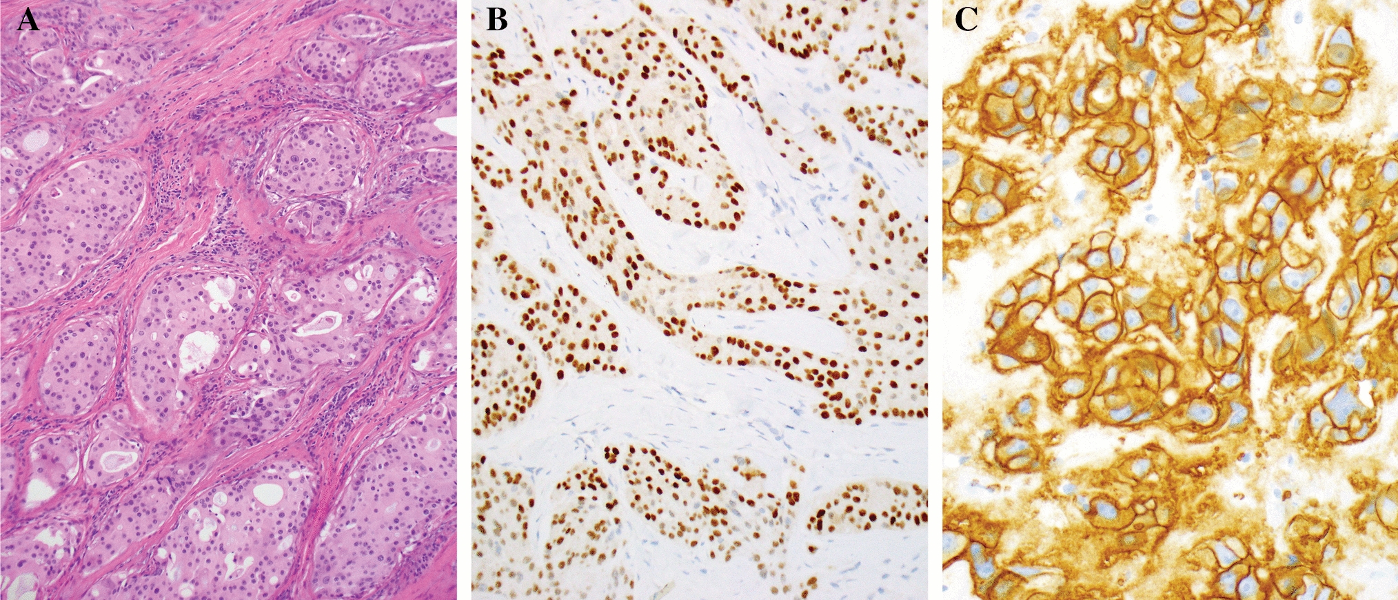
Sclerosing odontogenic carcinoma is an exceedingly rare gnathic malignancy first described by Koutlas et al. in 2008, and was only recently designated as a distinct pathologic entity by World Health Organization in the 2017 Classification of Head and Neck Tumors. To date, fewer than fifteen cases of this neoplasm have been reported in the English language literature. This tumor is characterized by thin cords, strands, and small nests of epithelium in a densely sclerotic stroma. In some tumor foci, the density of the stroma may be sufficient to compress the epithelial component beyond detection in the absence of immunohistochemistry, thus rendering this entity a particularly challenging diagnosis in small sample sizes.
Methods
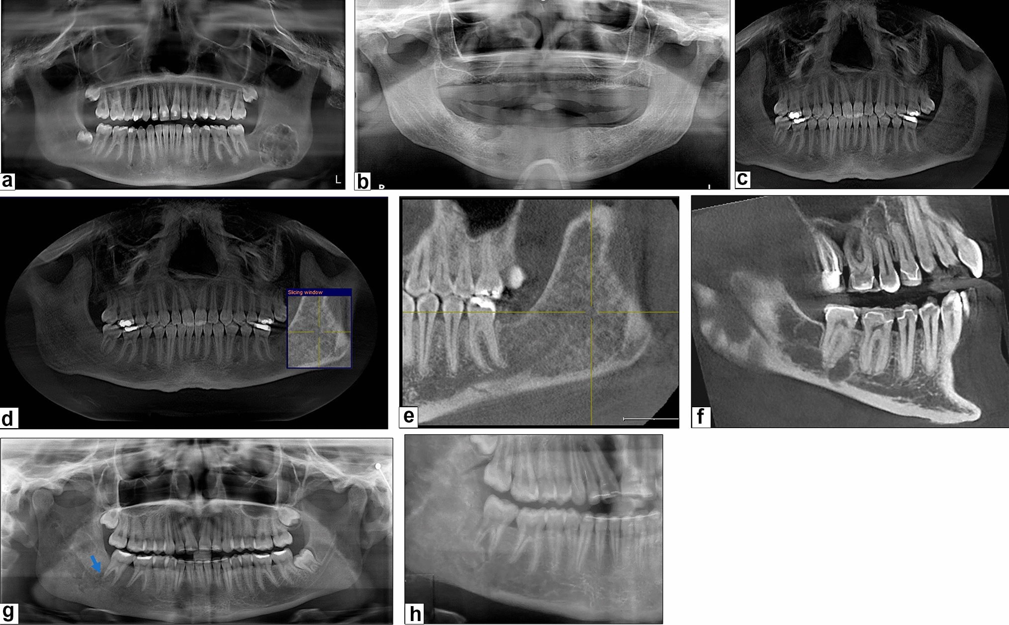
A 55-year-old male presented with an asymptomatic lesion of posterior left maxilla. Cone beam computed tomography (CBCT) demonstrated a large, well-defined bony lesion with scalloped border, spanning from canine to first molar. External root resorption of the adjacent teeth was also noted. Microscopic examination of the biopsy specimen revealed an odontogenic tumor with features consistent with sclerosing odontogenic carcinoma. Immunohistochemical staining was performed to confirm the diagnosis.
Results
The tumor was positive for CK5/6, CK19, E-cadherin, p63 and negative for CK20 and CK7.
Conclusion
Sclerosing odontogenic carcinoma is a rare, low-grade malignancy of odontogenic origin, which represents a diagnosis of exclusion in many cases. An immunohistochemical profile demonstrating positivity for markers including CK5/6, CK19, p63, and E-cadherin, in addition to a set of pertinent negative findings, can aid in the diagnosis of this tumor. This entity appears to lack metastatic potential despite its locally destructive behavior and a common histologic finding of perineural invasion.


留言 (0)