
記住我
Extravascular necrotizing granuloma is pathologically one of the most characteristic findings of eosinophilic granulomatosis with polyangiitis (EGPA).
A 46-year-old man presented to the rheumatology clinic with a 2-week history of arthralgia and 4-day history of dysesthesia of the feet. History taking revealed hyperlipidemia and asthma, which first appeared in his 30s. His medications history included rosuvastatin and inhaled salmeterol and fluticasone. Physical examination revealed polyarthritis and a painful papule on the left hand (Figure 1) and the left knee. Laboratory tests revealed an elevated absolute eosinophil count of 9160/μL and positivity for myeloperoxidase antineutrophil cytoplasmic antibody. HbA1c was normal. Hepatitis B surface antigen was negative. Nerve conduction study showed a mononeuritis multiplex pattern. Computed tomography of the lungs and sinuses was unremarkable. Histopathology of the papule on the left hand and knee found cutaneous extravascular necrotizing granuloma with eosinophils (Figure 2). EGPA was diagnosed based on 4 of 6 American College Rheumatology (ACR) criteria1: prodromal asthma, eosinophilia, eosinophilic tissue inflammation, and polyneuropathy. This patient also met the 2022 ACR/European Alliance of Associations for Rheumatology classification criteria for EGPA.2 A colonoscopy revealed enteritis. Although it was unclear whether the enteritis was responsible for EGPA, a glucocorticoid and cyclophosphamide regimen was administered based on this assumption; the treatment ameliorated the symptoms.
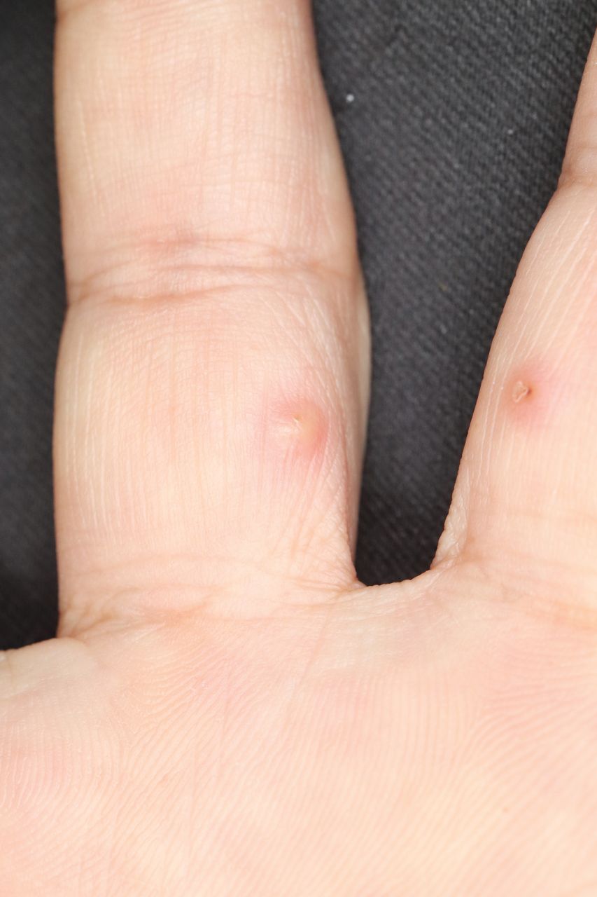 Figure 1.
 Papule on the left hand.
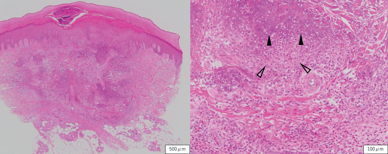 Figure 2.
 Histopathology of the hand papule shows cutaneous extravascular necrotizing granuloma. Necrotic basophilic collagen (black arrow) surrounded by histiocytes (outlined arrow) in lower dermis. Numerous eosinophils infiltrate around necrotizing granuloma (H&E stain).
A previous report identified extravascular necrotizing granuloma from 13 patients with EGPA; 12 specimens were from the elbow.3 Extravascular necrotizing granuloma is usually found on the extensor surface, particularly the elbows and legs.4 However, it can be found on hands. If EGPA is suspected, careful examination of the total body surface area is required.
ACKNOWLEDGMENTWe thank Dr. James R. Valera for his assistance in editing the manuscript and Dr. Takahiro Kiriu in interpreting pathology.
Copyright © 2022 by the Journal of Rheumatology
留言 (0)