First described in 1938 by Pearson et al. DNM remains a serious, aggressive disease often associated with fatal outcome [3, 5, 17, 18]. Even though it is a rare disease DNM should not be underestimated. It can still lead to sepsis and death, while once high mortality rates from 49% in 1938 decreased during the last years to 17.5% described in a review by Prado-Calleros in 2016 as diagnostics and multimodal therapy concepts improved [4, 5, 19,20,21]. Deep neck infections, DNI, develop from odontogenic or oropharyngeal infections and rapidly spread via deep fascial planes downward into mediastinum resulting in DNM [2, 11, 22, 23]. Based on its pattern of spreading Endo et al. developed a classification of CT findings: (I) focal form: localized to the upper mediastinal space above the carina. (II) Diffuse form reaches out below the tracheal bifurcation and is subdivided into (IIA): lower anterior mediastinum and (IIB): lower posterior mediastinum [16].
Common literature agrees that a diagnosis at early stage of DNM as well as a prompt initiation of appropriate medical and radical surgical treatment are imperative [2, 4, 9, 11, 17, 24,25,26,27,28,29].
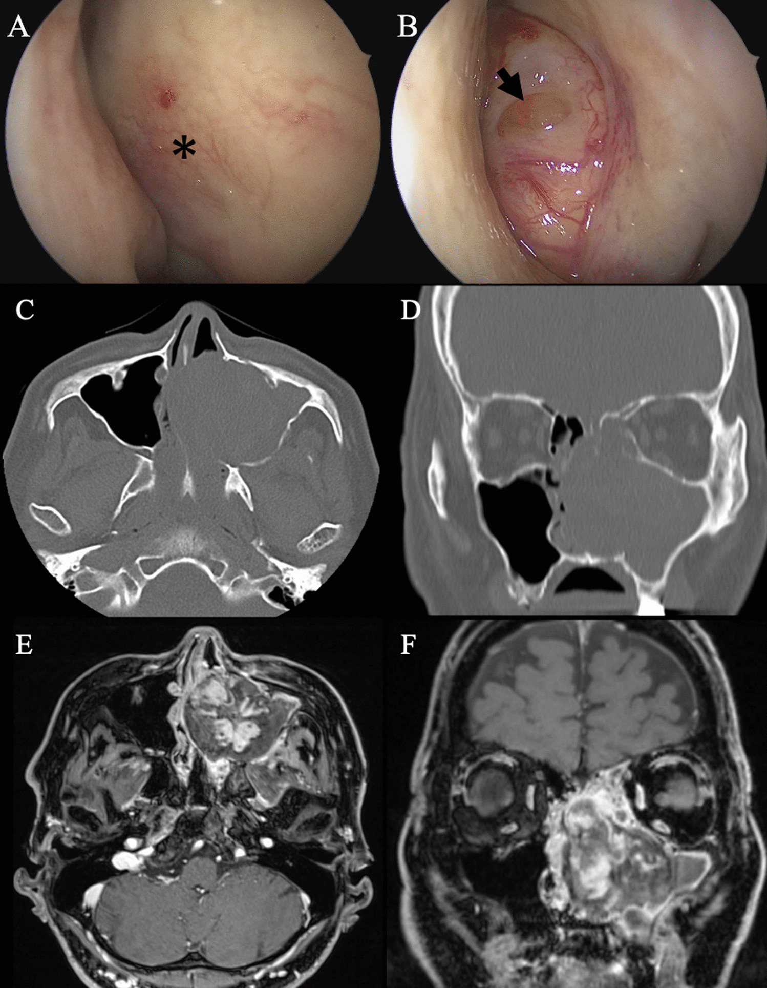
However, an immediate recognition of DNM can be challenging due to the following reasons: DNM is a rare disease not only ENT experts and general surgeons but also family doctors, pediatricians and general practitioners should keep in mind. Though only 2–3% of deep space neck infections develop to more serious infections, such as mediastinitis, one should always be aware of such a severe course as it can progress very fast [7, 30, 31]. Furthermore, it can affect all age groups as the results of our investigation, age range 3–84 years, show in concordance with the literature. In doubt the clinical picture is sufficient to suspect DNM and the patient should promptly be admitted to a hospital with ENT specialist for thorough examination of the pharynx and larynx including fiberoptic transnasal endoscopy and efficient initiation of imaging diagnostics [4, 32]. Moreover, symptoms may not be distinct. The infection is often clinically silent especially at the beginning and may be veiled by analgesics delaying the diagnosis. Therefore, a clear association between symptom, severity and extent of the disease is difficult [4, 12, 13, 32, 33].
In our study most common clinical symptoms, pain, swelling and odynophagia, at the time of presentation are related to common oropharyngeal infections and DNIs. However, disorders correlating explicitly with mediastinitis such as chest pain and mediastinal emphysema are less frequent in our patients [15, 27, 33, 34].
The clear relation between infections of an odontogenic or pharyngeal source and the development of DNM is undoubted [9, 17, 35]. The anatomic continuity of the posterior pharyngeal, parapharyngeal and submandibular spaces with the mediastinum explains this smooth transition. Once an infection enters one of these spaces a spreading downward is promoted because of gravity, respiration, intrathoracic negative pressure and absence of barriers in fascial planes [4, 35, 36]. The so-called danger space lies posterior to the alar fascia, runs from skull base to diaphragm and allows even a contralateral spread of the infection [9].
Also, in our patient group previous infection of odontogenic or pharyngeal origin is often mentioned. Four patients even reported a post-radiogenic situation of the neck. This underlines the importance of a diligent execution of medical history as these patients are more likely to suffer from wound healing disorders [37].
Several predisposing risk factors are well-recognized referring to patients with DNM, such as diabetes mellitus, poor dental or oral hygiene, immunosuppression, renal and liver failure, high blood pressure and recent steroids. Moreover, chronic nicotine, alcohol and IV drug abuse happens to appear frequently in patients with DNM [9, 11, 12, 15, 38,39,40]. The description that patients with DNM suffer significantly more often from comorbidities compared to patients with DNI underlines the high impact of these pre-existing risk factors to develop DNM [10, 12]. Our analyses turned the attention especially to comorbidities with a reduced tissue oxygenation as these expedite the development of DNM [4]. More than half of our patients suffered from pre-existing diseases associated with impaired tissue oxygenation, e.g., cardiac or respiratory insufficiency, diabetes mellitus, adiposities per magna and post cervical radiation. This coincides with the observations of Kocher et al. [2]. Nonetheless also young, healthy patients with no medical history can suffer from DNM [41,42,43].
The Charlson Comorbidity Index, CCI, is a well-approved statistical test to predict the mortality of patients based on underlying comorbidities and has widely been used since its first description in 1987. Patients are divided in four groups (0 points, 1–2 points, 3–4 points and 5 points) correspondent to an increasing mortality risk. We used the age adjusted variant [44, 45]. Only 23.9% of our patients had an index of 0 points. Park et al. observed a CCI ≥ 1 in 54.1% in a group of 135 patients with DNI [46]. The results of CCI in our study underlines even more the role of comorbidities in patients with DNM and indicates, that patients with pre-existing reduced health conditions may have a higher risk to develop DNM.
The polymicrobiological nature of DNM as often described in diverse studies makes sense considering its origin as an oropharyngeal infection that once it has penetrated the mucosa spreads downward to the mediastinum [2, 33]. Likewise, Palma et al. half of our patients had mixed anaerobic and aerobic infections [11]. Nonetheless no result regarding extensive microbiological examinations was obtained in 18 of our patients. This lack of microbiological results was observed as well by other authors. It might be due to the fact that quite a high number of patients, e.g., in our study 43.1%, has already been treated with antibiotics before microbiological examinations was obtained [4, 12, 32].
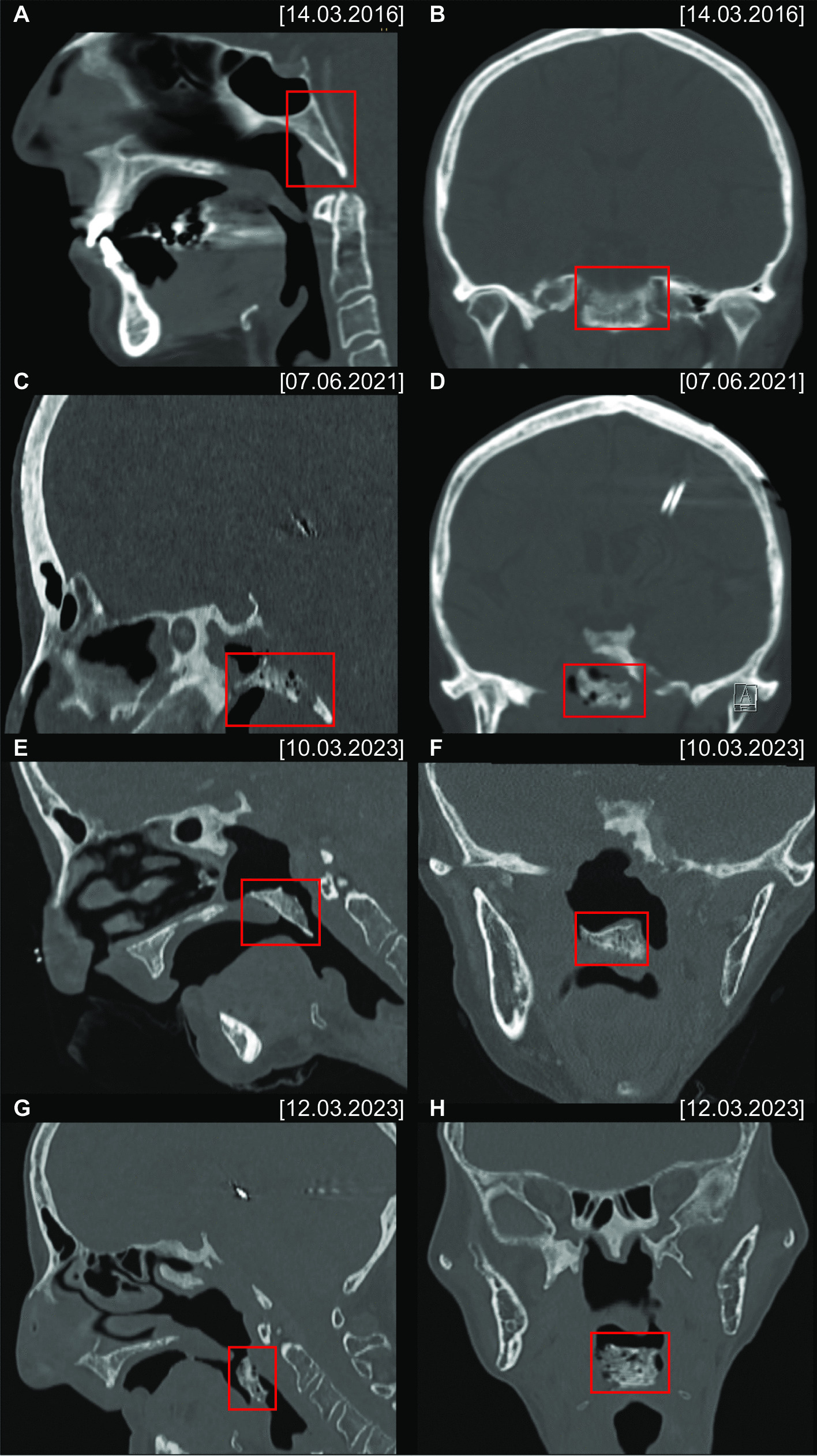
In more than three-quarter of our patients the upper part of mediastinum above the carina was affected corresponding to Endo Type I. CT scans are an early part of the diagnostic pathway and crucial in recognizing DNM before it spreads even deeper. Radiation exposure should be accepted in case of deep neck infection or just its clinical suspicion to exclude DNM yet at the beginning. Freitas et al. even suggests an algorithm with serial CT scans every 24–48 h to assess disease progression [32]. In line with that also most of our patients received CT scans in the follow-up to monitor the course of DNM and if applicable to act immediately. The expressiveness of CT scans after repeated surgical interventions with changed tissue textures can be hindered. Moreover, laboratory findings as WBC or CRP levels can be indistinct, e.g., if they do not decrease conspicuously. In case of doubt, we, therefore, favor a liberal decision to revision surgery.
The surgical drainage of the affected parts of mediastinum is without any doubt mandatory. However, there is still some controversy about the optimal approach. A proper approach should thoughtful be chosen according to patients condition, the extent of the disease and also the surgeons experience [2]. Responsible for the mortality of DNM is not only a delayed diagnosis but as well an inappropriate drainage of the mediastinum. Therefore, the latter should also carefully be focused on [11]. As most of our patients suffer from DNM Endo Type I, where infection is located in the upper part of the mediastinum, we are of the opinion that in these cases a sole transcervical drainage is sufficient for surgical debridement and necrotic tissue removal. A highly aggressive surgical treatment irrespective of the extent of DNM as formerly advocated by many authors can also implicate disadvantages and may go along with a higher complication rate [6, 13, 28, 33, 36]. In case of an advanced stage of DNM, Endo Type II, we also certainly support a thoracic approach via thoracotomy in combination with transcervical debridement for drainage of the upper and lower parts of mediastinum. Most accomplished is a posterolateral thoracotomy on the more affected side [5, 18]. As healing of DNM may often be protracted and revision surgery is commonly needed we usually perform incomplete closure and insertion of large tubes in the wounds combined with daily irrigation with antiseptic solutions.
The infectious process of DNM can frequently lead to pharyngolaryngeal edema and consequently cause dyspnea. We recommend in these cases of foreseeable difficult intubation an awake or at least video assisted intubation if necessary in preparedness of coniotomy. If upper airway is compromised or patients seem to need a long-term treatment because of severity of DNM we approve tracheotomy. Nevertheless, we refuse to overhasty decisions for routine tracheotomy in patients with DNM as spreading of cervical infection may occur [2, 5].
Amongst our patients mortality was significantly influenced by higher age and higher CRP-levels. Although not relevant to mortality in the present survey we do think other factors as comorbidities, extent of involved mediastinal site and duration of discomfort until treatment still have an impact on progress and outcome of DNM. Therefore, the overall impression of the patient must not be disregarded.
We are confronted with a modest number of questionnaires sent back by our patients who were also treated at ICU. Besides we face a heterogeneous distribution of time passed, since patients suffered from DNM. In addition, Gerth et al. report a loss of patients in follow-up controls in their review regarding changes in health-related quality of life after ICU [47]. Amongst others a reason to not sending back the questionnaire might be the long period of time of more than 20 years included. Patients suffering from DNM years ago not only became essentially older as well as their health condition presumably impaired and, therefore, precluded answering and sending back the questionnaires. Basically the SF-36 we used is well-approved and the most employed questionnaire to evaluate quality of life after critical illness. Despite the quantity our findings implicate a tendency that patients post DNM rather suffer from impaired physical than mental health in long-term. This is concomitant with the results of Gerth et al. [47]. Kramer et al. indeed reported that physical-functionary health-related quality-of-life subscales remain depressed even years after the acute illness in patients after ICU [48].
Our findings implicate that persisting dysphagia after DNM actually occurs hence two-thirds of our patients had above average high values in EAT-10 questionnaire. This corresponds to the observation that dysphagia is well-recognized as a late complication of DNM by several authors. We likewise subscribe to the advice of comprehensive long-term physiotherapy and logopedic support in patients with DNM not only to prevent aspirations but also to improve their quality of life [8, 17, 40].


留言 (0)