
記住我
The human hepatoblastoma cell line HepG2 was obtained from Chinese Academy of Sciences Cell Bank (Shanghai, China). Culture media and agents were purchased from Gibco (Waltham, USA). CellTiter-Glo® 2.0 cell viability assay kit was purchased from Promega (Madison, USA). Seahorse consumables were all purchased from Seahorse Biosciences (North Billerica, USA). The pure test compounds were purchased from Dalian Meilun Biotechnology Co., Ltd. (Dalian, China). Xian-Ling-Gu-Bao capsule was produced by Tongjitang Pharm Co., Ltd. (Guiyang, China), and the six TCMs in it were purchased from Tongling Hetian Herbal Pieces Co. Ltd. (Anhui, China). Acetonitrile and formic acid for HPLC were purchased from Sigma-Aldrich (St. Louis, USA). The methanol and ethyl acetate used in extracting the samples were all analytical grade.
Determination of the main compounds in Psoraleae FructusSample preparation1 g of XLGB powder was added into a 50 mL volumetric flask, added with 75% methanol, ultrasonicated (SY-800, 40KHZ, Shanghai Weimi Technology Co., Ltd., Shanghai, China) for 30 min, and then made to volume at room temperature. Then accurately measured 3 mL of the solution into a 10 mL volumetric flask, and added methanol to make up.
A 10 g fine-grinded powder sample of Psoraleae Fructus was dissolved in 100 mL of water, and extracted with ethyl acetate for three times, each 100 mL, to obtain 0.39 g of the extract, which was then diluted with methanol into a 5 mL volumetric flask.
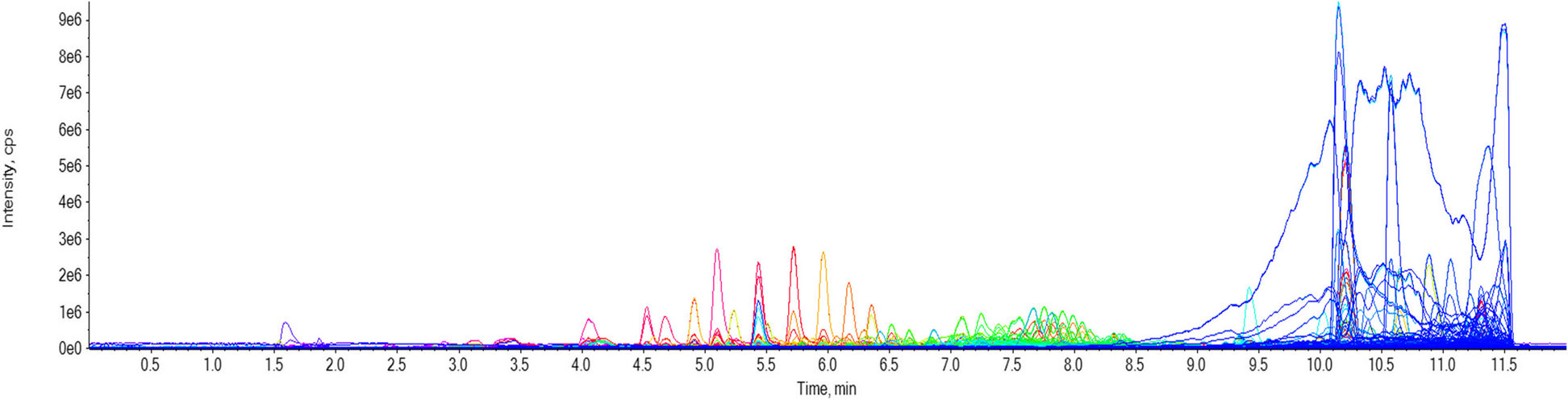
Fingerprint analysis of Psoraleae Fructus and XLGBThe Agilent 1200 series HPLC system (Agilent Technologies, USA) was used for the chromatographic fingerprint analysis. The equipment was equipped with a quatemary solvent delivery pump (G-1311C), an online degasser (G-1322A), a diode array detector (G-4212B DAD), a column temperature controller (G-1316A) and Agilent ChemStation. The column used was an Agilent Eclipse plus C18 column (4.6 mm × 250 mm, 5 μm), the flow rate was set to 0.8 mL/min, the sample volume was 10 μL, and the detection was performed at 270 nm and 35 °C. Acetonitrile (A)-0.1% aqueous formic acid (B) was used as the mobile phase for separation, and the elution gradient of the mobile phase was as follows (A%): 0–5 min, 2% → 2%; 5–80 min, 2% → 36%; 80–95 min, 36% → 48%; 95–110 min, 48% → 80%; 110–112 min, 80% → 100%.
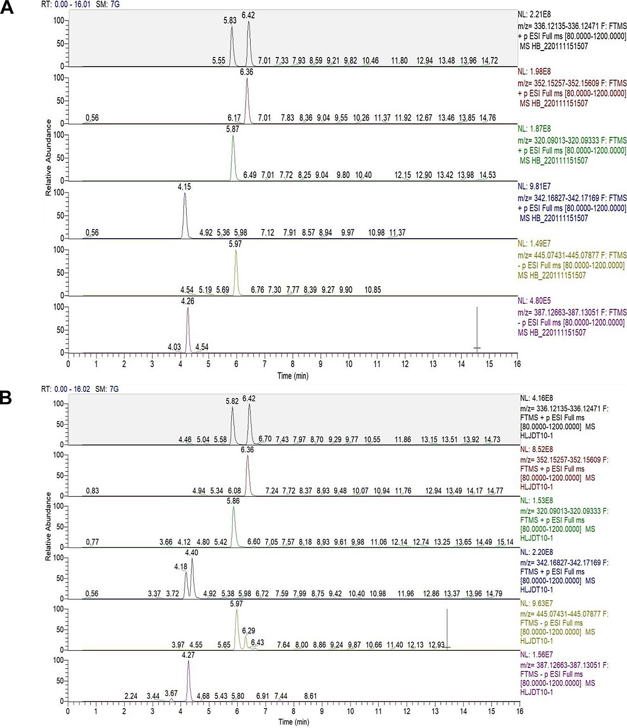
HPLC-TOF-MS analysis of Psoraleae FructusMass spectra in positive and negative ion mode were acquired on an Agilent 6538 UHD and accurate mass Q-TOF/MS. After separation on HPLC system (Agilent 1290 Infinity UPLC, Agilent Technologies, USA), mass spectra were achieved on Agilent 6538 UHD and Accurate-Mass Q-TOF/MS (Agilent Technologies, USA) with the following conditions: electrospray ion source (Dual ESI+); capillary pressure, 3500 V; atomized gas pressure, 45 psi; dry gas temperature, 350 °C; debris voltage, 120 V; data acquisition range, m/z100–1100. Before the experiment, TOF/MS automatically corrected the mass coordinate axis by real-time injection of reference solution (Agilent Technologies, USA), and the flow rate of reference solution was 100 μL/min; Negative ion mode detection: electrospray ion source (Dual ESI); other conditions are the same as positive ion mode.
Cell cultureHepG2 Cells were cultured in DMEM (L-Glutamine 4.00 mM, Glucose 4.50 mg/L, and Sodium Pyruvate 110 mg/L) added with 10% FBS, 100 U/mL penicillin and 100 μg/mL streptomycin, and then maintained in six-pore suspension culture plates at concentration of 10,000 cell/mL with 5% CO2 at 37 °C.
Cell viability assayDetermination of cell viability was by the Cell Titer−Glo® (CTG) 2.0 Assay (Promega, Madison, USA). 5000 cells per well were seeded in 96-well plates and adhered to the plates overnight. Cells were disposed with test compounds diluted at 0.002, 0.02, 0.2, 2, 20, and 200 μg/mL final assay concentrations in serum-free medium, and an equal amount of Cell Titer Glo reagent was supplemented 72 hours later. Transfer half the total volume of each well to 96-well opaque plates, and the emitted light was measured by Multi-Plate Reader (Biotek Synergy 2) after ten minutes.
Measurement of cell bioenergeticsExtraction of herbal medicines200 g of herbal powder was added to 2000 mL of water, extracted with rotary vacuum evaporator (N-1001, Shanghai EYELA Technology Co., Ltd., Shanghai, China) 3 times, 2 hours each time, combined with the extract, concentrated and dried (Vacuum drying oven, 9715, Changshu Pharmaceutical Marchinery Factory Co., Ltd., Jiangsu, China).
Seahorse assayThe bioenergetic function of HepG2 cells was measured by Agilent Seahorse XFe96 Analyzer (Seahorse Biosciences, USA). Day prior to assay, HepG2 cells were inoculated into the cell culture plates of Seahorse analyzer at the density of 20,000 cells per well, adhered and grew in 37 °C humidified incubator (containing 5% CO2), and hydrated the XF extracellular flux sensory cartridge. On the experimental day, the appropriate concentrations of herbal extracts and compounds were added and continued to incubate for 2 hours to make the cells reach about 80% confluency. Then, the cell plate medium was replaced with XF Base DMEM Medium (pH 7.4) containing 1.0 M glucose, 100 mM sodium pyruvate and 200 mM of glutamine solution, and incubated at 37 °C without CO2 for half an hour. Finally, Oligomycin (1.5 μM), FCCP (1 μM) and the mixture of Rotenone and Antimycin A (0.5 μM) were injected sequentially according to the instrument setting procedure, and then the data analysis was carried out [20].
The bioenergetic profile generated by the sequential addition of mitochondrial inhibitors can delineate the details of the respiratory chain (Fig. 1 a). From the bioenergetic profile, we can derive six parameters about mitochondrial function, which are basal respiration, ATP-linked production, proton leak, reserve capacity, maximal respiratory capacity and non-mitochondrial respiration [21]. Applying these mitochondrial function parameters allows the derivation of respiratory flux control ratios: Stateapparent, respiration control ratio (RCR), coupling efficiency and phosphorylating respiration (Fig. 1b). The classical State 3 and State 4 observed in isolated mitochondria cannot be replicated in intact cells, whereas an intermediate transition state is proposed in intact cells, termed State 3.5 (Stateapparent). The cells apparent respiratory state can be determined using Stateapparent [22]. Based on the assumption that State 3 respiration equals the rate determined after the addition of FCCP and State 4 equals the rate after the addition of oligomycin, the equation of Stateapparent and RCRmax can be obtained [23]. Coupling efficiency is calculated as the section of basal mitochondrial respiration used for ATP synthesis, and phosphorylating respiration is expressed as the fraction of respiration used to generate ATP under conventional conditions [22, 24].
Fig. 1
Overview of different respiratory parameters obtained by Seahorse assay. (A) This example is from control HepG2 cells with sequential additions of mitochondrial stressors Oligomycin, FCCP, and the mixture of Rotenone and Antimycin A, allowing evaluation of the mitochondrial function parameters: basal respiration, ATP-linked production, proton leak, maximal respiratory capacity, reserve capacity, and non-mitochondrial respiration. (B) Using the mitochondrial function parameters described above, the respiratory flux control ratios can be derived. In these equations, Basal stands for the basal respiration, Oligo stands for oligomycin-insensitive OCR, which is the proton leak OCR, and FCCP stands for FCCP-stimulated OCR, which is the maximal respiration OCR
Network pharmacology analysisThe canonical SMILES of the compounds were collected in the PubChem database (http://pubchem.ncbi.nlm.nih.gov) and then used in Swiss Target Prediction (http://www.swisstargetprediction.ch) to predict the molecular targets of the compounds. The UniProt database (https://www.uniprot.org) and Comparative Toxicogenomics Database (http://ctdbase.org/) further validated the targets of the compounds. Targets associated with mitochondrial toxicity were obtained using GeneCards (https://www.genecards.org) and Comparative Toxicogenomics Database with the keyword “mitochondrial dysfunction”. Uploading selected targets to the online Venn diagram (https://bioinfogp.cnb.csic.es/tools/venny/) yielded common targets for compounds and mitochondrial toxicity. Protein-protein interactions (PPIs) were identified using the STRING database (https://string-db.org). GO and KEGG pathway enrichment analysis was performed using WebGestalt (http://www.webgestalt.org). Network Visualization was conducted with Cytoscape software (version 3.7.1).
Western blot analysisHepG2 cells were rinsed with PBS and then lysed in lysis buffer (containing Tris-HCl 62.5 mM, DTT 100 mM, 10% glycerol, 2% SDS, and pH 6.8). The protein concentration was determined by Pierce BCA protein assay kit (Thermo Scientific, USA), and equal amounts of protein was separated by SDS-PAGE (Yamei Biotec, China) and then electrophoretically transferred to Immobilon-E PVDF Membrane (merck millipore, Germany). After blocking using 5% BSA (Thermo Scientific, USA) for 1 hour at room temperature, the transferred membranes were incubated overnight with primary antibodies (1:1000) at 4 °C. Next, the membranes were washed by TBST (Tween-20 in Tris-buffered saline), added with secondary antibodies (1:1000), and incubated for 2 hours at room temperature. Protein bands were visualized by ODYSSEY CLx (LI-COR, USA) with IR700/IR 800 labeled secondary antibody and analyzed using Image Lab software (Image Studio Lite v3.1, LI-COR, USA).
Statistical analysisData were statistically analyzed using SPSS 23.0 (SPSS, USA) and Graphpad Prism® 9 software (GraphPad, USA). Statistically significant differences were evaluated with Student’s paired t-test, one-way Analysis of Variance (ANOVA). A p-value < 0.05 is statistically significant. The IC50 values were obtained using a nonlinear regression curve fit in Graphpad Prism.
留言 (0)