An angiogenesis of a brain is a tightly controlled process [1]. In general, blood vessels are hardly formed in the mature brain, unless under specific pathological conditions (e.g. hypoxia/ischemia) [1, 2]. Meanwhile, a transient ischemic attack (TIA), a brief episode of brain dysfunction with clinical symptoms typically lasting less than one hour and without infarction, has a high possibility of proceeding to a permanent cerebral ischemia (i.e. ischemic stroke) [3], [4], [5], [6]. Previous studies have shown that even though without current infarction in brain, the early implementation of secondary stroke prevention strategies, such as inducing angiogenesis, anticoagulation, and stents implantation, reduce the risk of stroke after TIA up to an 80% [6], [7], [8], [9]. Among them, the induction of angiogenesis, which have evaluated to have better prognosis [10], serves to restore oxygen and nutrient supply needed for cellular infiltration and metabolic support [11, 12]. Since the angiogenesis requires a complex and multi-step process with timely essential various angiogenic growth factors (GFs), the exogenous administration of angiogenic GFs could also effectively regulate the angiogenesis mechanism [11], [12], [13], [14]. Thus, the delivery of exogenous angiogenic GFs have been emerging as a promising strategy for inducing blood vessels formation [15].
The angiogenic GFs that promote the initial and maturation phases of angiogenesis have been well identified [14, 16]. The co-administration of pro-angiogenic factors of each phase has also been recognized as a beneficial method to promote regeneration of the blood vessels [11, [17], [18], [19], [20], [21]]. In particular, vascular endothelial growth factor (VEGF) is one of the critical factors for promoting the initial phase of angiogenesis and proliferating the endothelial cells to induce immature vessel sprouts. However, concerns about the clinical usage of VEGF have been raised due to the adverse effects of VEGF, such as pro-inflammatory response that includes increased vascular permeability [11]. Hepatocyte growth factor (HGF)—another significant angiogenic factor that promotes the maturation phase of angiogenesis—plays synergistic effects with VEGF via decreasing side effects of VEGF and promoting vascular endothelial cells. Thus, this sequential process can induce more robust angiogenic responses through prevention of vessel regression or formation of the leaky vessels [11, 12]. Therefore, consecutive administration of these multiple pro-angiogenic factors, following the process of native angiogenesis mechanism, is imperative for promoting blood vessels formation effectively [18, [22], [23], [24]].
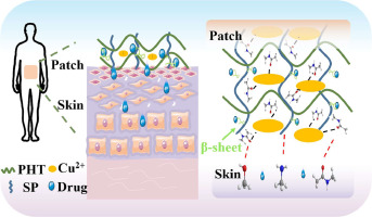
However, the conventional use of exogenous GFs, usually administrated by intravenous injections, is stunted by both safety and efficacy concerns. High dosages or repeated delivery are often required to achieve a desired effect, which results in several side effects and lower the efficacy [25], [26], [27], [28]. Effectively inducing angiogenesis by administrating the angiogenic factors in a manner of physiologically proper times (temporal modulation) and at the proper site (site-specific spatial modulation) targeting remain imperative challenges for engineered drug delivery platforms [29]. Thus, it is necessary to develop a hydrogel patch-type drug delivery system having the tunable release profiles of angiogenic factors, while with soft and flexible properties for the local application in the delicate and fragile brain area.
To fabricate the patch, 3D printing technology could be adopted due to its versatility for manufacturing controllable and customizable patch-type delivery systems in a single and successive process using various types of biomaterials [30], [31], [32]. Many printing parameters (e.g., dimensions and design of the system) and material properties (e.g., synthetic and naturally-derived biomaterials, crosslinking density, and concentration) can be implemented with a desired shape to customize the release profiles [33], [34], [35], [36]. As for the biomaterials, tissue-mimetic decellularized extracellular matrix (ECM)-based hydrogels and hyaluronic acid (HA, also an ECM-based hydrogel as a major component of brain ECM), which affects angiogenesis [37, 38], have been suggested as a viable option [30, 31, 39]. Particularly, we recently demonstrated a vascular tissue-derived decellularized extracellular matrix (VdECM) with capability as a drug delivery carrier. However, the deficiency of printability of single VdECM has made it a challenge to be used as a printable biomaterial ink [29, 40, 41].
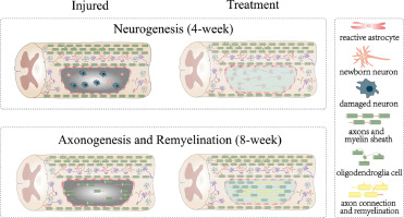
Herein, we fabricated a spatiotemporally compartmentalized cerebral angiogenesis inducing (SCAI) hydrogel patch using a biocompatible and biodegradable ECM-based hybrid hydrogel ink [42, 43] with 3D printing technology for printing sophisticated and spatiotemporally separated patch at once with capability of sequential and sustained release of dual angiogenic GFs (Figure 1). To develop the biomaterial ink, we used an effective chemical crosslinking mechanism at mild conditions, named as aza-Michael addition reaction, to promote the printability of VdECM ink through combining with methacrylated hyaluronic acid (HAMA). Methacrylates (Michael acceptors) in HAMA and amines (Michael donors) in VdECM undergo chemical reaction to form a hybrid ink (HAVEM ink). The aza-Michael addition reaction is an ideal mechanism for fabricating SCAI patch owing to the unnecessary of exogenous catalysts and the facilitating to tailor the printability and release profile of angiogenic GFs by simply changing the chemical crosslinking density. Using two types of HAVEM inks with different chemical crosslinking densities, we were able to fabricate a structure with spatially separated outer and inner layer by 3D printing technique. In addition, we investigated the efficacy for the combination of both GFs (VEGF and HGF), enabling the mimicking of angiogenesis for synergistically inducing more robust blood vessels. Therefore, the outer layer of the SCAI patch was printed with the initial early angiogenic factor VEGF laden HAVEM ink with lower chemical crosslinking density. Moreover, the inner layer was printed using the late angiogenic factor HGF laden HAVEM ink with higher chemical crosslinking density. After printing, the vitrification process to the SCAI patch was applied to make a thin, soft, flexible, and easily manipulable patch for grafting at the brain site. The superior in vitro and in vivo efficacy for the promoted neovascularization were demonstrated for the SCAI patch-based approach. We particularly observed time-dependent angiogenesis behaviors of cerebral microvessels for 14 days after implantation using a label-free, optical-resolution photoacoustic microscopy (OR-PAM) system [44], [45], [46], [47], [48], [49], [50]. The quantified results showed significant neovascularization performance at the brain site. Our dECM based hydrogel patch type approach with spatiotemporally separated dual GFs leads to excellent neovascularization in the brain area, and it could potentially contribute to clinical studies in ischemic diseases requiring induction of the angiogenesis.


留言 (0)