


記住我

A.S., conceptualization, software, investigation, resources, and writing—original draft; Z.W., investigation and writing—original draft; W.Z., writing—review and editing; W.S., investigation and resources; R.C., investigation and resources; J.J., investigation and resources; Z.N., investigation and resources; X.Q., investigation and resources; H.Q., investigation and resources; G.F., project administration and supervision; Y.L., funding acquisition and project administration. All authors have read and agreed to the published version of the manuscript.
Figure 1. Experimental modeling process.
Figure 1. Experimental modeling process.
Figure 2. UPLC-Q-TOF chromatograms of water extract of HP. (A) Variable wave length ultraviolet detector (VWD) chromatogram of water extract of HP in ESI+ mode. (B) Total ion chromatogram (TIC) chromatogram of water extract of HP in ESI+ mode. (C) Extracted ion chromatograms (EIC) chromatogram of water extract of HP in ESI+ mode.
Figure 2. UPLC-Q-TOF chromatograms of water extract of HP. (A) Variable wave length ultraviolet detector (VWD) chromatogram of water extract of HP in ESI+ mode. (B) Total ion chromatogram (TIC) chromatogram of water extract of HP in ESI+ mode. (C) Extracted ion chromatograms (EIC) chromatogram of water extract of HP in ESI+ mode.
Figure 3. Network pharmacology map of the major active components of HP and depression. (A) Venn diagram of related targets of HP and depression. (B) The active components of HP and their corresponding anti-depression targets were imported into Cytoscape software to construct the network diagram of the target of the active component. (C) PPI network of overlapping targets between drug and disease; the size of the circle represents the target degree. (D) KEGG pathway analysis of intersection targets between the main components of HP and depression.
Figure 3. Network pharmacology map of the major active components of HP and depression. (A) Venn diagram of related targets of HP and depression. (B) The active components of HP and their corresponding anti-depression targets were imported into Cytoscape software to construct the network diagram of the target of the active component. (C) PPI network of overlapping targets between drug and disease; the size of the circle represents the target degree. (D) KEGG pathway analysis of intersection targets between the main components of HP and depression.
Figure 4. The EIC chromatogram of components in plasma from rats after administration of HP. (A) The EIC chromatogram of quercetin (C11) in plasma from rats after the last administration. (B) The EIC chromatogram of quercetin (C11) in water extract of HP. (C) The EIC chromatogram of Hyp (C7) and isoquercetin (C8) in plasma from rats after the last administration. (D) The EIC chromatogram of Hyp (C7) and isoquercetin (C8) in water extract of HP.
Figure 4. The EIC chromatogram of components in plasma from rats after administration of HP. (A) The EIC chromatogram of quercetin (C11) in plasma from rats after the last administration. (B) The EIC chromatogram of quercetin (C11) in water extract of HP. (C) The EIC chromatogram of Hyp (C7) and isoquercetin (C8) in plasma from rats after the last administration. (D) The EIC chromatogram of Hyp (C7) and isoquercetin (C8) in water extract of HP.
Figure 5. HP’s main active components (Hyp, isoquercetin, and quercetin) and depression: a network pharmacology map. (A) Venn diagram of the main active components of HP and depression-related targets. (B) The main active components of HP and their corresponding antidepressant targets were imported into Cytoscape software to construct an active component-target network diagram. (C) KEGG pathway analysis of intersection targets for Hyp and depression.
Figure 5. HP’s main active components (Hyp, isoquercetin, and quercetin) and depression: a network pharmacology map. (A) Venn diagram of the main active components of HP and depression-related targets. (B) The main active components of HP and their corresponding antidepressant targets were imported into Cytoscape software to construct an active component-target network diagram. (C) KEGG pathway analysis of intersection targets for Hyp and depression.
Figure 6. Effects of Hyp on depressive-like behaviors of chronic stress-induced mice. (A) Compared with the control group, the CUMS group had a slower rate of body weight growth. Body weight growth was faster in the CUMS+paroxetine and CUMS+Hyp groups than in the CUMS group. (B) Hyp and paroxetine significantly increased the sucrose preference rate of CUMS mice in the sucrose preference test. (C) Compared with the control group, the CUMS group increased the immobility time of TST. TST immobility time was significantly reduced by CUMS+paroxetine and CUMS+Hyp. (D) The immobility time of the FST was increased in the CUMS group compared with the control group. CUMS+paroxetine and CUMS+Hyp significantly decreased the immobility time of FST. Results are shown as the mean from multiple experiments, n = 5, one-way ANOVA, followed by Dunnett multiple comparison test; * p < 0.05, ** p < 0.01, and *** p < 0.001.
Figure 6. Effects of Hyp on depressive-like behaviors of chronic stress-induced mice. (A) Compared with the control group, the CUMS group had a slower rate of body weight growth. Body weight growth was faster in the CUMS+paroxetine and CUMS+Hyp groups than in the CUMS group. (B) Hyp and paroxetine significantly increased the sucrose preference rate of CUMS mice in the sucrose preference test. (C) Compared with the control group, the CUMS group increased the immobility time of TST. TST immobility time was significantly reduced by CUMS+paroxetine and CUMS+Hyp. (D) The immobility time of the FST was increased in the CUMS group compared with the control group. CUMS+paroxetine and CUMS+Hyp significantly decreased the immobility time of FST. Results are shown as the mean from multiple experiments, n = 5, one-way ANOVA, followed by Dunnett multiple comparison test; * p < 0.05, ** p < 0.01, and *** p < 0.001.
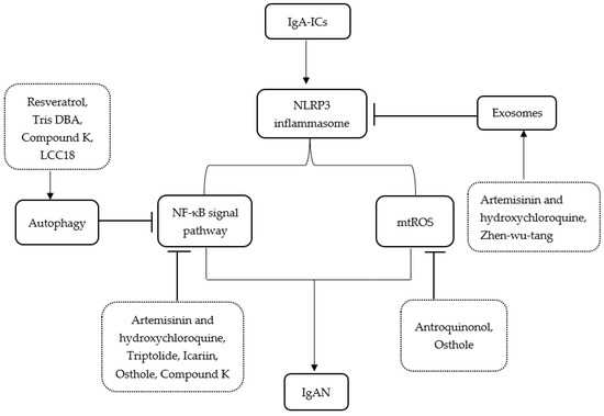
Figure 7. Hyp reduced NLRP1 inflammasome activation and inflammatory response in the hippocampus of chronic-stress-induced mice. (A–C) Statistical results show that Hyp decreased the mRNA expression of (A) NLRP1, (B) ASC, and (C) caspase-1 in the hippocampus of chronic stress-induced mice. (D–G) Statistical results show that Hyp reduced the mRNA levels of (D) IL-6, (E) IL-18, (F) IL-1β, and (G) TNF-α in the hippocampus of chronic-stress-induced mice. Results are shown as the mean from multiple experiments, n = 5, one-way ANOVA, followed by Dunnett multiple comparison test; * p < 0.05, ** p < 0.01, and *** p < 0.001.
Figure 7. Hyp reduced NLRP1 inflammasome activation and inflammatory response in the hippocampus of chronic-stress-induced mice. (A–C) Statistical results show that Hyp decreased the mRNA expression of (A) NLRP1, (B) ASC, and (C) caspase-1 in the hippocampus of chronic stress-induced mice. (D–G) Statistical results show that Hyp reduced the mRNA levels of (D) IL-6, (E) IL-18, (F) IL-1β, and (G) TNF-α in the hippocampus of chronic-stress-induced mice. Results are shown as the mean from multiple experiments, n = 5, one-way ANOVA, followed by Dunnett multiple comparison test; * p < 0.05, ** p < 0.01, and *** p < 0.001.
Figure 8. Hyp reduced CXCL1/CXCR2 mRNA levels and increased BDNF mRNA levels in the chronic-stress-induced mice. (A) Hyp reduced CXCL1 mRNA level expression in chronic-stress-induced mice. (B) Hyp reduced CXCR2 mRNA level expression in chronic-stress-induced mice. (C) Hyp prevents the downregulation of BDNF mRNA levels in chronic-stress-induced mice. Results are shown as the mean from multiple experiments, n = 5, one-way ANOVA, followed by Dunnett multiple comparison test; ** p < 0.01, and *** p < 0.001.
Figure 8. Hyp reduced CXCL1/CXCR2 mRNA levels and increased BDNF mRNA levels in the chronic-stress-induced mice. (A) Hyp reduced CXCL1 mRNA level expression in chronic-stress-induced mice. (B) Hyp reduced CXCR2 mRNA level expression in chronic-stress-induced mice. (C) Hyp prevents the downregulation of BDNF mRNA levels in chronic-stress-induced mice. Results are shown as the mean from multiple experiments, n = 5, one-way ANOVA, followed by Dunnett multiple comparison test; ** p < 0.01, and *** p < 0.001.
Table 1. The PCR primer sequence.
Table 1. The PCR primer sequence.
GeneForward Primer, 5′–3′Reverse Primer, 5′–3′NLRP15-GCTGAATGACCTGGGTGATGGT-35-CTTGGTCACTGAGAGATGCCTG-3ASC5-CTTGTCAGGGGATGAACTCAAAA-35-GCCATACGACTCCAGATAGTAGC-3Caspase-15-TCCGCGGTTGAATCCTTTTC-35-CCTTTCCAACAGGGCGTGAA-3CXCR25-CTCTATTCTGCCAGATGCTGTCC-35-ACAAGGCTCAGCAGAGTCACCA-3CXCL15-ACTGCACCCAAACCGAAGTC-35-TGGGGACACCTTTTAGCATCTT-3BDNF5-TCATACTTCGGTTGCATGAAGG-35-AGACCTCTCGAACCTGCCC-3ACTIN5-TTCCTTCCTGGGTATGGAAT-35-GAGGAGCAATGATCTTGATC-3IL-65-CTGCAAGAGACTTCCATCCAG-35-AGTGGTATAGACAGGTCTGTTGG-3IL-185-TATCGACCGAACAGCCAACG-35-GATAGGGTCACAGCCAGTCC-3IL-1β5-GAAATGCCACCTTTTGACAGTG-35-TGGATGCTCTCATCAGGACAG-3TNF-α5-CCTGTAGCCCACGTCGTAG-35-GGGAGTAGACAAGGTACAACCC-3 Table 2. Main chemical constituents of water extract of HP [28,29,30]. Table 2. Main chemical constituents of water extract of HP [28,29,30]. NO.NameFormulaRT/MinCal [M-H]-[M-H]-Diff (ppm)Fragment IonsC1Quinic acidC7H12O61.18 191.0560191.0561 0.52173.0452,127.0396,111.0083C2PyrocatecholC6H6O22.42109.0294109.0292 −1.8390.0111,80.0262,79.0184C3Chlorogenic acidC16H18O93.28 353.0878353.0875 −0.79191.0562,173.0454,135.0447C4Caffeic acidC9H8O44.17 179.0350179.0349 −0.34135.0450,134.0372,111.0079C5CatechinC15H14O64.77 289.0717289.0712 −1.87245.0811,123.0448,125.074C6RutinC27H30O168.78 609.1461609.14640.53343.0466,300.0278.301.0347C7HyperosideC21H20O128.83 463.0882463.08830.26300.0279,.0346,271.0247C8Isoquercetin C21H20O129.28463.0882463.0883 0.26300.0275,301.0342,271.0239C9Kaempferol 3-O-glucosideC21H20O1110.65447.0933447.0933 0.09300.0281,301.0350,271.0246C10Guaijaverin C20H18O1111.05433.0776433.07811.13300.0278,301.0348,271.0247C11Quercetin C21H20O1111.72447.0933447.09411.88300.0283,301.0342,271.0247C12ProtohypericinC30H18O814.72 505.0971505.0980 1.78300.0268,301.0345,271.0243
留言 (0)