
記住我


Our inclusion criteria are single thoracolumbar burst fracture with AO type A3, A4, B1, and B2 with/without neurological deficit, treated by short-segment fixation, by ligamentotaxis, of the vertebra above the injured one and the one below it between September 2006 and August 2021 in five institutions. We excluded patients with AO type B3 and C, and multiple thoracolumbar burst fractures. Two hundred and fifty-three patients met these criteria. The protocol for our retrospective study was approved by the institutional review boards of all hospitals where the procedures took place, and all methods were carried out in accordance with relevant guidelines and regulations. The requirement for informed consent was waived because of its retrospective and observational study, and it is approved by institutional review boards.
The patients consisted of 140 males and 113 females with an average age of 43 years (range, 13 to 69 years). Their injuries were caused by falls from a significant height (169 patients), traffic accidents (64 patients), being hit by a falling object (9 patients), falling from horses (8 patients), and skiing accidents (3 patients). Thus, all sustained high-energy injuries. The level of spine involvement was T11 in 8 patients, T12 in 33, L1 in 110, L2 in 68, and L3 in 34.
Outcome measuresRadiographic assessment was performed using supine anteroposterior and lateral roentgenograms and computed tomography (CT) scans before surgery. All patients were monitored radiographically by the use of standing or sitting anteroposterior and lateral roentgenograms and CT scans within 1 week after surgery. Five independent spine surgeons evaluated all radiographs and CT scans. The sagittal plane contour was assessed by measuring the vertebral body angle (VBA), which was the Cobb angle between the superior and inferior endplates of the injured vertebra, as we used in previous studies [9,10,11]. From these data, we calculated the reduction angle (preoperative VBA − postoperative VBA) and the reduction rate (%) using the following formula:
$$\left[ }\;}}\;}} \right)/}\;}} \right] \, \times \, 100$$
When the reduction rate exceeded 100%, we counted that as 100% (Fig. 1).
Fig. 1
a, b Lateral radiographs from a 56-year-old man with an L1 burst fracture, showing changes before and after surgery. a The vertebral body angle (VBA) was 31° before surgery. b Surgery corrected the VBA to 3°. Therefore, the reduction angle was 28°, and the reduction rate was calculated as being 90% ([31 − 28/31] × 100). c, d Lateral radiographs from a 38-year-old man with an L1 burst fracture. c The VBA was 9° before surgery. d Surgery corrected the VBA to 0°. Therefore, the reduction angle was 9°, and the reduction rate was calculated as being 100% ([9 − 0/9] × 100). Thus, the reduction rate is suitable for patients with a minor deformity before surgery
Canal compromise was determined using CT scanning by directly measuring the anteroposterior canal dimension (in millimeters) at the maximum area of the retropulsed osseous fragment or fragments. This value was then compared with the average of similar dimensions measured above and below the injury. The result of this comparison was recorded as the canal compromise ratio (%) at the injured vertebra. Fracture severity was calculated using the load-sharing classification [6] and the AO classification [12].
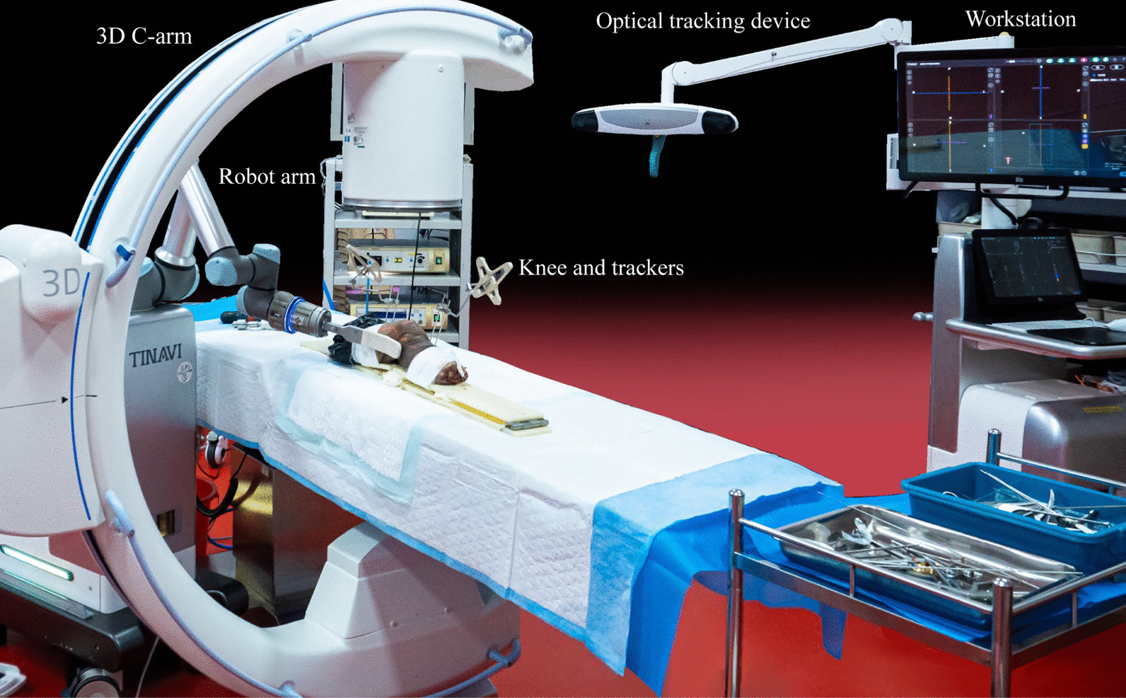
Surgical techniquesAll surgical procedures were performed with the patients under controlled general anesthesia. Patients were placed in a prone position; initial postural reduction was then obtained. Pedicle screws were placed down into the pedicles of the bilateral vertebrae above and below the fracture. When we used Schanz pedicle screws, posterior wall decompression by indirect reduction via ligamentotaxis was performed, and for all 253 patients, segmental distraction by screws was performed. The surgical techniques used have been described in detail elsewhere [9, 10]. We performed additional vertebroplasty in 116 patients (46%). For fixation, we used Schanz pedicle screws (AO Universal Spine System, DePuy Synthes, West Chester, PA, USA) in 203 patients, the CD Horizon Longitude fixation system (Medtronic Sofamor Danek, Memphis, TN, USA) in 33, and the ES2 spinal system (Stryker, Kalamazoo, MI, USA) in 17.
Statistical analysisWe used SPSS statistical software (version 21.0; IBM, Armonk, NY, USA) for all analyses; statistical significance was set at a p value of < 0.05. Group comparisons were conducted using Welch’s exact test for dichotomous variables. The correlation coefficient between two continuous or ordinal variables was analyzed using Spearman’s rank correlation coefficient test. Guilford [13] describes correlation coefficients of < 0.20 as being interpreted as “slight, almost negligible relationships”; correlations of 0.20 to 0.40 as “low correlation”; of 0.40 to 0.70 as “moderate correlation”; of 0.70 to 0.90 as “high correlation, marked relationship”; and of > 0.90 as “very high correlation, very dependable relationship.” To determine predictors of insufficient reduction, which was measured as the reduction angle and the reduction rate, we performed multiple linear regression analyses with stepwise selection. Before performing those analyses, we confirmed that the correlation coefficient between any two independent variables was < 0.7.
The correlated factors studied were age, gender, time elapsed between injury and surgery, affected level (T11–L1 vs L2 and L3), combination of vertebroplasty with surgery, AO classification (types A3 and A4 vs type B), LSS, preoperative VBA, and the ratio of canal compromise before surgery.
留言 (0)