
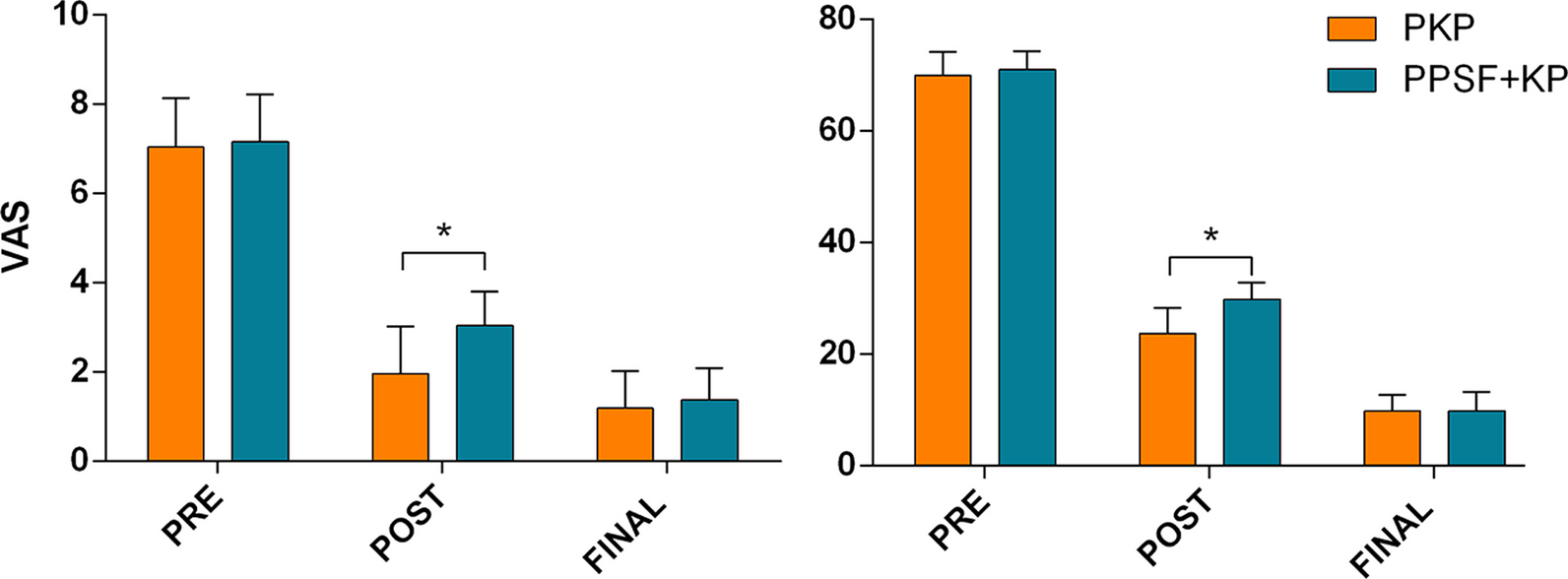
記住我


Navigation technology has developed rapidly in recent years. Current evidence suggests that the accuracy of robotic screws placement or intraoperative CT navigation is higher than free-hand or intraoperative X-ray navigation [19]. However, this navigation equipment is expensive, and most spinal surgeons still place pedicle screws free-hand or with intraoperative X-ray. The accuracy of free-hand screw placement is not ideal, especially for L5. An increasing body of evidence suggests that during placement of L5 segment, it should be directed outwards and upwards [5, 20]. Measurement and analysis of CT images suggest that the entry points for L5 have a tendency to be inward, requiring either the “^”-shaped crest or Magerl technique.
At present, the commonly used screw placement entry points use the superior articular process and transverse process as the reference. However, the transverse process of the lower lumbar spine is deep and sometimes blocked by the iliac crest and pulled by the erector spine muscle; accordingly, it is difficult to fully expose the root of the transverse process during surgery. Therefore, the morphology of the superior articular process and its position relative to the spinal canal directly affect the accuracy of pedicle screw placement. When the superior articular process faces inward, the insertion point is also inward relative to the spinal canal. In this case, screw placement at an angle of 10–15° can cause the screw to penetrate the inner wall of the pedicle (Fig. 5). Importantly, we found that when the MCD was less than 6 mm, the screw placement failure rate increased significantly. Evaluation of the preoperative CT showed that the STAmax of screw placement was only 1–7°. For these patients, the angle of insertion should be abducted if the screws are placed at commonly used entry points.
Fig. 5
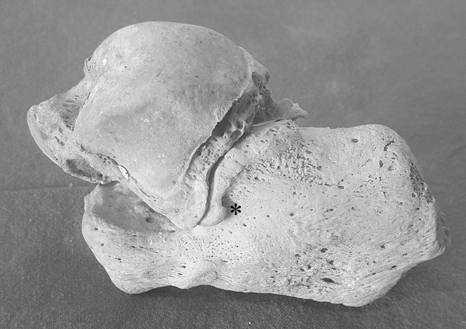
The vertical line through the mammillary process is medial to the midpoint of the pedicle
In fact, such cases are not uncommon. More than 10% of the 299 patients (n = 36) presented with a low MCD, which may be attributed to selection bias caused by the smaller ZJA [21].
All 299 patients included in this study were patients with lumbar disc herniation or lumbar spinal stenosis. Although patients with severe facet arthritis were excluded at the time of inclusion, changes in facet joint morphology could affect the final measurements [22, 23]. The ZJA at L5 is generally about 45° and does not exhibit significant heterogeneity across different populations. The reported average ZJA in Indian [24], Chinese [25], and French [26] populations is 48.32°, 47.7°, and 44.4°, respectively. In the present study, the average ZJA of L5 (n = 598) was 46.3°, consistent with the literature. However, the ZJA in group A decreased significantly to 33.7°. Morphologically, this type of facet joint is more similar to the superior facet of L4. When the L5 superior facet became more sagittal than horizontal, it increased susceptibility to degenerative lumbar diseases, including lumbar disc herniation and degenerative lumbar spondylolisthesis [22, 27]. Excessively small vertebral bodies and steep facet joints were associated with lumbar instability, while excessively steep facet joints could accelerate the rate of disc degeneration [28].
Moreover, we found that the small distance between superior articular processes of L5 was associated with a small MCD. A study by Santiago et al. where the sagittal diameters of lumbar vertebrae and articular processes of 39 healthy Spaniards were measured showed the distance between L5 superior articular processes was 22.2 mm ± 0.40 mm [29]. Consistently, Oguz et al. measured the lumbar vertebrae of 24 healthy Turks, and the distance between L5 superior articular processes was 21.7 mm ± 2.8 mm [30]. In this study, the inter-facet distance in group A was only 17.8 mm ± 1.4 mm, indicating that the superior articular processes of group A are closer to each other than in other groups. In addition, the SAW was also smaller in group A compared with BCD, although this reduction was not statistically significant on one side. Compared with the normal superior articular process, the superior joint of group A exhibited a steeper and sharper variation. The steep superior articular process tended to narrow the spinal canal compared to the gentle superior articular process. The L5 spinal canal of this type of superior articular process typically exhibits a “clover” shape, with the superior articular process extending inward (Fig. 6).
Fig. 6
Illustration demonstrating the difference between the two types of superior articular processes. In groups with low MCD (B), the superior articular processes are closer to each other and rotate inward compared to groups with normal MCD (A). At this point, the spinal canal exhibits a "clover" shape
IPD is also a factor that affects the relative position of the entry point. However, the IPD in group A was only 28.3 ± 2.2 mm and did not increase significantly. Meanwhile, the overall mean IPD was 28.4 mm ± 2.3 mm, which was not significantly different from the reported average transverse diameter in Chinese (29.61 mm ± 0.63 mm) and Indian (28.02 mm ± 0.37 mm) populations in the literature [31].
Mitra, Datir, and Hou et al. observed a sequential outward migration of the central entry point of the lumbar pedicle from L1–L4 [14, 32]. At L5, the entry point is located lateral to the lateral boundary of the facet joint. However, if the angle of the superior facet of L5 is small with no “abduction,” the usual Magerl entry point is inward-facing. In some medical centers, to avoid this kind of situation, the root of the transverse process is adopted for screw insertion, which is more lateral than Margel entry point, with an accuracy rate of > 98% [33]. However, a more external entry point means more muscle dissection, bleeding, and longer surgical time. Although screw placement failure was avoided in a small number of patients, the cost-effectiveness of this approach needs further study. Based on the current Magerl or “^”-shaped crest insertion methods, preoperative estimation of excessive orientation of the L5 superior articular process is a cost-effective method. When the MCD is less than 6 mm, the internal angle of screw placement should be reduced, and vertical entry into the vertebral body may be required.
留言 (0)