Colorectal cancer is the second most common cause of cancer death in the world, resulting in approximately 935,173 deaths in 2020 alone (https://gco.iarc.fr/). The incidence of colorectal cancer is steadily rising in developing countries and in younger patients [1], [2], [3]. Traditional treatments such as surgery, chemotherapy, and radiotherapy are the standard care therapies used for colorectal cancer [4]. However, most lethal colorectal cancer is diagnosed at advanced stages, after suitable times for surgical resection, and presents with unresectable tumors or those that recur after surgery [4]. Chemotherapy and radiotherapy are also challenged by the unsatisfactory efficacy as well as potential serious side effects [4].
In recent years, immune checkpoint blockade therapy, especially the blockade of programmed death-1 (PD-1)/programmed death-ligand 1 (PD-L1) pathway, has emerged as a promising approach for colorectal cancer therapy [5], [6], [7]. By disrupting the inhibitory immune checkpoint signals, immune checkpoint inhibitors augment organism's own immune system to fight tumors [8, 9]. To date, two PD-1/PD-L1 inhibitors (Pembrolizumab and Nivolumab) have been approved by the Food and Drug Administration (FDA) for the treatment of mismatch repair deficient and microsatellite instability-high (dMMR/MSI-H) colorectal cancer [10, 11]. Despite the unprecedented durable response rates that have been observed with PD-1/PD-L1 blockade in some patients with colorectal cancer, many patients are still resistant to PD-1/PD-L1 inhibitors and some responders may relapse after a period of response [12, 13]. The most straightforward reason for this resistance to PD-1/PD-L1 inhibitors is the immunosuppressive tumor microenvironment [14]. The immunosuppressive tumor microenvironment is composed of immunosuppressive cells and immunosuppressive mediators, and is typically characterized by several elements, such as extensive exhaustion in CD8+T cells, poor infiltration of lymphocytes, and the accumulation of suppressive immune cells [14, 15]. Hence, strategies aimed at remodeling the tumor microenvironment may help evoke anti-tumor responses and improve the efficacy of PD-1/PD-L1 inhibitors in the treatment of colorectal cancer.
Photothermal therapy (PTT), which utilizes photothermal agents to convert optical energy into heat energy for tumor ablation, is a remotely controlled and minimally invasive tumor therapeutic strategy [16, 17]. Recently, the emergence of “photothermal immunotherapy” has expanded its application [18], [19], [20]. Tumor-associated antigens (TAAs) released by tumor cells after photothermal ablation can be recognized by dendritic cells (DCs) and thus enhance the chemotaxis and activation of immune cells [21, 22]. Additionally, local heating in tumor sites increases tumor vascular permeability and tumor perfusion, which promotes the adequate infiltration of immune cells [23]. Overall, these effects of PTT tend to reverse the immunosuppressive tumor microenvironment, which are helpful for enhancing the efficacy of immune checkpoint inhibitors. Therefore, the combination of PD-1/PD-L1 blockade and photothermal ablation may have synergistic anti-tumor effects. Of note, previous studies have indicated that enhancing the accumulation of photothermal agents in tumor sites can significantly improve therapeutic efficacy and minimize side effects; thus, identifying practical methods to avoid immune clearance, prolong blood circulation, and enhance tumor targeting of photothermal agents is also crucial [24].
Cell membrane-coated nanoparticles are a promising biomimetic approach for drug delivery, which integrate the biophysiological advantages of the host cell membrane with the engineering versatility of synthetic nanomaterials [25], [26], [27]. By fusing a layer of cell membrane onto a synthetic core, nanoparticles are endowed with the complex components and unique biological functions of original cell membrane [28]. Biomimetic nanoparticles that inherit biophysiological features of original cell membrane can be recognized as self-elements to evade immune clearance and prolong their systemic circulation [28]. Additionally, by utilizing natural or engineered targeting ligands from an original cell membrane, biomimetic nanoparticles can bind to cells that overexpress corresponding antigens [29]. Moreover, taking advantage of the fact that most signal transmission begins with ligand-receptor interactions on cell membrane, biomimetic nanoparticles have been applied to block interactions by coating with cell membrane that overexpress neutralizing antibodies, corresponding ligands, or specific receptors [30], [31], [32].
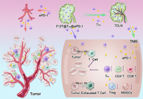
Inspired by cell membrane-coated nanoparticle delivery systems, we designed functionalized biomimetic nanoparticles that combined PD-1/PD-L1 blockade with photothermal ablation for the synergistic treatment of colorectal cancer. Due to the large absorption surface, strong near-infrared (NIR) absorption, efficient photothermal conversion ability, and extensive photothermal stability, polydopamine-modified gold nanostar nanoparticles (PDA-GNS) were chosen as the core for these biomimetic nanoparticles [33]. Then, cell membrane isolated from anti-PD-L1 single-chain variable fragment (scFv) engineered cells was fused to the surface of these PDA-GNS cores to construct anti-PD-L1 functionalized biomimetic polydopamine-modified gold nanostar nanoparticles (PDA/[email protected] NPs) (Scheme 1). The resulting PDA/[email protected] NPs, which inherited anti-PD-L1 scFv from original cells, could specifically bind to the PD-L1 on tumor cells. Consequently, these PDA/[email protected] NPs could disrupt PD-1/PD-L1 immunosuppressive signals and actively accumulate in tumor sites. Additionally, PDA-GNS as the cores of these PDA/[email protected] NPs were carried to tumor sites, which were conducive to generate heat energy for tumor photothermal ablation after NIR irradiation. The photothermal ablation induced by PDA-GNS could trigger immune-stimulatory responses in DCs to further activate anti-tumor immune responses and alter the immunosuppressive tumor microenvironment. Meanwhile, hyperthermia increases the tumor vascular permeability and tumor perfusion, which was of benefit for increasing the infiltration of immune cells and enhancing the accumulation of nanoparticles at tumor sites. As a result, by remodeling the tumor microenvironment, PDA-GNS sensitized tumors to PD-1/PD-L1 blockade therapy.
In this study, PDA/[email protected] NPs showed good biocompatibility, efficient photothermal conversion ability, and enhanced tumor-targeting ability. In murine colorectal cancer models, a PDA/[email protected] NP-based therapeutic strategy could promote the maturation of DCs, increase the infiltration of CD8+T cells, decrease the number of immunosuppressive cells (such as regulatory T cells and myeloid-derived suppressive cells), and induce the release of a variety of cytokines (such as IFN-γ and TNF-α) to remodel the tumor microenvironment. Moreover, a PDA/[email protected] NP-based therapeutic strategy exerted enhanced therapeutic efficacy, which could significantly inhibit tumor growth and prolong overall survival. Taken together, our results demonstrated that a PDA/[email protected] NP-based therapeutic strategy had enhanced synergistic anti-tumor capabilities by combining PD-1/PD-L1 blockade with photothermal ablation, which highlights the potential of PDA/[email protected] NPs to remodel the tumor microenvironment for effective clinical colorectal tumor therapy.


留言 (0)