Bone metastasis is a common clinical impairment in patients with advanced breast cancer, which is a multistep process that involves escape from the primary tumor, entry into microvessels, movement in the circulation, colonization and proliferation in bone [1]. Once tumor cells metastasize to bone, they cooperate with local bone cells such as osteoclasts in bone microenvironment to promote the progression of bone metastasis [1, 2]. Commonly used therapeutic strategies such as chemotherapy are particularly effective in inhibiting tumor growth, but their ability to inhibit osteoclast activity is limited [3]. Anti-resorptive agents such as bisphosphonates can inhibit osteoclast-mediated bone resorption. However, they lack antitumor activity and even cause osteonecrosis and hypocalcemia of the jaw [4]. Therefore, it is very necessary to develop a strategy that can inhibit both tumor growth and osteoclast resorption. Compared with traditional therapeutic strategies, nanoparticles (NPs)-based photothermal therapy (PTT) have become an attractive therapeutic technology for solid tumors because of their minimal invasiveness, high precision, operational controllability and low side effects to normal tissues [5, 6]. Based on the advantages of PTT, various photothermal reagents had been developed for the treatment of bone tumors. In general, hyperthermia (over 50°C) induced by PTT agents can effectively hinder tumor growth. Nevertheless, accidental ambustion caused by hyperthermia to healthy tissue nearby may trigger inflammatory responses. Besides, nonspecific thermal diffusion that is induced by hyperthermia may destroy the surrounding blood vessel, leading to block the accumulation of therapeutic drugs in deep tumors [7]. Therefore, it is of great significance to design an advanced PTT nanoparticle to eliminate the treatment bottleneck of traditional PTT for bone metastasis.
To overcome the side effects of traditional PTT, mild-PTT (∼ 45°C) recently is developed for tumor treatment [8]. This mild temperature can kill tumor cells with few damage to the surrounding tissue. But the cell apoptosis caused by relatively mild temperature may be restored because tumor cells usually exhibit resistance to photothermal by varying degrees, resulting in ineffective treatment [9]. Besides, the laser strength attenuated with the increase of tumor depth [10], in which the penetration depth decreased as well. Furthermore, the mild heat treatment could up-regulate the expression of heat shock protein 90 (HSP 90) and programmed cell death ligand 1 (PD-L1) in tumor cells, which could lead to photothermal resistance and immunosuppressive microenvironment [11, 12]. Previous studies have indicated that photothermal resistance is mainly triggered by an intracellular self-protection mechanism named ‘autophagy’ [13], who is an evolutionarily conserved lysosomal pathway [14]. And the duration and severity of autophagy can result in different fates of tumor [15, 16]. Specifically speaking, the “mildly activated autophagy” induced by mild-PTT can trigger an intracellular self-protection mechanism through removing damaged organelles caused by therapeutic stress in tumor cells [17]. Although inhibition [18] or overactivation [19] of autophagy has been reported to amplify therapeutic efficacy of mild-PTT by reversing the photothermal resistance of tumor cells, the strategy of autophagy activation may be more effective in the treatment of metastatic bone tumors [20, 21]. Deng et al [22] found that restoring mitophagy function could effectively inhibit bone metastasis and prolong the survival time of tumor-bearing mice. Therefore, the design of an advanced mild-PTT nanoparticle with efficient autophagy activation and reversed immunosuppressive microenvironment was expected to be a highly attractive and advantageous treatment strategy for bone metastasis.
In order to improve the efficacy of mild-PTT, small molecule drugs [23] or small interfering RNA [24] have been explored as combined strategies to transform “mildly activated autophagy” triggered by mild-PTT to “overactivated autophagy”. Compared with small interfering RNA, small molecule drugs have multiple merits such as convenient in use, low price and long-term stability. Compared with other small molecule drugs with autophagy activation ability, Curcumin (Cur), a potential antitumor phytochemical isolated from Curcuma longa, also inhibited osteoclastogenesis and expression of PD-L1 protein, improving the bone microenvironment [25, 26]. Cur may be a candidate drug that can effectively enhance cell apoptosis caused by PTT agents upon irradiation, reverse immunosuppressive microenvironment and inhibit bone resorption. The ideal PTT system should have the characteristics of easy preparation, high photothermal conversion efficiency and good biocompatibility. At present, researches explored multiple PTT reagents, such as carbon nanotubes [27], graphene oxide [28] and gold-based NPs [29] for tumor treatment. However, they still have shortcomings such as high cost, difficult biodegradation and complex synthesis process, which usually limit their clinical large-scale use [30]. Gallium based-liquid metals (LMs), as new functional materials, have attracted extensive attention in biomedical applications due to their favorable biocompatibility, biodegradability and intriguing physiochemical properties [31], [32], [33], and have become potential PTT reagents in tumor therapy [34, 35]. However, naked LM NPs exhibited serious stability defects due to fluidity and biodegradation under a mild acid tumor microenvironment, diminishing photothermal performance with the extension of irradiation time [36, 37]. Although various strategies have been explored to improve the stability of LM, mainly including organic materials such as thiolate ligands [38] or inorganic materials exampling silica shell [37]. Low drug loading rate, lack of on-demand drug release performance or poor degradation was the main obstacles for their application. Zeolite imidazole frame-8 (ZIF-8), a metal-organic framework nanomaterial with high biocompatibility, pH-response drug release and high loading capacity, has been widely explored in the delivery of therapeutic reagents [39].
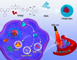
Herein, a ZIF-8 functionalized LM-based nanoparticle was successfully designed to both inhibit tumor growth and bone resorption by modulating the bone microenvironment and activating autophagy “on demand”. As shown in Scheme 1, a core-shell structure was constructed from zeolitic imidazolate framework-8 (ZIF-8) and LM NPs modified by silicon layer, in which the autophagy activator Cur was enclosed inside ZIF-8. Then, the acquired substance was functionalized with hyaluronic acid (HA)/alendronate (ALN) (obtained as CLALN). The ZIF-8 coating was designed to achieve specific release of Cur in tumors. And the purpose of HA/ALN functionalization was to overcome physiological barriers, prevent premature drug leakage, and then enhance the accumulation of CLALN in bone metastatic tumor region and ingestion of target cells. After intravenous injection, CLALN would be immediately decomposed and released autophagy activator Cur due to its pH-response, which was accompanied by triggering mild autophogy level. Next, the mild temperature was applied to rapidly induce “overactivated autophagy” through the combination of Cur and PPT, which would successfully realize autophagic cell death. Meanwhile, Cur would show anti-bone resorption effect in combination with ALN in bone microenvironment. More importantly, CLALN would inhibit the expression of PD-L1 protein and recruit more active immune T cells, thereby alleviating the immunosuppressive microenvironment induced by mild-PTT. This well-designed CLALN with efficient photothermal conversion efficiency and autophagy activation was discussed in detail in vitro and in vivo.


留言 (0)