1. IntroductionBecause of favorable mechanical properties and biocompatibility, nanomaterials have been applied to many aspects of our life and work, such as biomedicine [
1], the food industry [
2], and electronics [
3]. The toxicity of nanomaterials has been increasingly studied [
4,
5]. Titanium dioxide nanoparticles (TiO2 NPs) are one of the most widely used nanomaterials. A study detected the titanium particles in a human post mortem liver and spleen and found that more than 24% of TiO2 was nanoscale [
6]. More and more studies have found that TiO2 NPs can be cytotoxic [
7,
8] and genotoxic [
9]. Oxidative stress was induced through the stimulating redox interactions, leading to DNA damage and genomic instability [
10,
11]. Moreover, there is a growing interest in the in vitro epigenetic changes induced by TiO2 NPs [
12,
13]. TiO2 NPs are one of the most commonly used nanomaterials in food additives, pharmaceuticals, and personal hygiene products, such as toothpaste [
14], so oral exposure is more likely to happen. The liver is a multicellular organ that plays an important role in activating and eliminating many metabolites; therefore, the liver is the primary target organ of oral exposure to TiO2 NPs [
15,
16,
17,
18]. Many in vivo experiments found that oral exposure of TiO2 NPs can cause liver damage, hepatocyte necrosis, and liver function damage in mice [
19,
20]. Moreover, some studies concluded that acute toxicity of rats with TiO2 NPs induced adverse effects in the liver [
21,
22]. However, the key mechanism of hepatotoxicity induced by TiO2 NPs is not been fully understood and needs further study.In addition to cytotoxic and genotoxic effects, nanoparticle-induced epigenetic changes and the epigenetic mechanisms behind observed toxicity have also attracted increasing attention. Some studies have found that exposure to nanomaterials can lead to epigenetic changes [
23,
24]. Pogribna et al. investigated the effect of TiO2 NP exposure on histone modifications, a major epigenetic mechanism in human colorectal (Caco-2) and lung (NL20) epithelial cell lines, and found changes in several histone modifications after exposure to TiO2 NPs [
23]. Epigenetics is an important link between genotype and phenotype and plays a key role in the regulation of numerous cellular processes. The main mechanisms of epigenetics include DNA methylation, histone modification, and non-coding RNAs [
25]. Non-coding RNAs (ncRNAs) refer to functional RNA molecules that cannot be translated into proteins, among which common regulatory non-coding RNAs include microRNAs (miRNAs), PIWI-interacting RNAs (piRNAs), and long non-coding RNAs (lncRNAs). Many ncRNAs can regulate gene expression through interactions with other epigenetic processes, such as histone modification, chromatin remodeling, and DNA methylation [
26,
27].LncRNAs are non-coding RNAs whose transcript lengths range from 200 nt to 100 kb and are one of the key factors in gene transcriptional regulation, affecting all aspects of cellular homeostasis [
28]. LncRNAs affect nearly all fundamental processes in living cells, including chromatin formation, replication, transcription, splicing, translation, and post-translational modification, and constitute the richest part of the transcriptional genome [
29]. According to the position of lncRNA in the genome to nearby messenger RNAs (mRNAs), lncRNAs can be divided into the following five types [
30]: long intergenic noncoding RNAs (lincRNAs), natural antisense transcripts (NATs), overlapping, bidirectional, and sense intronic. LincRNAs are open chromatin structures with transcripts less than 10 kb in length and do not appear at any protein-coding site [
31]. LincRNAs are also the most numerous of the lncRNA types [
32]. NATs are lncRNAs that block splice-site recognition and recruit epigenetic modifiers [
33]. Overlapping transcripts are transcribed in the same direction as a protein-coding gene and contain one protein-coding gene [
32]. Bidirectional transcripts compete for transcription initiation and promote chromatin modification of target genes [
34]. Sense intronic transcripts are introns derived from a protein-coding gene and their transcription direction is the same as that of the neighboring protein-coding gene [
30]. A significant amount of evidence suggests that lncRNAs regulate gene expression in multiple ways on the levels of epigenetic, chromatin remodeling, transcription, and translation [
35] and they are potential biomarkers for diagnosing, prophesizing, and monitoring disease progression [
36,
37,
38]. Because of the lower levels of splicing, polyadenylation, and nuclear localization, it is more complex to detect and quantify lncRNAs [
35].
To fully assess the toxicity of TiO2 NPs, it is critical to assess the epigenetic role of TiO2 NPs. However, there is no report yet to explore the function of lncRNAs in the process of TiO2 NP-induced toxicity. Therefore, this study treated HepG2 cells with 100 μg/mL TiO2 NPs for 48 h and investigated the changes in lncRNAs.
2. Materials and Methods 2.1. Characterization of NanomaterialsThe TiO2 NPs used in this study were obtained from Shanghai Macklin Biochemical Co., Ltd. (Shanghai, China). The detailed characterization methods and physicochemical properties of TiO2 NPs were described in our published paper [
39]. JEM–1400 electron microscope (JEOL Company, Tokyo, Japan) was used to measure the equivalent diameter. X-ray powder diffractometry (XRD, PANalytical’s X’Pert PRO, X’Celerator, Almelo, The Netherlands) was used to test the crystal form. Dynamic light scattering instrument Zetasizer Nano ZS90 (Malvern Instruments Ltd., Malvern, UK) was used to measure the hydrated particle size and Zeta potential in the serum-free medium containing 1 mg/mL TiO2 NPs. 2.2. Cell Culture
Human hepatocellular carcinoma cells (HepG2), obtained from the National Biomedical Experimental Cell Resource Library of China, were routinely cultured in Minimum Essential Medium (MEM, HyClone, Thermo Scientific, Logan, UT, USA) supplemented with 10% fetal bovine serum (FBS, Hyclone, Thermo Scientific, Logan, UT, USA), 1% MEM Non-Essential Amino Acids Solution (100×) (NEAA, Gibco, Thermo Scientific, Logan, UT, USA), and 2% GlutaMAX-1 (Gibco, Thermo Scientific, Logan, UT, USA). For subculturing purposes, the cells were digested by 0.25% trypsin and seeded to 96-well plates at a density of 1 × 10⁴ cells per well or 60 × 15 MM plates with 5 × 10⁵ cells per well.
2.3. Cytotoxicity Assay Study
Cell Counting Kit-8 assay (CCK-8, Biotopped, Dojindo Laboratories, Kumamoto, Japan) was used to determine the cytotoxicity of TiO2 NPs, based on the measurement of the amount of methotrexate generated proportional to the number of living cells. After exposure to 0, 1.5625, 3.125, 6.25, 12.5, 25, 50, 100, and 200 μg/mL TiO2 NPs for 48 h, the cells in the 96-well plate were incubated with CCK-8 solution for 2 h. After collecting the supernatants, a microplate reader was used to detect the value of absorbance at 450 nm, taking 600 nm as a parameter. The computation formula is as follows: cell viability = (E − B)/ (C − B). E refers to the experimental hole (containing cell, culture medium, CCK 8, and different concentrations of TiO2 NPs), C refers to the control hole (containing cell, culture medium, and CCK 8), and B refers to a blank hole without any cells and TiO2 NPs.
2.4. Construction of cDNA Libraries and RNA Sequencing
Every control and treatment group set up three repeat samples and then the samples were collected for RNA extraction. The extracted total RNA was qualified by Agilent 2100 Bioanalyzer (Agilent Technologies, Santa Clara, CA, USA) and purified by RNAClean XP Kit (Cat A63987, Beckman Coulter, Inc., Kraemer Boulevard, Brea, CA, USA) and RNase-Free DNase Set (Cat#79254, QIAGEN, GmBH, Dusseldorf, Germany).
The purified total RNA was carried out with rRNA removal, fragmentation, first-strand cDNA synthesis, second-strand cDNA synthesis, end repair, 3′ end plus A, ligation joint, and enrichment. The cDNA was then sequenced with a high-throughput sequencer (Illumina Hiseq 2000/2500, San Diego, CA, USA).
2.5. Identification and Quantification of lncRNAsGffcompare (version 0.9.8) was applied to identify new transcripts that did not match known annotations and three types of transcripts were picked out with the conditions that transcription length was greater than or equal to 200 bp, the number of exons was greater than or equal to 2, and open reading frame (ORF) was less than 300 bp. Then, Contrastive Predictive Coding analysis (CPC), Coding-Non-Coding Index (CNCI) analysis, and Pfam protein domain analysis were performed to predict the lncRNAs. CPC used supervised machine learning to establish a classification model by learning peptide chain length, amino acid composition, protein homology, secondary structure, protein alignment, or expression [
40]. Its classification model was mainly based on the characteristics of sequence ORF length and protein homology; Pfam was a large database of protein family collections, represented by multiple sequence alignments and hidden Markov models (HMMs) [
41]. The assembled transcript sequence was annotated by the PfamScan tool. If the sequence matched the Pfam protein database, it was mRNA, and there was no comparison on lncRNA. CNCI identified coded and non-coding sequences by analyzing adjacent nucleotide triplets [
42]. Then the transcript with CPC score
http://www.noncode.org/, accessed on 15 November 2020) and the known lncRNAs in the Ensembl database to form the lncRNA sequence for subsequent analysis.
String tie (version: 1.3.0) was applied to quantify the expression of lncRNA sequences. Then edgeR was applied for differential lncRNA analysis between samples and the p-value was corrected through a multiple-hypothesis test, and the q-value was the corrected p-value by controlling FDR (False-Discovery Rate). The differential expression multiple fold change was calculated based on the FPKM value. The differential lncRNA filters were as follows: q-value ≤ 0.05 and fold change ≥ 2.
The structure of lncRNA and mRNA was compared and analyzed by comparing the differences in transcript length, exon number, and expression level of lncRNA and mRNA. The difference between lncRNA and mRNA molecules was obtained and the predicted lncRNA molecules were verified.
Trans regulation and Cis regulation were used for target gene prediction. Cis referred to how lncRNA regulated neighboring mRNAs (e.g., on the same chromosome) and trans referred to targets at the distal position of chromosomes after different chromosomes.
Finally, KEGG enrichment (
https://www.kegg.jp/, accessed on 15 November 2020) was used to analyze the target gene analysis of the differential lncRNA. The selected differentially expressed genes were mapped to each term of the KEGG database, the number of genes for each entry was calculated, and then a super geometric test was applied to a threshold of p-value ≤ 0.05 after correction by multiple-hypothesis tests, and the KEGG term that satisfied this condition was defined as the KEGG term that was significantly enriched in the differentially expressed genes. 2.6. Statistical Analysis
The numerical data were presented as mean ± standard deviation (m ± SD) of at least three determinations. The statistical analysis was performed by R 3.1.3. A p-value less than 0.05 was defined as statistical significance.
4. Discussion
TiO2 NPs are exposed to the human body through many pathways and have adverse effects on human health. Moreover, the liver is the target organ of oral exposure to TiO2 NPs. The objective of this study was to analyze the effects of TiO2 NP exposure on the expression profile of lncRNAs and we tried to understand the potential mechanism of hepatotoxicity through bioinformatics analysis. Through the differential lncRNA analysis and lncRNA–mRNA network, we found that TiO2 NPs could induce a change in the expression profile of lncRNAs and may interfere with the Hedgehog signaling pathway and Glutamatergic synapse, eventually leading to hepatotoxicity.
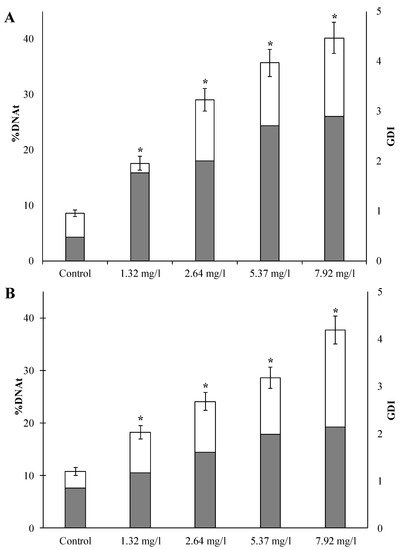
From the CCK-8 assay, TiO2 NPs can be slightly toxic to human liver cells. However, many researchers have found that hepatotoxicity is one of the target organ effects of oral exposure to TiO2 NPs [
17,
43]. Geraets et al. [
43] investigated the tissue distribution and blood kinetics of various TiO2 NPs in rats and found that the liver was identified as the main target tissue, followed by the spleen and lung. Another study found that the liver was the tissue most sensitive to TiO2 NP-induced oxidative stress [
44]. Many in vivo studies have found that TiO2 NPs may produce ROS and promote oxidative stress and liver inflammation [
44,
45,
46]. Sprague-Dawley rats were orally exposed to 0, 2, 10, and 50 mg/kg TiO2 NPs for 90 days and were found to induce tissue-specific oxidative stress and elemental imbalance in the liver [
44]. In addition, many in vitro studies have found that TiO2 NPs can induce damage to hepatocyte line cells [
47]. Current major toxicity mechanisms may exert cytotoxic effects on the structure and function of the liver by inducing oxidative stress, inflammation, and apoptosis [
16,
48,
49]. Oxidative stress, considered a common mechanism of the toxicity in NPs, can damage lipids, carbohydrates, proteins, and DNA, ultimately leading to hepatotoxicity [
50]. Azim et al. treated mice with anatase TiO2 NPs (21 nm, 150 mg/kg/day) for 2 weeks and then added three kinds of antioxidants (idebenone, carnosine, and vitamin E) for 1 month. They finally found that TiO2 NPs significantly injured liver function and can be alleviated after the use of antioxidants [
49]. This study attempted to further understand the new mechanism of hepatotoxicity from the perspective of epigenetics and found that lncRNAs may play an important role.In the study, some changes in lncRNAs and changes in the mRNAs matched with differential lncRNAs occurred, with statistical significance, implying that epigenetics may play a role in hepatotoxicity. Epigenetics is an important link in the regulation of genotype and phenotype. The regulation and dysregulation of genotype and phenotype often lead to the occurrence of diseases and have long-term negative effects. According to the 3R principle, epigenetics is also gradually being used in the toxicity study of nanomaterials. Some studies have also found that, in addition to genetic and cytotoxic effects, they can also affect the epigenome of target cells [
23,
51]. Lu et al. exposed human and murine macrophages (THP-1 and RAW264.7, respectively) and human small-airway epithelial cells (SAECs) to environmentally relevant concentrations of TiO2 NPs, resulting in modest alterations in DNA methylation [
51]. Another study also found that low concentrations of TiO2 NPs can alter the enzymes responsible for epigenetic modifications [
52]. Because their concentrations are well below sublethal levels, changes in DNA methylation can serve as good biomarkers of early exposure to TiO2 NPs. Therefore, epigenetic studies are critical for a complete assessment of potential risks from nanoparticle exposure.In recent years, lncRNAs have become an important class of regulators of gene expression and epigenetic regulation [
53]. Some reports found that lncRNAs play a role in cell-cycle regulation, apoptosis, and the establishment of cellular identity [
54,
55]. Changes in the expression of lncRNAs have been proven to be linked with cancer (e.g., prostate cancer) and several neurological disorders [
31,
56]. One study proposed that the use of electrochemical nucleic acid sensors is very sensitive to lncRNA HULC detection, providing a new alternative for clinical HCC diagnosis [
57]. The study did find that certain lncRNAs (such as NONHSAT256380.1 and NONHSAT173563.1) showed remarkable changes, which may be prevalent to the hepatotoxicity of TiO2 NPs. Therefore, lncRNAs can help to study the mechanism of hepatotoxicity in more depth and explore the role of epigenetic regulation in hepatotoxicity.In addition, small integral membrane protein 22 (SMIM22, CASIMO1), matched with the up-regulated lncRNA (NONHSAT173563.1), has been shown to play a key role in carcinogenesis, cell proliferation, and cell lipid homeostasis [
58]. The depletion of Jrk helix-turn-helix protein (JRK, JH8, jerky), matched with the down-regulated lncRNA (NONHSAT256380.1), inhibits the transcriptional activity of β-catenin and reduces cell proliferation, and it has been validated for carcinogenic effects in primary tumors [
59]. From the result of KEGG enrichment analysis, TiO2 NPs could interfere with the Hedgehog signaling pathway, which played a key role in tissue development and dryness. The imbalance in the Hedgehog signaling pathway was present in many different tumors, such as skin, brain, liver, and gallbladder [
60]. There are three homology genes for Hedgehogs in mammals: Sonic Hedgehog (SHH), Indian Hedgehog (IHH), and Desert Hedgehog (DHH) [
61]. Hedgehog signaling is controlled by two receptors, Patched (Ptc) and Smoothened (Smo), on the membrane of the target cell [
62]. These unique signaling molecules are highly expressed in most malignant tissues and have been considered biomarkers for progression and prognosis [
63,
64]. Additionally, many in vitro studies have found that chronic liver damage or liver cancer may activate the sonic hedgehog (SHH) pathway [
65,
66].
The main advantage of this article is the use of epigenetics to study the alterations in the lncRNA expression profile induced by TiO2 NPs in hepatotoxicity. In the future, the influence of oral exposure to nano-titanium dioxide on epigenetics and related mechanisms can be further studied. However, this study also has some drawbacks. Firstly, this study lacks more in-depth studies on screened lncRNAs and, secondly, the verification of this study is at the mRNA level, so there is a lack of PCR verification at the lncRNA level. Therefore, we will next conduct more in-depth studies on differential lncRNAs, such as knocking out relevant genes to study their impact on subsequent functions. We will also further focus on the effects of apoptosis or genetic damage of TiO2 NPs.


留言 (0)