
記住我


We first investigated the role of CCNY in hippocampal synaptic plasticity and cognitive functions by characterizing electrophysiological features in Ccny KO mice [29]. The frequency of action potential firing triggered by current injection in CA1 neurons was not altered in Ccny KO mice (Fig. 1a). The current-voltage relationship (Fig. 1b), input resistance (Fig. 1c), and sag ratio (Fig. 1d) remained unaffected, demonstrating that intrinsic neuronal excitability is normal in Ccny KO mice. Moreover, the paired-pulse ratio (PPR) was not significantly altered in Ccny KO mice (Fig. 1e), indicating that presynaptic release probability at Schaffer collateral-CA1 synapses in Ccny KO mice remains comparable to that of wild-type (WT) mice. This result is in agreement with the finding that CCNY expression is absent in the synaptic vesicle-enriched (LP2) presynaptic fraction, where a presynaptic protein synaptophysin exists in the mouse (Supplementary Fig. S1a) and rat forebrain [25]. CCNY was enriched in postsynaptic fractions of the mouse forebrain, including the synaptic plasma membrane (SPM) and the postsynaptic density (PSD) fractions, where postsynaptic proteins, such as GluA1, a subunit of AMPARs, and PSD-95, are located (Supplementary Fig. S1a).
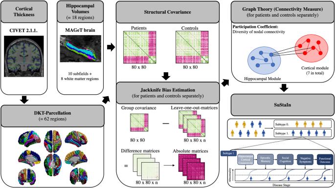
Fig. 1: Ccny knockout (KO) mice exhibit enhanced long-term potentiation (LTP) and moderate long-term depression (LTD).
a−i Electrophysiological characteristics of Ccny KO mice. a Neuronal intrinsic excitability is normal in Ccny KO mice. Data represent mean ± SEM of the number of action potentials triggered at each current step. The frequency of action potential firing was measured upon injecting a current step ranging from 0 to 330 pA in increments of 30 pA. ns, not significant compared to wild-type (WT) at each current step, Student’s unpaired t test. n = 15 and 16 cells from 4 WT and 3 KO mice, respectively. Representative action potentials recorded from WT and Ccny KO mice are shown. Scale bars, 20 mV, 100 ms. b–d The current-voltage relationship is not altered in Ccny KO mice. b Data represent mean ± SEM of the peak membrane potential measured at each current step. Membrane potential was measured upon injecting a current step ranging from 20 to −150 pA. ns, not significant compared to WT at each current step, Student’s unpaired t test. n = 15 and 16 cells from 4 WT and 3 KO mice, respectively. Representative voltage responses recorded from WT and Ccny KO mice are shown. Scale bars, 5 mV, 100 ms. Input resistance (c) and sag ratio (d) were calculated from the current-voltage relationship. Data represent mean ± SEM. ns, not significant, Student’s unpaired t test. e Paired-pulse ratio (PPR) remains unchanged in Ccny KO mice. Data represent mean ± SEM of PPR at each time interval. Two consecutive field excitatory postsynaptic potentials (fEPSPs) were evoked at Schaffer collateral-CA1 synapses with various time intervals (25, 50, 100, 200, and 400 ms). PPR was calculated by dividing the peak of the second fEPSP by that of the first one. ns, not significant, Student’s unpaired t test at each time interval. Representative fEPSP traces recorded from WT and Ccny KO mice are shown. Scale bars, 0.4 mV, 100 ms. f NMDA/AMPA ratio at hippocampal Schaffer collateral-CA1 synapses is normal in Ccny KO mice. Data represent mean ± SEM of NMDA/AMPA ratio. AMPAR-mediated currents recorded at −70 mV were measured at the peak amplitude, and NMDAR-mediated currents recorded at +40 mV were measured at 60 ms after stimulation. ns, not significant, Student’s unpaired t test. n = 9 and 11 cells from 4 WT and 3 KO mice, respectively. Representative excitatory postsynaptic currents (EPSCs) recorded at −70 mV (downward current) and +40 mV (upward current) from WT and Ccny KO mice. Scale bars, 100 pA, 100 ms. g Miniature EPSCs (mEPSCs) are not altered in hippocampal CA1 neurons from Ccny KO mice. (Upper) Representative recordings of AMPAR mEPSCs in hippocampal CA1 neurons from WT and Ccny KO mice. Scale bars, 40 pA, 5 s. (Lower) Both mEPSC amplitude (left) and frequency (right) are not altered by chronic Ccny KO. Cumulative distribution function plots of AMPAR mEPSC amplitude (left) and inter-event intervals (right). Insets, Data represent mean ± SEM. ns, not significant, Student’s unpaired t test. n = 8 and 9 cells from 5 animals each for WT and KO mice, respectively. h, i The input-output relationship is not altered at Schaffer collateral-CA1 synapses in Ccny KO mice. h fEPSPs were measured by stimulating the Schaffer collateral afferents with various stimulus intensity. (Upper) Representative fEPSP traces recorded from WT and Ccny KO mice with a series of increasing input stimulus ranging from 10 to 90 μA. Scale bars, 0.5 mV, 20 ms. (Lower) Data represent mean ± SEM of fEPSP slope at each stimulus intensity. ns, not significant, P = 0.669 (10 μA), P = 0.289 (20 μA), P = 0.430 (30 μA), P = 0.255 (40 μA), P = 0.403 (50 μA), P = 0.772 (60 μA), P = 0.935 (70 μA), P = 0.540 (80 μA), and P = 0.765 (90 μA) compared to WT at each stimulus intensity, Student’s unpaired t test. n = 11 and 12 slices from 6 WT and 5 KO mice, respectively. i Data represent mean ± SEM of the ratio of slope to volley. The ratio of fEPSP slope to the fiber volley amplitude at the stimulus strength that evokes 40% of the maximal fEPSP amplitude. ns, not significant, P = 0.662, Student’s unpaired t test. n = 11 and 12 slices from 6 WT and 5 KO mice, respectively. j, k LTP at Schaffer collateral-CA1 synapses is enhanced in Ccny KO mice. j High-frequency stimulation (HFS)-induced LTP is enhanced in Ccny KO mice. The magnitude of LTP was quantified as an increase in the fEPSP slope relative to the baseline. Representative fEPSP traces from WT and KO mice before (1) and after (2) LTP induction are shown. Scale bars, 0.2 mV and 10 ms. n = 8 and 8 slices from 5 WT and 6 KO mice, respectively. Data represent mean ± SEM of fEPSP slope. k Data represent mean ± SEM of averages of fEPSP slopes during the last 10 min (71−80 min) of the recordings in (j). *P < 0.05 as indicated, Student’s unpaired t test. l, m LTD at Schaffer collateral-CA1 synapses is reduced but still expressed in Ccny KO mice. l Ccny KO mice exhibit reduced hippocampal LTD induced by low-frequency stimulation (1 Hz, 900 pulses) at Schaffer collateral-CA1 synapses. The magnitude of LTD was quantified as a decrease in the fEPSP slope relative to the baseline. Data represent mean ± SEM. n = 7 and 7 slices from 7 WT and 6 KO mice, respectively. Representative fEPSP traces from WT and KO mice before (1) and after (2) LTD induction. Scale bars, 0.2 mV and 10 ms. m Data represent mean ± SEM of averages of fEPSP slopes during the last 10 min (66-75 min) of the recordings in (l). **P < 0.005 as indicated, Student’s unpaired t test. See also Supplementary Fig. S2.
We next examined the function of AMPAR and N-methyl-D-aspartate (NMDA) receptor (NMDAR). The ratio of NMDAR- to AMPAR-mediated synaptic transmission (NMDA/AMPA ratio) was normal in Ccny KO mice (Fig. 1f), and the amplitude and frequency of miniature excitatory postsynaptic currents (mEPSCs) at Schaffer collateral-CA1 synapses were not altered (Fig. 1g). In addition, the input-output relationship between presynaptic stimulus intensity and the slope of the resulting field excitatory postsynaptic potentials (fEPSPs) against the presynaptic stimulus intensity (Fig. 1h) and the ratio of fEPSP slope to the fiber volley amplitude (Fig. 1i) were also not affected in Ccny KO mice. These results together indicate that basal excitatory synaptic transmission remained unchanged in Ccny KO mice. In support of these findings, the postsynaptic subcellular localizations of GluA1 and PSD-95 were not altered in the forebrain of Ccny KO mice (Supplementary Fig. S1b, c). The unaltered basal excitatory synaptic transmission in Ccny KO mice is rather contradictory to that reported in a previous study showing that a short hairpin RNA (shRNA)-mediated knockdown of CCNY increases the amplitude but not the frequency of mEPSCs in cultured rat hippocampal neurons [26]. This difference could be due to the use of different systems; the chronic depletion of CCNY in Ccny KO mice might be systemically complemented in vivo, whereas 3- to 4-day knockdown of CCNY in cultured neuronal system rather exhibits an acute response to reduced CCNY levels. Together, these results suggest that the complete depletion of CCNY does not affect intrinsic neuronal excitability and basal excitatory synaptic transmission in the hippocampus.
CCNY regulates hippocampal LTP and LTD at Schaffer collateral-CA1 synapsesHippocampal synaptic plasticity, including LTP and LTD, is widely considered as a fundamental mechanism underlying cognitive brain functions of learning and memory [5, 9, 10, 30,31,32,33]. We found that the magnitude of LTP induced by high-frequency stimulation (HFS) at Schaffer collateral-CA1 synapses was slightly but significantly enhanced in acute hippocampal slices prepared from Ccny KO mice compared to WT littermates (Fig. 1j, k). Furthermore, LTD induced by low-frequency stimulation (900 pulses, 1 Hz, 15 min) at Schaffer collateral-CA1 synapses was expressed with reduced magnitude in Ccny KO mice compared to WT littermates (Fig. 1l, m). In contrast, both voltage-gated calcium channel (VGCC)-dependent LTP (Supplementary Fig. S2a, b) and metabotropic glutamate receptor (mGluR)-dependent LTD (Supplementary Fig. S2c, d) were normal in Ccny KO mice. Collectively, these results indicate that Ccny KO mice possess a shifted spectrum of synaptic plasticity toward potentiation in the hippocampus and specifically affect the NMDAR-dependent form of synaptic plasticity, thereby potentially affecting hippocampus-dependent cognitive functions, such as spatial learning and memory flexibility.
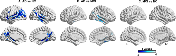
Hippocampus-dependent spatial learning is enhanced in Ccny KO miceWe next investigated whether CCNY regulates hippocampus-dependent spatial learning and memory using the MWM task equipped with surrounding spatial cues (Fig. 2a). The latency to platform was significantly shorter on days 3, 4, and 5 in Ccny KO mice compared to WT littermates during the five consecutive training days (Fig. 2b), indicating an enhanced learning ability in Ccny KO mice. In addition, during the probe test on day 6, the latency (Fig. 2b) and travel distance (Fig. 2c) to the platform zone where the platform was originally located was significantly shorter in Ccny KO mice. Even though the number of entries to the target quadrant was not significantly different between WT and Ccny KO mice (Fig. 2d), the time spent in the target quadrant was increased in Ccny KO mice compared to WT littermates (Fig. 2e). Furthermore, the frequency of passing the platform zone was increased in Ccny KO mice compared to WT littermates (Fig. 2f). These results together indicate an enhancement of spatial memory ability in Ccny KO mice. In the visible platform test, which is a hippocampus-independent non-spatial learning task, the performance of Ccny KO mice was comparable to that of WT mice in terms of the latency to platform during the training sessions (Fig. 2g) and to the platform zone during the probe test (Fig. 2h), suggesting that the improved spatial learning and memory of Ccny KO mice in the MWM task (Fig. 2b−f) were not due to the enhanced motivation for escape or improved vision and/or motor skills.
Fig. 2: Spatial learning and memory flexibility are improved in Ccny KO mice.
a Schematic diagram for the Morris water maze (MWM) task, including original (Fig. 2b−f) and reversal (Fig. 2i−m) learning and visible platform (Fig. 2g, h) tests. b Learning curve of latency to platform during the original training sessions, Day 1 to 5, and latency to platform zone during the probe test session on Day 6 performed in the MWM after platform removal (gray shaded area). Data represent mean ± SEM. **P < 0.005, ***P < 0.0005 as indicated, Student’s unpaired t test. Data obtained from three cohorts. c Travel distance to platform zone during the probe test on Day 6 performed in the MWM after platform removal. *P < 0.05, Student’s unpaired t test. d, e The number of entries to (d) and time spent in (e) each quadrant during the probe test on Day 6 performed in the MWM after platform removal. TQ, target quadrant; LQ, quadrant left to the TQ; RQ, quadrant right to the TQ; OQ, quadrant opposite to the TQ. *P < 0.05, **P < 0.005, ***P < 0.0005 as indicated, Student’s unpaired t test. f The number of passing the platform zone during the probe test on Day 6 performed in the MWM after platform removal. *P < 0.05, Student’s unpaired t test. g, h Latency to platform during the visible sessions, V-Day 1 to 3 (g) and latency to platform zone during the visible probe test on V-Day 4 (h). Data represent mean ± SEM. ns, not significant, P = 0.223 (V-Day 1), P = 0.100 (V-Day 2), and P = 0.143 (V-Day 3) in (g), P = 0.203 in (h), Student’s unpaired t test. See also Supplementary Fig. S3. i−m Ccny KO mice exhibit an improved memory flexibility in reversal learning of the MWM task. i Latency to platform during the reversal learning training sessions, R-Day 1 to 3, and latency to platform zone during the reversal learning probe test on R-Day 4 (gray shaded area). Data represent mean ± SEM. *P < 0.05, **P < 0.005 as indicated, Student’s unpaired t test. Data obtained from three cohorts. j Travel distance to the platform zone during the reversal learning probe test on R-Day 4 performed in the MWM after platform removal. *P < 0.05, Student’s unpaired t test. k Time spent in each quadrant during the reversal learning probe test on R-Day 4 performed in the MWM after platform removal. Data represent mean ± SEM. PQ, previous target quadrant; TQ, target quadrant; LQ, quadrant left to the TQ; RQ, quadrant right to the TQ. *P < 0.05 as indicated, Student’s unpaired t test. l The number of entries to the previous target (PQ) and current target quadrant (TQ) during the reversal learning probe test on R-Day 4 performed in the MWM after platform removal. Data represent mean ± SEM. *P < 0.05, ***P < 0.0005 as indicated, Student’s unpaired t test. m The number of passing the platform zone during the reversal learning probe test on R-Day 4 performed in the MWM after platform removal. Data represent mean ± SEM. *P < 0.05, ***P < 0.0005 as indicated, Student’s unpaired t test. n Ccny KO mice exhibit improved memory flexibility in a delayed nonmatch to place (DNMTP) T-maze task. Data represent mean ± SEM of percentage of correct choices. *P < 0.05, Student’s unpaired t test. ##P < 0.005 as indicated, repeated-measures two-way ANOVA, the effect of genotype, F (1, 29) = 9.874. See also Supplementary Fig. S4. o Memory flexibility in GluN2B-C456Y-mutant mice is comparable to the littermate WT in a DNMTP T-maze task. Data represent mean ± SEM of percentage of correct choices. Student’s unpaired t test. ns, not significant, repeated-measures two-way ANOVA, the effect of genotype, F (1, 15) = 2.244. See also Supplementary Fig. S4.
In addition, the total distance moved, the time spent in the center, and the number of center entries in an open field task were not different between WT and Ccny KO mice (Supplementary Fig. S3a−h), further supporting that basal locomotion, anxiety, and willingness to explore were not affected in Ccny KO mice compared to WT littermates. Moreover, no significant difference was observed between WT and Ccny KO mice in a novel object recognition task (Supplementary Fig. S3i−k) used to test non-spatial memory [34, 35] or in a passive avoidance task (Supplementary Fig. S3l, m) used to assess aversive associative memory [36]. These results together demonstrate that hippocampus-dependent spatial learning and memory is specifically improved in Ccny KO mice and suggest future studies exploring the roles of CCNY in other spectrums of learning behaviors and/or other behavioral/non-behavioral functions in other brain regions.
Memory flexibility is improved in Ccny KO mice and normal in Grin2b +/C456Y miceLTD is associated with behavioral and memory flexibility [3,4,5,6, 9, 10, 27, 28]. Because hippocampal LTD was significantly altered in Ccny KO mice (Fig. 1l, m), we further investigated whether CCNY is involved in the regulation of memory flexibility. To this end, a spatial reversal learning task was conducted by moving the hidden platform to a new location (i.e., the quadrant opposite to the previously memorized quadrant) following the original learning task in the MWM. The latency to platform was significantly shorter on days 2 and 3 during the training sessions in Ccny KO mice (Fig. 2i). In addition, on day 4 of the reversal learning probe test, the latency (Fig. 2i) and travel distance (Fig. 2j) to the platform zone were also significantly reduced in Ccny KO mice, and the time spent in the target quadrant was significantly increased in Ccny KO mice compared to WT littermates (Fig. 2k). Furthermore, both the time spent in the previous target quadrant (Fig. 2k) and the frequency of entries to the previous target quadrant during the reversal learning probe test (Fig. 2l) were significantly decreased in Ccny KO mice compared to WT littermates. Both WT littermates and Ccny KO mice more frequently entered to the target quadrant (Fig. 2l) and target platform zone (Fig. 2m) than to the previous quadrant (Fig. 2l) and previous platform zone (Fig. 2m), respectively, but the degrees of increase in the frequency of entries to the target quadrant and target platform zone were much higher in Ccny KO mice than in WT littermates (Fig. 2l, m). Altogether, these results indicate that memory flexibility (or new learning and memory ability) is improved in Ccny KO mice.
We further investigated whether the lack of CCNY also influences the performance in the DNMTP T-maze task as an independent test to examine hippocampus-dependent memory flexibility [3, 27, 28, 37]. Similar to the reversal learning of MWM task which requires memory flexibility to learn and memorize new spatial information, the location of rewards for the choice runs in the DNMTP T-maze task alternates between the right and left arms in each trial, thus requiring memory flexibility to make a correct choice. Consistent with the enhanced reversal learning (Fig. 2i−m), Ccny KO mice also showed improved learning flexibility in the DNMTP T-maze task (Fig. 2n). In agreement with this finding, both travel distance (Supplementary Fig. S4a) and travel time (Supplementary Fig. S4b) taken to make a correct choice in the DNMTP T-maze task were shorter in Ccny KO mice compared to WT littermate. The body weights of the mice subjected to this task were maintained at approximately 80% of the initial body weights during the handling period throughout the experiments (Supplementary Fig. S4c). Because the animal behaviors in the open field task were not significantly different between WT and Ccny KO mice (Supplementary Fig. S4a−h), the improved memory flexibility in Ccny KO mice was not due to a change in basal locomotion, anxiety, or willingness to explore. Collectively, our findings indicate that even reduced LTD in Ccny KO mice (Fig. 1l, m) supports memory flexibility of both reversal learning in MWM (Fig. 2i−m) and the DNMTP T-maze task (Fig. 2n).
To further examine the notion that reduced LTD is sufficient to support memory flexibility, we performed a DNMTP T-maze task using GluN2B-C456Y-mutant mice (Grin2b+/C456Y) that exhibited reduced LTD induced by low-frequency stimulation but normal LTP at Schaffer collateral-CA1 synapses (i.e., presenting an altered spectrum in synaptic plasticity) along with normal original and reversal learning in MWM [38]. Despite the reduced hippocampal LTD [38], the GluN2B-C456Y-mutant mice showed memory flexibility comparable to WT littermates in the DNMTP T-maze task (Fig. 2o), consistent with the normal reversal learning of MWM observed in GluN2B-C456Y-mutant mice [38], confirming that reduced LTD is sufficient to support memory flexibility. In agreement with this finding, both travel distance (Supplementary Fig. S4d) and travel time (Supplementary Fig. S4e) taken to make the correct choice in GluN2B-C456Y-mutant mice were comparable to WT littermates. The body weights of the mice were maintained at approximately 80% of their initial body weight during the handling period throughout the experiments (Supplementary Fig. S4f). Considering that the GluN2B-C456Y-mutant mice showed a difference in terms of normal LTP and normal original learning when compared to Ccny KO mice, the improved memory flexibility in Ccny KO mice seems to be accounted for by the enhanced LTP and improved original learning ability.
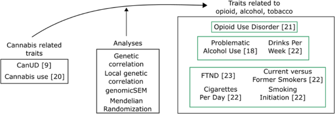
To further support the requirement of residual LTD in Ccny KO mice for the improved memory flexibility, the residual LTD in Ccny KO mice was completely blocked using a selective GluN2B antagonist and a specific LTD inhibitor, Ro25-6981 [4, 39]. Ro25-6981 treatment did not affect LTP (Fig. 3a, b), whereas it completely removed residual LTD in both WT and Ccny KO mice (Fig. 3c, d). In agreement with LTP results, Ro25-6981 administration to Ccny KO mice did not affect spatial learning in the MWM task, as it resulted in no significant changes in the latency to the platform (zone) (Fig. 3e), travel distance (Fig. 3f), number of entries to the target quadrant (Fig. 3g), time spent in the target quadrant (Fig. 3h), and frequency of passing the platform zone (Fig. 3i). In agreement with the notion that the requirement of the residual LTD in Ccny KO mice is required for the improved memory flexibility, latency to the platform (zone) was significantly delayed (Fig. 3j) and travel distance to the platform zone tended to increase with Ro25-6981 treatment (Fig. 3k). Both Ro25-6981-treated and untreated Ccny KO mice spent more time in the target quadrant than in the previous quadrant (Fig. 3l) and more frequently passed the target platform zone than the previous target platform zone (Fig. 3m). However, the degrees to which both the time spent in the target quadrant and the frequency of passing to the target platform zone increased were significantly lower in Ro25-6981-treated Ccny KO mice than in vehicle-treated Ccny KO mice (Fig. 3l, m). These data indicate that the enhanced memory flexibility in Ccny KO mice is dependent on the residual LTD. Taken together, these findings suggest that to improve memory flexibility, it is critical to facilitate not only the extinction or weakening of the previously encoded spatial memory, but also the formation of new spatial memory. The facilitation of the weakening of previously acquired memory information and the formation of new spatial memory are presumably mediated by weakened LTD and enhanced LTP, respectively, in Ccny KO mice.
Fig. 3: The specific LTD inhibitor Ro 25-6981 inhibits LTD and memory flexibility but does not affect LTP and spatial learning in Ccny KO mice.
a, b Ro25-6981 (0.5 μM) treatment does not affect the pairing-induced LTP in both WT and Ccny KO mice. a Data represent mean ± SEM of the peak EPSC amplitude relative to the baseline. LTP was induced by pairing stimulation (2 Hz for 90 s with 0 mV holding) at Schaffer collateral-CA1 synapses. n = 8, 8, 7, and 6 slices from 4 mice in each of 4 experimental groups. Representative evoked EPSC traces before (1) and after (2) LTP induction are shown. Scale bars, 50 pA and 40 ms. b Data represent mean ± SEM of evoked EPSC amplitudes during the last 5 min (35−40 min) of the recordings in (a). ns, not significant, ***P < 0.0005 as indicated, one-way ANOVA followed by Tukey’s multiple comparisons test. c, d Ro25-6981 (0.5 μM) treatment inhibits the pairing-induced LTD in both WT and Ccny KO mice. (c) Data represent mean ± SEM of the peak EPSC amplitude relative to the baseline. LTD was induced by a pairing protocol (5 Hz for 3 min with −40 mV holding) at Schaffer collateral-CA1 synapses. n = 7, 6, 6, and 7 slices from 4 mice in each of 4 experimental groups. Representative evoked EPSC traces before (1) and after (2) LTD induction are shown. Scale bars, 50 pA and 40 ms. d Data represent mean ± SEM of the evoked EPSC amplitudes during the last 5 min (35−40 min) of the recordings in (c). ns, not significant, ***P < 0.0005 as indicated, one-way ANOVA followed by Tukey’s multiple comparisons test. e−i The intraperitoneal injection of Ro25-6981 (6 mg/kg) into Ccny KO mice did not affect original learning in the MWM task. e Learning curve of latency to platform (zone) during the MWM task. Data represent mean ± SEM. colored ns, not significant, Student’s unpaired t test at individual days. ns, not significant as indicated, two-way ANOVA. f Travel distance to platform zone during the probe test on Day 6. ns, not significant, Student’s unpaired t test. g, h The number of entries to (g) and time spent in (h) each quadrant during the probe test on Day 6. i The number of passing the platform zone during the probe test on Day 6. ns, not significant, Student’s unpaired t test. j−m Intraperitoneal injection of Ro25-6981 (6 mg/kg) into Ccny KO mice inhibited memory flexibility in reversal learning of the MWM task. j Latency to the platform (zone) during the reversal learning. Data represent mean ± SEM. *P < 0.05, **P < 0.005 as indicated, Student’s unpaired t test. ***P < 0.0005 as indicated, two-way ANOVA. k Travel distance to the platform zone during the reversal learning probe test on R-Day 4. ns, not significant, *P < 0.05 as indicated, Student’s unpaired t test. Bars on the left side of the dotted line are the averaged data of T1 (Trial 1), T2 (Trial 2), and T3 (Trial 3). l Time spent in each quadrant during the reversal learning probe test on R-Day 4. Data represent mean ± SEM. ***P < 0.0005, ****P < 0.0000005 as indicated, Student’s unpaired t test. m The number of passing the platform zone during the reversal learning probe test on R-Day 4. Data represent mean ± SEM. *P < 0.05, ***P < 0.0005 as indicated, Student’s unpaired t test.
Original learning upregulates genes related to the actin cytoskeleton, synaptic plasticity, or learning in WT mice with similar changes in untrained Ccny KO miceSpatial learning and memory are correlated with spine morphology and plasticity [2, 40, 41], which requires actin remodeling [42,43,44,45,46,47,48]. CCNY is an actin-binding protein and regulates spine plasticity induced by LTP through the cofilin-actin signaling pathway [26], implying the functional significance of CCNY in modulating spatial learning and memory through the actin signaling pathway.
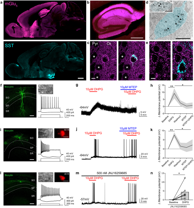
We found that the expression levels of Ccny mRNA detected by quantitative real-time polymerase chain reaction (qRT-PCR) (Fig. 4a) and CCNY protein detected by immunoblot analysis (Fig. 4b) were significantly reduced in the hippocampus after original learning in WT mice. Notably, improved spatial learning and memory ability in Ccny KO mice (Fig. 2b−f) and the decreased CCNY levels in WT mice following the original learning and memory task (Fig. 4a, b) together suggest a possible correlation between CCNY expression levels and cognitive function. Therefore, we aimed to determine whether Ccny KO mice in the naive state share molecular changes similar to those in WT mice subjected to original learning and memory. The potential candidates of molecular processes associated with original learning were examined by analyzing the transcriptome using high-throughput RNA sequencing. A total of 560 genes were differentially expressed when comparing WT mice under basal state with WT mice after original learning, in which 265 genes were upregulated and 295 genes were downregulated in the WT mice subjected to original learning (Fig. 4c; WT_Basal vs WT_OL). In untrained naive Ccny KO mice (KO_Basal), the expression levels of 314 genes were differentially regulated compared to untrained naive WT mice (WT_Basal), with 95 upregulated and 219 downregulated genes (Fig. 4c). Importantly, 29 genes that were commonly upregulated in both WT mice subjected to original learning and in untrained Ccny KO mice compared to WT mice under basal state (Fig. 4d, e) were significantly associated with the regulation of the actin cytoskeleton and the regulation of neuronal synaptic plasticity and learning, as revealed by the Kyoto Encyclopedia of Genes and Genomes enrichment analysis and the Gene Ontology analysis, respectively (Fig. 4f). These results strongly suggest that actin signaling plays a crucial role in mediating both the learning in WT mice and more improved learning ability in Ccny KO mice.
Fig. 4: Molecular changes induced by spatial learning training in WT mice are similarly associated with those in untrained Ccny KO mice.
留言 (0)