In this study, the scRNA-seq data of omentum metastasis sites from 6 ovarian cancer patients, GEO microarray data of 3769 patients, TCGA-OV bulk RNA-seq data of 379 patients and clinical information were combined to construct a prognostic prediction model of ovarian cancer composed of a 6-OMAGs signature and 3 clinicopathological features.
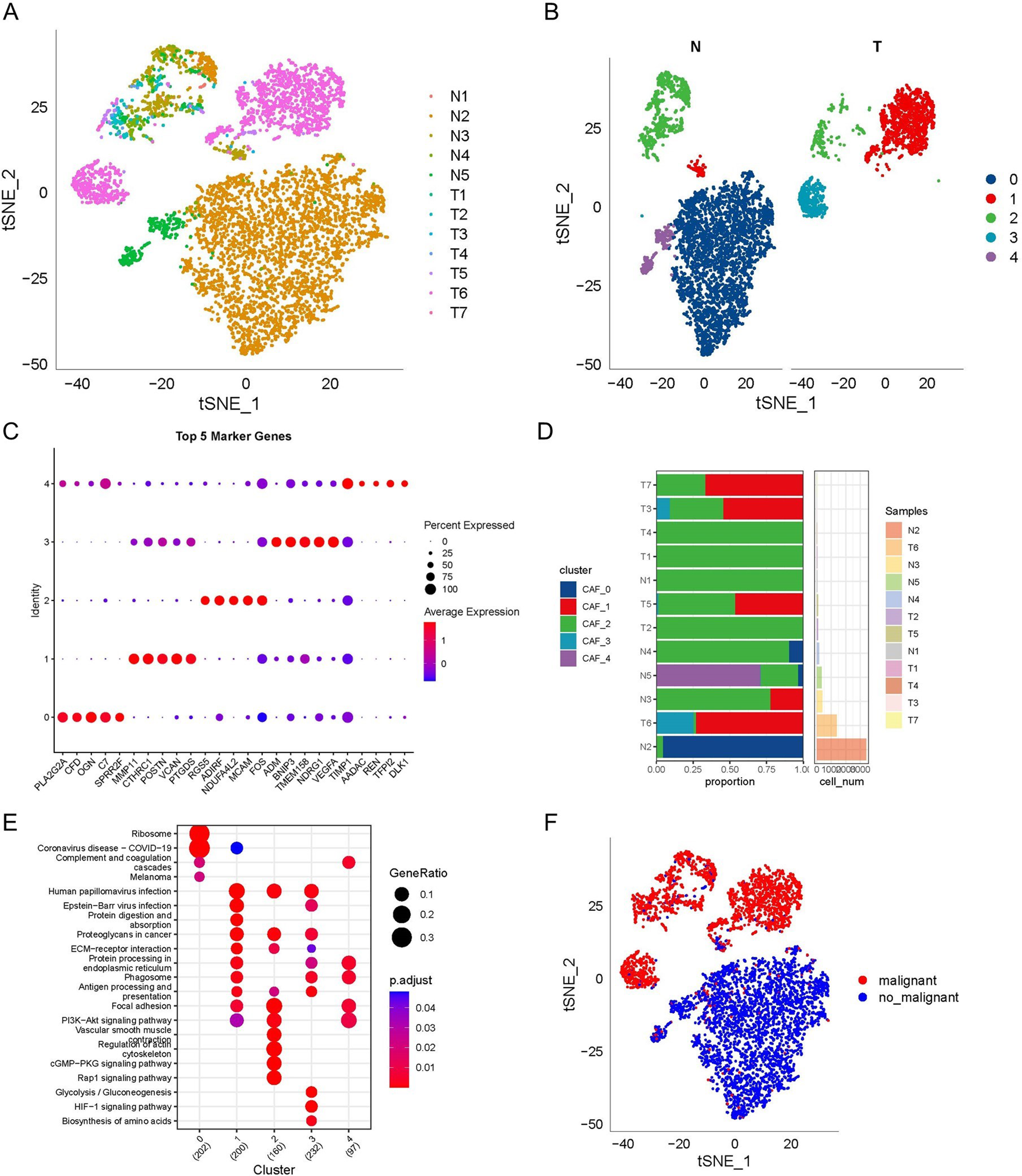
In comparison with the original authors [12], they performed the hierarchical clustering with a resolution of 0.2, PCs varied from 10 to 20 depending on the sample and UMAP dimension reduction, with 12 clusters detected. While we selected a PC value of 18, a resolution of 0.5, and t-SNE dimensionality reduction analysis identified the single cells into 18 clusters. The difference between these parameters and methods will change the cluster distribution and mapping. The original authors adopted a sophisticated way of cell annotation, integrating canonical genes, functional categories, and cell line correlation. Finally, they annotated the 12 clusters into nine types of cells and three unidentified clusters. We mainly used the SingleR package and adjusted the annotation results according to the marker genes in the literature, and obtained 14 cell types. In our cell annotation results, six cell types are the same as those of the original author, including epithelial cells, fibroblasts, mesenchymal stem cells, endothelial cells, B cells, and plasma B cells. But there are also some differences. For example, the original authors only annotated T cells, but we divided T cells into two different clusters, they annotated macrophages, but we only had monocyte. Interestingly, in our results, the epithelial cells (cancer cells) were separated into three clusters. This may be attributed to the heterogeneity of tumors. It may be valuable to further investigate the differences between these epithelial cell clusters in our follow-up research, which may provide some ideas for the precise treatment of ovarian cancer.
The differentiation trajectory analysis of ovarian cancer omentum metastasis sites revealed that each cell type was not necessarily in only one state, and each state contained multiple cell types. Cells might express diverse sets of genes during different states; when cells move between states, some genes might be silenced, while some might be newly activated to carry out their work. Hence, these 781 characteristic genes might have a connection with the composition, function and state of cells in the TME and are likely related to the progression of ovarian cancer.
When the patients from GSE132342 were grouped into 3 clusters in accordance with the expression of 74 cell state marker genes, patients in cluster 1 had relatively low expression in the downregulated genes of states 2, 3, 4, and 5 and relatively high expression in the upregulated genes, and it was speculated that the samples’ TME might be in states 2, 3, 4, and 5. Similarly, it could be inferred that the TME of samples in cluster 2 might be in state 1, and the TME of samples in cluster 3 might be in states 2, 3, and 4. Comparing this conjecture with the conclusion of the original author of scRNA-seq data, they found high T cell infiltration group had an anti-tumor response. Our results showed that T cells were mainly concentrated in state 5, while patients with cluster 1 had similar expression characteristics to those with state 5, and the prognosis of patients with cluster 1 was significantly better than that of patients with cluster 2, which was also consistent with the original author’s conclusion.
The clustering results according to the marker genes of cell states were supplemented and examined by CIBERSORT and TME analysis. This was helpful to further analyze the relationship between the omental metastasis sites’ characteristic genes of cell state and ovarian cancer prognosis. Cluster 2 had a higher percentage of neutrophils, which proved to be one of the cells facilitating the formation of omentum PMNs [4]. Among the immune cells with a higher proportion in cluster 3, follicular helper T cells [32] and macrophage M1 [33] were recognized to have antitumor effects, similar to plasma cells. The original author of the scRNA-seq data discovered a unique plasmablast and plasma B cell clusters that may contribute to the immune response within the TME [12], which might be the reason why patients in cluster 3 had a better prognosis.
The immune checkpoint analysis displayed seemingly contradictory results between expression level and prognosis; however, research has also shown that high expression of PD1 and PD-L1 is associated with favorable outcome in lung cancer of early-stage but an adverse outcome in late-stage [34]. The ratio of stage I-II patients in cluster 3 was much higher than that in cluster 2, and it might be the dual effect of PD1 and PD-L1 on prognosis that leads to this result.
The TME analysis also suggested that there might be a correlation between the cell states and the sample clustering. As presumed previously, the cells of cluster 1 samples were mainly in states 2, 3, 4, and 5; cluster 2 was mainly in state 1; and cluster 3 was mainly in states 2, 3, and 4. The expression characteristics of state 5 were unique to cluster 1, and state 5 contained a large quantity of immune cells. The expression characteristics of state 1 were unique to cluster 3, which consisted of many stromal cells. In comparison, cluster 3 indeed had the highest tumor cell content. This result implied that the progression and prognosis of tumors not only depended on the characteristics of tumor cells but also relied on interactions within the niche.
The 6-OMAGs finally screened were TIMP3, FBN1, IGKC, RARRES1, RPL21 and UCHL1. TIMP3 (tissue inhibitors of metalloproteinase 3) was shown to be associated with metastasis and poor survival in serous ovarian cancer by regulating TGF-beta signaling [35]. Our results also found that TIMP3 was positively correlated with TGF beta; in addition, TIMP3 was positively related to EMT, extracellular matrix (ECM), angiogenesis, apoptosis, the P53 pathway, ECM degradation, and collagen formation, which are the potential mechanisms of its tumor-promoting effect. TIMP3 had a diverse expression level among 45 human ovarian cancer cell lines, which might be due to their different characteristics. In single-cell samples, TIMP3 had a significantly high expression in mesenchymal cells, and studies demonstrated that TIMP3 can promote tumor progression through EMT [36, 37], which also verifies our conclusion.
FBN1 (fibrillin-1) has been reported to enhance the cisplatin resistance of ovarian cancer by being involved in angiogenesis and glycolysis [38]. Our study also found a strong correlation between the expression of FBN1 and angiogenesis with a ρSpearman of 0.76. Our results inferred that it is related to EMT with a ρSpearman of 0.75, which might be the reason why it is highly expressed in cells annotated as mesenchymal. In addition, we also found a considerable correlation of its expression with ECM, apoptosis, inflammatory response, PI3K/AKT/mTOR pathway, P53 pathway, TGFβ, IL-10 anti-inflammatory signaling pathway, collagen formation and ferroptosis. It also had a diverse expression level in different cell lines, and its complex mechanism in cancer progression remains to be further elucidated.
IGKC (immunoglobulin kappa C), expressed in plasma cells, has been identified as one of the top genes of a prognostic B cell metagene in breast cancer, correlated with favorable prognosis and response to chemotherapy [39]. A study showed that plasma cell infiltration in epithelial ovarian cancer has a significant impact on tumor progression and prognosis [40], and high expression of IGKC is associated with good outcome [41]. In our study, although it was widely expressed in a variety of cell types, it was only significantly overexpressed in B cells: plasma cells. Our pathway correlation analysis also did not find a significant positive correlation between IGKC and 20 tumor-related pathways, and its expression level in ovarian cancer cell lines was generally low, indicating that its high expression in ovarian cancer may be detrimental to the proliferation of ovarian cancer cells.
Ribosomal proteins (RPs) are involved in the cellular process of translation, and in recent years, RP dysfunction has been found in tumors, such as mutation, expression level changes and correlation with differentiation [42,43,44]. RPL21 (ribosomal protein gene 21) has been found to be associated with the proliferation of pancreatic cancer cells [45] and may be used as a potential marker for cervical intraepithelial neoplasia [46], but there are few reports in ovarian cancer. As a cellular translation process-related gene, it is not surprising that RPL21 is widely and highly expressed in various cell clusters and 45 ovarian cancer cell lines; however, our study revealed significantly low expression in the cluster of epithelial cells (CSCs), suggesting that the change in its expression may be related to the increased stemness of ovarian cancer cells.
UCHL1 (ubiquitin carboxyl-terminal esterase L1) is an oncogene encoding a deubiquitinating enzyme that adjusts the balance between mTOR complexes [47] and plays a significant role in the ubiquitin system as well as different cellular processes [48]. It has been widely studied and confirmed to be related to tumor progression, such as prostate cancer, lymphoma, lung cancer, breast cancer, neuroblastoma, etc. [48,49,50,51]. In ovarian cancer, UCHL1 may be one of the markers related to early stages of high-grade serous carcinoma, immunogenicity [52, 53], and cisplatin resistance [54, 55]. Our results showed that it was specifically overexpressed in epithelial cells (CSCs), while the expression levels varied significantly in different ovarian cancer cell lines. Interestingly, no significant correlation was found between UCHL1 and tumor-related functions. This suggests that UCHL1 is related to the stemness of ovarian cancer cells, but its specific mechanism requires further investigation.
RARRES1 (retinoic acid receptor responder 1) is one of the common methylated loci in several cancers and is believed to be a putative tumor suppressor gene [56, 57]. Promoter hypermethylation and loss of RARRES1 seem to facilitate cancer progression [58]. In this study, RARRES1 was found to be highly expressed in epithelial cells (CSCs) and fibroblasts, and studies have revealed the possibility that RARRES1 can be used as a carcinoma-associated fibroblast (CAF) marker gene in breast cancer and may lead to chemotherapy resistance [59, 60]. It could be speculated that fibroblast cells with high RARRES1 expression might be transformed into CAFs after remodeling by tumor cells, making the omental microenvironment easier for tumor cell migration/invasion. Compared with the other five OMAGs, the overall expression level of RARRES1 in ovarian cancer cell lines was not high. Moreover, although it has a significant correlation in multiple cancer-related pathways, the correlation coefficients are between 0.3 and 0.5, which does not show a strong correlation. This may be related to the stemness of tumor cells, which still needs further research.
Although there are various models related to the prognosis of ovarian cancer, few focus on omentum metastasis. Our study has proven to a certain extent that there is a correlation between the cell states of omental metastasis sites and the prognosis of ovarian cancer. For ovarian cancer patients who have not yet metastasized, the marker genes of cell states also have prognostic value. This may be because the omentum microenvironment with these characteristics is more likely to form PMN during the interaction with residual tumor cells, making it for proliferation and colonization. There are still many deficiencies and limitations in our research. Although GSE132342 contains considerable samples for analysis, one of its nonnegligible shortcomings is that it only has the expression information of 513 genes, and many valuable characteristic genes of cell states are excluded. The number of state marker genes decreased from 781 to 74. On the other hand, these 513 genes are also supported by high-level evidence that they are related to the prognosis of ovarian cancer. When we intersected these 513 genes with the characteristic genes of cell states, the 74 genes obtained were more likely to be related to both the state of ovarian cancer omental tumor cells and the prognosis of ovarian cancer. Apart from this, the study lacks first-hand data, and the potential mechanism has not been verified by experiments, so more clinical trials and laboratory experiments are needed in follow-up research.


留言 (0)