Microbial infection is an escalating medical issue that has posed severe threats to human health. Wound infection is resulted from the breakage of skin integrity when exposure to surgery, trauma, temperature extreme, or chemicals. Bacterial infection may cause persistent inflammation and the intensified oxidative stress, obviously impedes the natural progression of healing stages, decreases the quality of wound healing or even develops into hard-to-heal chronic wounds. More seriously, the development into systemic infection may induce sepsis or shock even multiorgan failure and results in long hospitalization and high mortality rate [1]. Traditionally wound infection is examined through visual observation of wounds and confirmed by bacterial culture. Clinical symptoms, such as pain, purulent discharge, swelling, and tissue damage arise only when the bacterial load is over 105 colony forming units (CFU) per gram of tissues [2]. Bacterial culture involves swabbing the wound site and testing the swabs in a microbiology lab in terms of colony counting, morphological analysis, and biochemical identification. These detection methods always require complex experimental procedure and time-consuming processes, and the significant waiting stage may increase the risk of infection spreading and miss the best time for treatment [2]. Therefore, there is an urgent need to develop an early-stage, sensitive and point-of-care diagnosis platform for management of wound infections.
To address these issues, strategies have been developed to monitor the infection status from bacteria-induced microenvironment changes (e.g., pH and temperature) and bacteria-secreted substances (e.g., enzymes and toxins). Lebègue et al. designed liposomes with encapsulated potassium ferrocyanide as a redox probe, and the interaction with bacterial toxins led to the release or electrolysis of the encapsulated probe for electrochemical sensing. A submicromolar concentration of rhamnolipid was monitored, but these electronically based technologies face challenges from disturbances by the complex wound environment of proteins, chemokines and electrolytes [3]. Alternatively, optimal imaging is increasingly demanded to monitor bacterial infection without the aid of trained operators and advanced instruments [4]. Luan et al. developed nanoprobes through self-assembly of amphiphilic fluorescent molecules into dense aggregates, which were disasembled upon encountering with bacteria and then assembled into cell walls. The metabolism-driven turn-on fluorescencerealized visual detection and imaging of bacteria [5]. In addition, the integration of biomaterials has created functional wound dressings with diagnostic and therapeutic capacities against bacterial infections. Among them, hydrogels are potential wound dressings with inherent functions to maintain a moist environment, absorb excess tissue exudate, allow oxygen permeation, and carry various bioactive factors [6]. Zhou et al. constructed dual-layered photocrosslinkable methacrylated gelatin hydrogels to encapsulate liposomal vesicles containing antimicrobials in the lower layer and vesicles containing self-quenching dye carboxyfluorescein in the upper layer. Bacterial toxins or enzymes would damage the vesicle membrane to release the payloads for bacterial inhibition and fluorescent imaging [7]. These detection systems are derived from the leakage of fluorescent indicators from dressing materials, leading to a subsequent decline in imaging effectiveness and potential cytotoxicity. The traditional strategies require frequent dressing changes to achieve long-term monitoring, but these dessging changes are always associted with pain and secondary tissue damage, especially for chronic wounds [8]. Therefore, challenges remain in the rational construction of clinically adaptable dressings for life-cycle monitoring of wound infection or possible infection recurrence [9].
Antibiotics have been widely accepted to combat bacterial infections but are confronted with low bioavailability, adverse side effects and drug resistance in clinical disinfection therapies [10]. Enormous efforts have been made to develop novel antibacterial agents to mitigate these problems, including bacteriophages, cationic polymers and metal nanoparticles, but their safety concerns limit the potential application [11]. Alternatively light-assisted antibacterial methods, including photothermal (PTT) and photodynamic therapies (PDT) have shown high spatial control and low risks in acquiring bacterial resistance [12]. PTT offers thermal inactivation of bacteria, but the elevated temperature (over 50 °C) may damage the surrounding healthy tissues and even produce heat shock proteins to generate resistance to PTT [13]. PDT produces reactive oxygen species (ROS) to cause oxidative stress, lipid peroxidation, protein dysfunction, and DNA damage [14]. However, ROS usually has an extremely short lifespan (∼ 3 µs) and short diffusion distance (< 0.02 µm) [15]. In addition, hydrogel dressings are difficult to fit the wound size perfectly, and injectable hydrogels offer an easy-to-operate solution for repairing wounds with irregular shapes. The sol−gel transformation of the injectble hydrogels is derived from crosslinking building blocks through physical interactions or chemical reactions. Physical crosslinking involves hydrogen bonding, hydrophobic, electrostatic, and host-guest interactions induced by pH, temperature or specific ligands, and always shows relatively weak mechanical stability and uneven crosslinking density [16]. Chemical crosslinking usually involves click reactions (e.g., thiol-ene, azide-alkyne, and Diels-Alder) and photoinitiated polymerization of hydrogel precursors with carbon−carbon double bonds [17]. Clickable hydrogels remain challenges including uncontrolled cross-linking rates and poor tunability, and photocrosslinkable hydrogels have limitations in the toxicity of photoinitiators and tissue penetration of ultaviolet light [18]. Therefore, it is still a challenge to construct injectable, light-attainable and robust hydrogels as wound dressings.
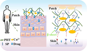
Herein, injectable theranostic hydrogels were developed for reversible naked-eye diagnosis and on-demand PDT of wound infections. Scheme 1a illustrates the preparation process of ε-polylysine (ePL)-based hydrogels through free radical crosslinking of methacrylated ePL (mPL) and their derivatives. Tetrakis(4-carboxyphenyl) porphyrin (TCPP) photosensitizers were conjugated on mPL (mPL-TCPP) to produce free radicals under irradiation by a 660 nm laser, and phenol red chromophores were conjugated on mPL (mPL-Pr) to exhibit color changes in response to the acidic microenvironment of infected wounds. As shown in Scheme 1b, laser irradiation plays a “one-stone-two-birds” role, i.e., PDT-produced ROS initiate sol−gel transformation and exhibit bactericidal effect. ROS-initiated colpolyerization of mPL, mPL-TCPP and mPL-Pr pregel mixtures (mPL-Pr-TCPP) generates [email protected] hydrogels without using any additional photoinitiators. As a typical antimicrobial peptide with abundant L-lysine residues, ePL not only changes the metabolic situation of bacteria, but also adsorbs bacteria through electrostatic interactions [19]. The bacterial capture alleviates the short lifetime and diffusion distance of ROS to achieve a synergistic bactericidal efficacy with PDT. In addition, chromophores and photosnsitizers are fixed in hydrogels, which not only provide robust imaging on infected wounds, after bacterial elimination, and upon infection recurrence, but also lauch on-demand antibacterial PDT. Thus, this study demonstrates the injectable theranostic hydrogels for real-time diagnosis and synergistic antibacterial treatment.


留言 (0)