The emerging requests for precise and minimal-invasive treatments in regenerative medicine have brought a paradigm shift to the design of biomaterials and tissue engineering strategies [1], [2], [3]. Aiming this, injectable and fit-to-defect adaptable hydrogel biomaterials have drawn increasing attention due to their superior properties, including facile administration via minimally invasive surgery, on-target cargo delivery, and high efficiency in nutrient/waste exchange for the loaded cells [4, 5]. However, conventional injectable hydrogels typically employ a triggered chemical reaction to enable gelation, resulting in compromised cell viability and heterogeneous hydrogel network due to the harsh and nonuniform crosslinking [6]. In addition, cells loaded in the hydrogel scaffolds can confront the complicated microenvironment at the defect site (e.g., inflammation, immune-clarence against the allogenic cells) [7], or suffer from limited nutrient/waste exchange upon encapsulation [8]. More importantly, although the traditional tissue engineering approaches for tissue mimics have progressed, the precise control of cell density and single-cell resolution inside 3D engineered tissues remains a challenging problem [9]; these limitations significantly restrain further bench-to-clinic translation of injectable hydrogel-based tissue engineering therapies.
Granular gels consisting of packed microgels as building blocks to assemble into an integral particulate network have recently drawn increasing attention for applications as injectable scaffolds for tissue engineering [5, 10]. Specifically, the particulate feature provides inherent porosity and a larger specific surface area for cell attachment, thereby enhancing the efficiency of mass transportation through the scaffold [10]. However, granular gels typically present poor mechanical strength and structural integrity due to the limited cohesion between microparticles [11, 12]. Besides, cells are usually loaded on the microgels, wherein they are directly exposed to the shearing forces during the injection [13], or the host immune-clearance against the allogenic or endogenic cells [14]. Moreover, the fabrication techniques of microgels used in most of the previous studies are traditional emulsion techniques, leading to low productivity, wide size distribution, low cargo loading efficiency, and significant batch differences [15]. All these drawbacks significantly compromise the therapeutic efficacy of granular gel-based strategies in tissue engineering. Therefore, it remains a challenge to develop a new strategy for the design and fabrication of granular gels with improved mechanical properties and material integrity, retained cell viability, controllable cellular functionality, and cost-efficient fabrication techniques.
Aiming this, microgels with cells loaded inside have been recognized as a more efficient approach for minimal-invasive and in situ tissue-engineering therapy, which can shield the loaded cells from the external injection force or protect them from rapid immune-clearance by the host [14, 16]. Previous studies reported the use of cell-laden microgels as injectable tissue-engineering modules for the regeneration of bone [17, 18], skin [19], and cartilage tissues [20, 21]. However, the overall mechanical properties and structural integrity of these cell-laden microgel fillers are poor, which can be attributed to the insufficient interlocks between the neighboring microgel interfaces and the low specific surface area of the microgels with hundreds of microns in diameter [10, 15]. A potential solution for this issue can be the use of substantially smaller cell-laden microgels with a size close to a single cell; this can improve the bulk mechanical strength of the granular gels by enhancing the microgel's packing density meanwhile shielding the entrapped cells from the external stress [14, 17, 22]. More importantly, this can enable the construction of tissue mimics with single-cell resolution and high cell density [7], which can outperform more traditional strategies of injectable bulk hydrogels by allowing more efficient nutrient/waste exchange, and a more controllable microenvironment via designing the microgel morphological and mechanical properties [23], [24], [25]. All these requests highly controllable and high-throughput microfabrication techniques for generating microgels with cell encapsulation even at the single-cell level.
Recent progress in microfluidic droplet-templated microgel fabrication has shown the efficacy and versatility of this technique for cell encapsulation with regard to the high loading efficiency, high-throughput, and enhanced controllability of microgel properties [26]. Hydrogels applicable for this purpose should exhibit desirable biocompatibility, low precursor viscosity to facilitate droplet pinch-off, mild in-drop gelation process, and tailorable mechanical properties [27], [28], [29], [30], [31]. However, currently available hydrogels are limited to ionically crosslinked alginate, thermally crosslinked gelatin, or photocrosslinkable hydrogels based on free-radical polymerization (FRP) such as poly (ethylene glycol) diacrylate (PEGDA) or methyacrylated gelatin (GelMA) [32]. Especially, FRP-based hydrogels are the most widely-used candidates for cell microencapsulation, since they can allow non-invasive photo-crosslinking, and no need for a specific combination of water/oil phases [33, 34]. Hence, one major challenge in using photopolymerizable microgels for droplet-based microfabrication is the limited fabrication accuracy and poor network homogeneity due to the oxygen inhibitory effect on FRP [35]. This effect becomes more pronounced upon the generation of substantially small microgels with diameters of tens of microns and an oxygen-permeable emulsion system [36]. Specifically, the abundant oxygen in cell culture media can rapidly react with photoinitiator and propagate monomer radicals to form peroxides [37], thereby inhibiting polymerization and resulting in a heterogeneous gel network and compromised structural integrity and encapsulation efficiency [38, 39]. Methods to counteract the oxygen inhibitory effect have been developed including adding oxygen scavengers (e.g. 9, 10-dimethylanthracene) or reduction agents (e.g. thiols), increasing irradiation intensity and photoinitiator concentration [40], or protection by oxygen-free gas [36], which, however, are not applicable for live-cell encapsulation owing to cytotoxicity [41], high gelation temperature, and short shelf-life [35]. Therefore, a new polymerization scheme for droplet-templating microgel production to overcome the oxygen inhibitory effect is imperative for further applications of microgel-based biomaterials.
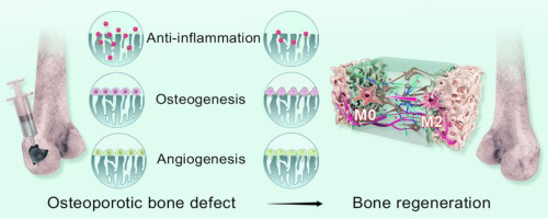
Herein, we developed the granular gels via bottom-up assembly of single cell-laden microgels via phototriggered imine-crosslinking (PIC) chemistry based on a microfluidic droplet-based fabrication technique (Figure 1). Specifically, PIC chemistry between o-nitrobenzene-functionalized hyaluronic acid (HANB) and GelMA, and FRP chemistry of methyacrylated hyaluronic acid (HAMA) and gelatin (GelMA) was simultaneously employed to induce the formation of a dual crosslinking GelMA/HANB/HAMA (GHH) composite microgels (Figure 2A). This strategy effectively alleviated the oxygen inhibitory effect and allowed the generation of GHH microgels with precisely controlled particle size, accelerated gelation process, and improved network strength. Moreover, single cell-laden, monodisperse GHH microgels were prepared using a high-throughput microfluidic fabrication technique with a production rate up to 3.7 × 10⁸ microgels/hr. As such, these cell-laden microgels are further packed to form granular gels with shear-thinning and self-healing behavior, thereby serving as an injectable and adaptable cellularized scaffold for bone tissue regeneration. Further in vitro and in vivo studies demonstrated that the hydrogel matrix can support normal functionality of the encapsulated mesenchymal stem cells. Our study may open up a new avenue for the design and fabrication of granular gels for biomedical applications such as cell niche remodeling [9, 14], cell delivery [17, 42], and tissue regeneration [25, 27].


留言 (0)