
記住我
Sir,
Alopecia areata is a common condition characterised by sudden onset of non-scarring patchy hair loss. Alopecia areata is considered as an organ-specific, T cell-dependent autoimmune disease with a genetic background. The exact aetiopathology remains unclear. It is presumed that alopecia areata results from the collapse of the immune privilege of the anagen hair follicles with the subsequent assault on the follicle bulb by CD8+ T lymphocytes. The term Renbök phenomenon was first described in 1991 by Happle et al.1 They reported retained hair growth localised to psoriasis plaques in patients with co-existing psoriasis and alopecia areata. Subsequently, many other diseases displaying Renbök phenomenon have been reported. Here, we report a case of severe alopecia areata with extensive involvement of the scalp showing retained hair growth in co-existing nevus flammeus. This case might provide further evidence for the occurrence and potential aetiopathology of the Renbök phenomenon in alopecia areata.
A 20-year-old Chinese woman presented with hair loss for eight years. She had patchy hair loss eight years ago, which was diagnosed as alopecia areata and resolved spontaneously. However, diffuse hair loss started suddenly three years ago without any trigger and gradually progressed to alopecia universalis. She was treated with oral prednisone 20 mg once daily and intralesional betamethasone once a month for three months, with minimal hair regrowth. Interestingly, throughout the course of the disease, the hair on a born nevus flammeus on her occipital scalp was mildly involved. She had a past history of allergic rhinitis. Her family history was noncontributary. Physical examination revealed an extensive loss of hair on her scalp with a SALT score of 85 per cent [Figure 1a]. Nevus flammeus on the occipital area was covered with a fair density of terminal hairs [Figure 1b]. A dermoscopic examination of the clinically spared area revealed reticular vessels and terminal hairs [Figure 1c].
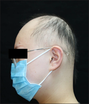
Figure 1a:: Extensive alopecia areata
Export to PPT

Figure 1b:: Obvious terminal hair growth within the area of a nevus flammeus on the occipital scalp
Export to PPT
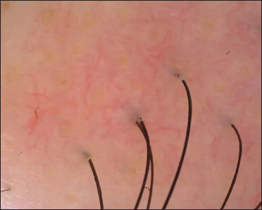
Figure 1c:: Dermoscopy under polarized light (CBS-907; 50× magnification) of the clinically spared area showing reticular vessels and terminal hairs
Export to PPT
There have been several case reports and studies describing alopecia areata sparing nevus flammeus, which suggest a possible association. A case-control study showed that the prevalence of nuchal nevus flammeus was significantly increased in patients with alopecia areata, especially in severe and chronic forms.2 However, the pathophysiological mechanism of the Renbök phenomenon for nevus flammeus in alopecia areata is still unknown.
Nevus flammeus is a disorder caused by genetic mosaicism, which may explain the sparing of hair loss on nevus flammeus.3 The mosaic skin might express altered immunomodulatory proteins in protected skin which are distinct from that in overlapping inflammatory skin.4 Previous findings have illustrated that the Th1 alopecia areata inflammatory response and the Th17 psoriatic inflammation could oppose each other presumably through changing local cytokine milieu.4 The dominant inflammatory profile might ultimately determine the final cytokine phenotype. In this case, extensive hair loss of alopecia areata spared the occipital area with nevus flammeus, suggesting that the hair follicles within the nevus lesion could reverse its vulnerability to the autoimmune reaction initiated by T cells.
Nevus flammeus is also a congenital vascular or capillary malformation. The persistent lesions in adults can show ectasia of subpapillary capillaries.2 Vascular endothelial growth factor has also been proved to play an important role in the development of nevus flammeus which may promote angiogenesis and vasodilatation.5 Interestingly, vascular endothelial growth factor produced by hair follicles is significantly reduced in the skin of alopecia areata patients, resulting in a loss of vascular support to the affected scalp tissue.6 Therefore, in this patient vascular endothelial growth factor production within the nevus flammeus may increase vascular support and protect itself from hair loss. Diphenylcyclopropenone, also known as diphencyprone, which is currently considered effective contact immunotherapy for severe alopecia areata, has been proven to upregulate vascular endothelial growth factor in hair follicle keratinocytes of alopecia areata patients.7 The study suggests that there are different factors regulating vascular endothelial growth factor expression, like epidermal growth factor, transforming growth factor-α and transforming growth factor-β, the proinflammatory cytokine tumour necrosis factor-α, interleukin-1β and interleukin-6.7 It could be postulated that some cytokines also play a critical role in the hair follicle keratinocytes of an alopecia areata -sparing nevus flammeus, which may be involved in the immune reaction to some extent.
In conclusion, we report a case of Renbök phenomenon in extensive alopecia areata with retained hair growth in co-existing nevus flammeus and discussed the possible pathophysiological mechanism of this phenomenon. Further study of this sparing phenomenon may shed new light on underlying mechanisms involved in alopecia areata, potentially leading to the development of new therapeutic strategies.
留言 (0)