
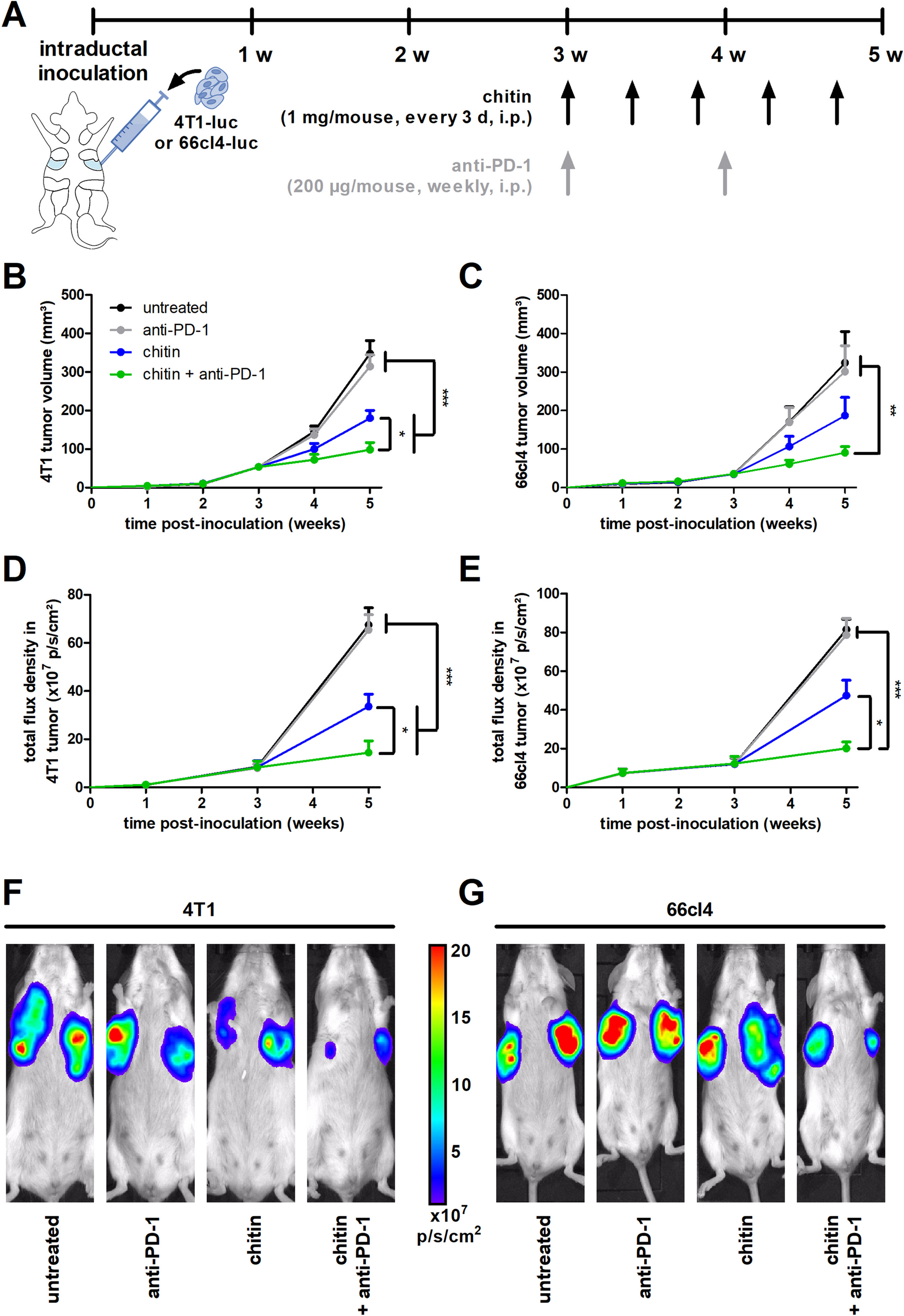
記住我
Among the 928 tumor samples analyzed, 46 patients (5%) carried BRCA 1/2 mutations (32 with germline mutations and 14 with somatic mutations). Mean HRD score in the whole cohort was 25.80 \(\pm\) 19.87. Using the classical cutoff value of 42 [16], high HRD score was found in 133/882 (15%) BRCA 1/2 WT patients and in 31/46 (67%) BRCA 1/2-mutated tumors (24 with germline mutations and 7 with somatic mutations) (Additional file 1: supplemental table 1).
We first considered pathological subtypes, namely estrogen receptor-positive/HER2 negative (ER + /HER2-; n = 606 patients, 66%), HER2 + (n = 157 patients, 17%) and triple negative (n = 165 patients, 17%) early breast cancer (eBC). We compared HRD score and S3 expression according to these pathological subtypes (Fig. 1A and 1C, respectively). A significant association was found between the median HRD score and BRCA 1/2 mutational status (median HRD score, respectively, 57.2 for patients with BRCA 1/2 mutation and 20 for patients with WT status, p value < 0.001) (Additional file 2: supplemental figure 1A (S1A)). The same observation was made in each BC pathological subtype (Fig. 1B). Using the classical cutoff value of 0.3 [19], a high level of S3 was found in 100/882 (11.3%) BRCA 1/2 WT tumors and in 27/46 (58.7%) BRCA 1/2-mutated tumors (22 with germline mutations and 5 with somatic mutations). A significant association was found between median expression of mutational S3 and BRCA 1/2 mutational status (median S3 score, respectively, 0.35 for patients with BRCA 1/2 mutation and 0 for patients with WT status, p value < 0.001) (Additional file 2: supplemental figure 1C (S1C)). Higher expression of mutational S3 was found in BRCA 1/2-mutated tumors, in ER + /HER2- (p < 0.001), HER2 + (p < 0.001) and in triple negative subtypes (p = 0.002) (Fig. 1D).
Fig. 1
Distributions of HRD score and S3 proportion according to BRCA 1/2 mutational status and breast cancer subtypes (standard pathological classification or PAM50 subtypes) considering the whole cohort (n = 928). A-C Violin plots representing the distribution of HRD score (A) and signature 3 proportion (C) according to breast cancer standard pathological classification. B-D Violin plots representing the distribution of HRD score (B) and signature 3 proportion (D) according to breast cancer standard pathological classification and BRCA 1/2 mutational status. *Wilcoxon p-value < 0.05. E–G Violin plots representing the distribution of HRD score (E) and signature 3 proportion (G) according to PAM50 subtypes. *Wilcoxon p-value < 0.05. F–H Violin plots representing the distribution of HRD (F) and signature 3 proportion (H) score according to PAM50 subtypes and BRCA 1/2 mutational status
Next, we considered intrinsic molecular subtypes (according to PAM50), namely basal-like (164 patients, 18%), HER2-enriched (77 patients, 8%), luminal A (492 patients, 53%) or luminal B (195 patients, 21%) tumors. Of note, tumors normal-like subtypes (n = 36) were excluded from all the analyses. We aimed to compare HRD score and S3 expression according to intrinsic molecular subtypes (PAM50), and ER expression among the different intrinsic subtypes. The highest levels of HRD score and S3 were found in the basal-like PAM50 subtype, while the lowest level were found in the Luminal A subtype (Fig. 1E and 1G). No significant difference was found in HRD scores between ER + and ER− tumors in the majority of PAM50 subtypes, except for the HER2 subtype (respectively, mean HRD score of 28 for patients with ER + status and 34 for patients with ER- status, p value = 0.03) (Additional file 2: supplemental figure 1B (S1B)). There was no difference in S3 levels between ER + and ER- tumors among the different PAM50 intrinsic subtypes (Additional file 2: supplemental figure 1D (S1D)). Higher levels of HRD score and S3 were found in BRCA 1/2-mutated tumors in each PAM50 intrinsic subtype (Fig. 1F and 1H).
In order to differentiate between BRCA1/2 germline or somatic mutations, same analyses were performed concerning the distribution of the HRD score and S3 proportion according to BRCA 1/2 mutational status (germline or somatic) considering the whole cohort (n = 928) (Additional file 3: supplemental figure 2E–F (S2 E–F)) or by breast cancer subtypes (standard pathological classification or PAM50 subtypes) (Additional file 3: supplemental figure 2A–D (S2 A–D)).
Fig. 2
Association of genomic features quantifying tumor HRD and immunological characterization in ER + /HER2- tumors (n = 606). Violin plots representing the distribution of HRD, TAI, LST and LOH scores (A), signature 3 proportion (B), and immune signatures (C) according to HRD level and BRCA 1/2 mutational status in ER + /HER2- tumors. *Wilcoxon p-value < 0.05
Finally, we considered exclusively ER + /HER2- tumors (pathological subtype), distributed according to PAM50 subtypes as follows: basal-like (17 patients, 3%, including 1 BRCA 1/2-mutated tumor), HER2-enriched (5 patients, 1%, including 1 BRCA 1/2-mutated tumor), luminal A (426 patients, 70%, including 16 BRCA 1/2-mutated tumors) or luminal B (158 patients, 26%, including 7 BRCA 1/2-mutated tumors): higher levels of HRD score and S3 were only found in luminal B BRCA 1/2-mutated tumors compared to WT tumors (Additional file 4: supplemental figure 3A–B (S3 A–B), Additional file 1: supplemental table 1).
Fig. 3
Gene set enrichment analysis in ER + /HER2- tumors (n = 606). A-C Gene set enrichment analysis (GSEA) barplots for HALLMARK collection. Bars represent the normalized enrichment score (NES), using genes differentially expressed between: (A) patients with BRCA-mutated tumors vs patients with BRCA WT HRD-low tumors, (B) patients with BRCA-mutated tumors vs patients with BRCA WT HRD-high tumors, and (C) patients with BRCA WT HRD-low tumors vs patients with BRCA WT HRD-high tumors. Red bars: patients with BRCA-mutated tumors, green bars: patients with BRCA WT HRD-high tumors, blue bars: patients with BRCA WT HRD-low tumors. Light colors represent non-significant p values and dark colors significant p values (< 0.05)
Taken together, these results make it possible to describe the HRD and S3 level in the different subtypes (pathological or molecular intrinsic) of breast cancer, according to the expression of hormone receptors, and BRCA deficient or proficient status. The distribution of HRD score, PAM50 intrinsic subtypes, BRCA 1/2 mutations, Signature 3 proportion, and ER status in whole cohort is summarized in Additional file 5: supplemental figure 4A (S4A). As expected, HRD-high tumors essentially comprise tumors of the basal-like subtype, the majority (but not all) of tumors with BRCA mutation, and with a high 3 signature.
Fig. 4
Mutational landscape in ER + /HER2- tumors (n = 606). A Barplots representing the frequencies of most frequent mutated driver genes, according to HRD and BRCA 1/2 mutational status. * Fisher's exact test p value < 0.05. B Landscape of pathogenic or likely pathogenic somatic mutations of genes involved in the homologous recombination pathway, BRCA1/2 excluded. C Pie charts of number of pathogenic or likely pathogenic somatic mutations of genes involved in the homologous recombination pathway according to HRD and BRCA 1/2 mutational status
A subset of BRCA-proficient ER + /HER2- breast cancers harbored high HRD scoreIn metastatic ER + /HER2- BC, we recently described a subgroup of BRCA-proficient tumors, but with high HRD score [18]. Therefore, we investigated whether such a BC subtype also existed in early stage BC. Considering exclusively ER + /HER2- tumors (n = 606) from the TCGA, we found 24 tumors with BRCA 1/2 mutation (4%), and also a group with high HRD score (> 42), but without BRCA 1/2 mutation (44 tumors, 7.3%). This enabled us to study these 3 distinct groups of ER + /HER2- tumors for the remainder of the experiments, i.e., BRCA mutated, BRCA-proficient (WT) HRD high, and BRCA-proficient (WT) HRD low. Considering molecular intrinsic subtypes according to the PAM50 classification, these 3 subgroups showed a significantly different distribution of molecular intrinsic subtypes (Additional file 5: supplemental figure 4B–E (S4 B–E)): BRCA-mutated tumors were mainly luminal A (62.5%) and luminal B (29%). BRCA WT HRD-low tumors were mainly luminal A (74%), and basal-like in only 2% of case. Conversely, in BRCA WT HRD-high tumors, we found 16% of basal-like, 54.5% of luminal B but only 29.5% of luminal A tumor intrinsic subtypes. Accordingly, we found a significantly positive correlation between HRD score and expression of a proliferation signature [20] (Additional file 6: supplemental figure 5A (S5A)). Tumors with high HRD score have higher proliferation signature (Additional file 6: supplemental figure 5B (S5B)), and these differences were seen in each molecular subtype (PAM50) (Additional file 6: supplemental figure 5C (S5C)).
Fig. 5
Progression-free interval and overall survival according to BRCA 1/2-mutated status and HRD score level in ER + /HER2- tumors (n = 606). A, B Kaplan–Meier curves of overall survival (A) and progression-free interval (B) according to BRCA 1/2 mutational status and HRD score level. Red curves: patients with BRCA-mutated tumors, green curves: patients with BRCA WT HRD-high tumors, blue curves: patients with BRCA WT HRD-low tumors. Ticks denote censored data. C, D Forest plots of hazard ratio (HR) for the association of the clinical variables and immune scores with overall survival (C) and progression-free interval (D) according to BRCA 1/2 mutational status and HRD score level. Red lines: patients with BRCA-mutated tumors, green lines: patients with BRCA WT HRD-high tumors, blue lines: patients with BRCA WT HRD-low tumors. Horizontal lines represent 95% CI. Each point represents estimated HR. The dashed vertical line indicates HR = 1. *Wald-test p-value < 0.05
In BRCA-proficient (WT) HRD-high tumors, HRD scores were even higher than those in BRCA-mutated tumors (Fig. 2A and Additional file 7: supplemental figure 6A (S6A)), driven by higher TAI and LOH scores. These BRCA WT HRD-high tumors had S3 levels comparable to BRCA-mutated tumors, and higher than BRCA WT HRD-low tumors (Fig. 2B and Additional file 7: supplemental fig 6B (S6B)). We next compared BRCA 1/2 expression between patients with BRCA-mutated tumors, BRCA WT HRD-high and BRCA WT HRD-low tumors. We did not find a significant difference in BRCA2 expression between WT HRD-high and WT HRD-low tumors, whereas BRCA2 expression was logically significantly lower in BRCA-mutated tumors (Additional file 8: supplemental figure 7 (S7)). Thus, the difference in HRD score between these 2 groups does not seem to be explained by epigenetic regulation of BRCA2 gene. Same analyses were performed within the different molecular subtypes to investigate the expression of BRCA1 and BRCA2 among the BRCA-mutated, WT HRD-high, and WT HRD-low groups (Additional file 9: supplemental figure 8 (S8)). Here again, no differences of BRCA1 or BRCA2 expression were seen between BRCA WT HRD-high or HRD-low tumors, whatever PAM50 subtype.
Immunological tumor landscape of ER + /HER2- breast cancers according to BRCA and HRD statusSince different immune microenvironments have been described between BRCA WT and BRCA-mutated BC [21], we next evaluated the immunological landscape of our 3 different groups of eBC even within ER + /HER2- tumors (Fig. 2C and Additional file 7: supplemental figure 6C (S6C)). No significant difference was found in any of our analyses between BRCA 1/2-mutated and WT HRD-high tumors. These results indicate that this small subset of HRD high BRCA 1/2 proficient ER + /HER2- eBC actually had immunological features comparable to those of their BRCA 1/2-mutated counterparts. On the contrary, several significant differences in tumor immune profile were found between WT HRD-high and WT HRD-low tumors, notably T cell abundance, CD8 T cell abundance, cytotoxic T cell abundance, and cytotoxicity expression signature, but also TIL abundance (evaluated by RNA TILS score signature), Th1 lymphocyte gene expression signature, interferon-γ expression signature, were all significantly higher in BRCA WT HRD-high compared to WT HRD-low tumors, suggesting a different immunological cellular landscape between these two groups, with WT HRD-high tumors having higher tumor immune infiltrate of cytotoxic lymphocytes. Moreover, these WT HRD-high tumors also appeared to have significantly higher tumor mutational burden (TMB), and higher CD274 (PD-L1) expression than WT HRD-low tumors. Concerning macrophages subpopulations, M1 were found to be higher in BRCA-mutated tumors and BRCA WT HRD-high when compared to BRCA WT HRD-low cases (with no difference regarding M2 subpopulation). Of note, no significant difference was observed in terms of parameters associated with B or NK cell lineage (not shown), or in terms of other (than PD-L1) inhibitory immune checkpoint expression. No difference was noted among BRCA-mutated tumors, when HRD-high and HRD-low cases were compared (Additional file 7: supplemental figure 6 (S6)), but the number of BRCA-mutated tumors with low HRD score was very limited.
Collectively, these results therefore show that among ER + / HER2- tumors, BRCA-proficient HRD-high tumors have an immunological landscape comparable to that of BRCA-mutated tumors, and apparently more favorable to therapeutic approaches using immune checkpoint blockers, compared to BRCA WT HRD-low tumors.
Tumor cellular pathways of ER + /HER2- breast cancers according to BRCA and HRD statusTo explore the underlying mechanisms of our findings, we next aimed to determine whether different cellular/oncogenic pathways could be associated, and differentially expressed in these 3 different subtypes of ER + /HER2- tumors. Pathway enrichment analysis using the Hallmark gene sets from MSigDB revealed strong differences between the 3 different HRD-status eBC (Fig. 3A–C). Briefly, compared to BRCA WT HRD-low BC, BRCA-mutated tumors were enriched in proliferation module gene sets (E2F target, G2M checkpoint, MYC targets, mitotic spindle signatures), but also in immune pathway (allograft_rejection signature), and mTORC1 pathway (Fig. 3A). Conversely, BRCA WT HRD-low were significantly more enriched in molecular signature modules related to estrogen response (early and late response).
Interestingly, BRCA WT HRD-high tumors also differed from BRCA-mutated tumors, but via different biological cellular processes (Fig. 3B): For example, BRCA WT HRD-high tumors appeared to be enriched in immune modules (allograft_rejection signature, interferon gamma, and interferon alpha response), proliferation module genes (E2F target, G2M checkpoint, MYC targets, mitotic spindle signatures), but also DNA repair (thereby confirming our previous results obtained for HRD scores), and other potential therapeutic targets like estrogen response or MTORC1, or KRAS signaling.
Finally, the main differences in molecular pathway signatures between BRCA WT tumors (HRD high vs HRD low) were statistically significant enrichment in numbers of immune pathways in WT HRD-high tumors (allograft_rejection signature, interferon gamma, and interferon alpha response, IL6_JAK_STAT3, inflammatory response, IL2_STAT5 signaling) thereby confirming our previous results concerning the tumor immune landscape, but also MTORC1, and logically DNA repair signaling in WT HRD-high tumors, compared with their WT HRD-low counterparts (which remained enriched in some classical pathways involved in luminal tumor biology, like estrogen response) (Fig. 3C).
Collectively, these results show that in addition to differences in immunological tumor profile, there seems to be a wider range of biological processes that differ between these 3 subtypes of ER + /HER2- BC. These results also provide a biological rationale for selecting appropriate pharmacological targeting in each of these different subtypes.
Tumor mutational landscape of ER + /HER2- breast cancers according to BRCA and HRD statusWe then aimed to determine the tumor mutational landscape in these 3 groups of ER + /HER2- tumors. Only driver genes were considered in this analysis. The most frequent mutated driver genes in each group are presented in Fig. 4A. Patients with WT HRD-high tumors have a different mutational landscape to that of BRCA 1/2-mutated tumors or WT HRD-low tumors. We found that BRCA WT HRD-high tumors presented a higher rate of TP53 mutations compared to BRCA WT HRD-low tumors or BRCA-mutated tumors. Conversely, PIK3CA, GATA3, MAP3K1, CDH1 mutations appeared to be more frequent in BRCA WT HRD-low tumors compared to BRCA WT HRD-high tumors.
Considering exclusively genes involved in homologous recombination (HR) (except BRCA1 and BRCA2), we found that 33 (6%) of the 541 samples with mutations available presented at least one somatic gene alteration (Fig. 4B). However, no significant difference was found in the number of somatic mutations in HR-associated genes between the BRCA 1/2-mutated, WT HRD-high, and WT HRD-low groups (Additional file 10: supplemental figure 9 (S9)).
A complete description of the mutations found in each group is presented in Fig. 4B.
Only 2 BRCA-mutated tumors had a second HR-gene mutation (Fig. 4C, upper panel). Similarly, in BRCA WT HRD-high tumors, we found only 2 cases (5% of these tumors) harboring a single HR-gene mutation (Fig. 4C, middle panel). In BRCA WT HRD-low tumors, we found the same proportion (n = 29; 6%) of cases with at least one HR-gene mutation (Fig. 4C, lower panel). In the vast majority of cases, it was a single mutation (n = 25), but we also found 4 tumors with ≥ 2 HR-gene mutations.
Outcome of ER + /HER2- early breast cancers according to BRCA and HRD statusWe finally aimed to determine whether these 3 groups of ER + /HER2- tumors, stratified according to HRD status, would have different prognosis, and how this new biological segmentation could account for the risk of relapse and death, compared to previously described, classical prognostic factors, and biological factors related to the tumor microenvironment.
We first sought to define the biological variables associated with the progression-free interval (PFI) in the ER + /HER2- tumors cohort (Table 1).
Table 1 Factors associated with progression-free interval by univariate and multivariate analysis using Cox models with lasso penaltyBy univariate analysis, patients with BRCA-mutated tumors had poorer PFI compared to patients with BRCA WT HRD-low tumors (HR: 2.56 [1.1; 5.93], p = 0.05). No significant difference was found between BRCA WT HRD-low and BRCA 1/2 WT HRD-high tumors (HR: 0.76 [0.27; 2.1], p = 0.61). (By the same, the direct comparison between BRCA-mutated and BRCA WT HRD-high cases found no significant difference in terms of PFI (not shown).) Two HRD-related variables were associated with shorter PFI, namely TAI (HR = 1.94 [1.21; 3.11], p = 0.02) and LST (HR = 1.95 [1.21; 3.13], p = 0.02). 13 biological variables, mainly immune variables (interferon gamma, T cell receptor (TCR), cytotoxicity (CYTOX), TH1, cytotoxic T cell lymphocytes (CTL), and inhibitory immune checkpoint (ICK)) signatures, T cells, CD8 T cells, cytotoxic lymphocytes, natural killer cells, B lineage, myeloid dendritic, and neutrophils abundances and TILS score, were associated with better PFI. Conversely, fibroblast abundance (HR = 1.64 [1.02; 2.63], p = 0.07) tended to be associated with shorter PFI.
By multivariate analysis, using a LASSO model, we found that TAI score (HR = 1.38 [1.34; 1.42]), LST score (HR = 1.19 [1.18; 1.20]), and fibroblast abundance (HR = 2.35 [2.25; 2.45]) were associated with shorter PFI. Inhibitory immune checkpoint (ICK) signature (HR = 0.48 [0.47; 0.50]), T cells (HR = 0.89 [0.88; 0.90]), cytotoxic lymphocytes (HR = 0.65 [0.65; 0.65]), myeloid dendritic cells (HR = 0.75 [0.74; 0.76]), IFNg (HR = 0.98 [0.97; 1.00]), and neutrophil (HR = 0.67 [0.66; 0.69]) abundance were associated with better PFI. Same trends in these exploratory analyses were observed when overall survival (OS) was studied (Additional file 11: supplemental table 2).
Concerning survival in our 3 groups of patients, median overall survival (OS) was not reached for patients with BRCA 1/2-mutated tumors or for patients with WT HRD-high tumors and was 130 months for patients with WT HRD-low tumors (Fig. 5A). Regarding PFI, the median PFI was 98 months for patients with BRCA 1/2-mutated tumors, not reached for patients with WT HRD-high tumors, and 168 months for patients with WT HRD-low tumors (Fig. 5B). There was no statistically significant difference in terms of OS or PFI between the 3 groups of tumors.
We then investigated in an exploratory analysis, whether OS and PFI were associated with different prognostic factors within these three tumor groups (Fig. 5C-D). Neither HRD score nor any its 3 components (LST, TAI, and LOH) were associated with different outcome in any of the 3 groups of tumors. The same was true for S3 level, except for BRCA WT HRD-high tumors, in which high S3 levels appeared to be associated with better PFI (but no difference for OS). Tumor immune profile seems to have a greater influence on the prognosis of BRCA-mutated and BRCA WT HRD-high tumors (compared to BRCA WT HRD-low cases), since high expression of B cell lineage, dendritic cells, cytotoxic T lymphocytes, T cells, and CD8 T cell signatures appear to be more strongly associated with favorable survival in these groups of tumors (Fig. 5C-D). Other cellular components of the tumor microenvironment such as fibroblasts or endothelial cells seem to be associated with particularly poor prognosis in BRCA WT HRD-high tumors, compared to other ER + /HER2- eBC. However, it should be noted that the prognostic impact of these biological factors remains of lesser magnitude than that of clinical variables (T and N stage), and molecular intrinsic subtypes according to PAM50, as classically described. Same exploratory analysis of variables associated with PFI and OS is also presented in Additional file 12: supplemental figure 10A–B (S10 A–B), but by adjusting each variable on T and N stage.
留言 (0)