
記住我

In Ayurvedic medicine, resin from C. mukul, sometimes refered as “guggul,” for centuries was used in arthritis treatment. Resin extract from C. mukul in a clinical study showed significant improvement in osteoarthritis after treatment with 500 mg TID for 1 month (Singh et al. 2007). A significant inhibition of NO formation by methanol resin extract of C. mukul in lipopolysaccharide (LPS) activated murine macrophages was exhibited using IC50 = 15 mg/mL (Meselhy 2003; Matsuda et al. 2004a). Also, MeOH extract demonstrated an anti-inflammatory property against LPS-induced inflammation (Cheng et al. 2011). Isolated compounds, polypodane triterpenoids, cembrane diterpenoids, lignans, and steroids have been studied for COX inhibitory activity and NO production. Prevention of NO production by E and Z-guggulsterones (22 and 21), myrrhanol A (23), and myrrhanone A (24) has been observed having IC 50 = 1.1, 3.3, 21.1, and 42.3 mM, respectively (Meselhy 2003). In lipopolysaccharide activated mouse peritoneal macrophages, myrrhanol A (23) and mukulol (25) were reported to have inhibitory action nitric oxide synthase (iNOS) induction (Matsuda et al. 2004a). E-guggulsterone (22) and Cembrene (26) were reported to be the most active in COX inhibition. They inhibited COX-1 by 79% and 67%, and COX-2 by 83% and 54% individually at 100 ppm (Francis et al. 2004). The anti-inflammatory properties of Z-guggulsterones and E-guggulsterones (21 and 22) were reported, eliciting their anti-inflammatory activity by subduing NF-kB activation and NF-kB regulated gene products expression (Shishodia and Aggarwal 2004; Lv et al. 2008). Kimura et al. (2001) reported C. mukul resin extract and pure compounds anti-inflammatory potential (23 and 24) using the model, air pouch granuloma induced by an adjuvant and observed that 23 was 7 × , 5 × , and 3 × more effective on carmine content, granuloma, and pouch fluid weight more than the standard drug (hydrocortisone) used as control. It, therefore, has the prospect of being developed into an anti-inflammatory drug. Furthermore, myrrh was reported to have significant inhibitory activity on the activator of transcription-1, transcription-3 (STAT-1 and STAT-3), and signal transducer leading to reduced production of cytokines through the pathway of janus kinase/STAT (Lv et al., 2008). Also, it suppresses down-regulation of cytokine synthesis, a reaction to a decreased interferon-gamma and interleukin-beta production. This auto-regulates JAK/STAT pathway through the control of transcription by transcriptions activator that also inhibits activation of pathway (Su et al. 2011).
Su et al. (2012) reported that myrrh water extract and combined water extract (CWE) at doses of 3.9 g/kg and 5.2 g/kg exhibited formalin-induced paw edema inhibition with an inhibition rate of 30.44% and 23.50%. Individually, a significant (P < 0.01 or P < 0.05) inhibition of PGE2 production was observed in samples tested. However, CWE was stronger in suppressing carrageenan-induced mice paw edema at 2 and 3 h after drug was administered. Reports showed the anti-inflammatory activities of extracts from C. molmol and Commiphora pyracanthoides. Inhibiting the release of IL-6 and IL-8 stimulated by IL-b in human gingival fibroblasts cells stimulated IL-b on the administration of C. molmol volatile oil has also been reported (Tipton et al. 2003). C. molmol resin pet. ether extract inhibited carrageenan-induced inflammation and cotton pellet granuloma. Extract from the stem of C. pyracanthoides was reported to be the most active with an IC50 = 27.86 mg/mL among all the Commiphora species tested for anti-inflammatory activity applying a 5-lipoxygenase (5-LOX) assay method (Paraskeva et al. 2008). Friedelin, an isolated from Commiphora berryi and its pet. ether bark extract, was reported to have shown inhibitory activity on soybean lipoxygenase with IC50 values of 35.8 mM and 15.3 mg/mL (Kumari et al. 2011). Commiphora erythraea resin hexane extract was reported to inhibit edematous response, reducing it by 84% at 1000 mg/cm2 in mice ear edema caused by croton oil. These compounds, Myrrhone (27), rel-3R-meth-oxy-4S-furanogermacra-1E,10 (15)-dien-6-one (28), and rel-2R-methoxy-4R-furanogermacr-1(10)E-en-6-one (29) present in the extract were proposed to have exhibited the anti-edematous activity of the plant (Fraternale et al. 2011). Ethanol leaf extract of Commiphora caudate administered orally at 250 mg/kg was reported to have inhibited carrageenan-induced paw edema response by 67% in rats (Annu et al. 2010). In vivo anti-inflammatory activity of isolated compounds, mansumbinoic acid (30) and 2a,3b,23-trihydroxyolean-12-ene (31), were reportedly studied (Fourie and Snyckers 1989; Duwiejua et al. 1993). Evidence from literatures showed C. mukul resin was the most investigated, showing promising anti-inflammatory property both in vitro and in vivo. This further establishes application in Ayurvedic medicine. Steroids (21 and 22) and triterpenoids (23 and 24) present in C. mukul are the active principles bringing about anti-inflammatory effects. The extracts and compounds mechanistic activity from the genus Commiphora as it relates to signaling pathways, multiple inflammation-related proteins were highlighted. Potential anti-inflammatory targets such as NO formation, COX, ROS, TNF-a, PGE2, MAPK, and NF-kB were identified and tested.
AntioxidantsCompounds of the class diterpenes, sesquiterpenoids, sterols, and triterpenes present in high quantity in myrrha extracts that may serve as electron donors react with free radicals converting them to a more stable product thereby terminating the radical chain reactions. This is a corroborated (Fraternale et al. 2011) research work where they showed myrrha resin hexane extract as having the best DPPH radical scavenging activity unlike to its oils. The same authors made suggestion that the action could be attributed to three compounds, 2-methoxy-furanogermacren-6-one myrrhone and 3-methoxy-furano germacradien-6-oneall of the furano-sesquiterpenoids family. Their DPPH radical scavenging potential had IC 50 values of 1.08, 4.29, and 2.56 mg/mL, respectively (Mohamed et al. 2014).
Triterpenes (ursolic and oleanolic acid) and essential oils in the resins of C. myrrh and Boswellia serrata were reported as having potent antioxidant activity in sunflower oil, although, with negative result in DPPH scavenging activity. C. myrrha essential oil (EO) inhibited lipid peroxidation in sunflower oil. It is then safe to conclude that essential oil of C. myrrh could be applied in functional foods, pharmaceutical, and cosmetic preparations mainly due to their antioxidant activity in oil substrate (Assimopoulou et al. 2005).
Antimicrobial activityBiological activity of myrrh on viruses and bacteria has been reported in literatures. Empirical evidences have shown that myrrh extracts possess effects on virus by virtue of which these extracts possess antibacterial and antiviral activities on different virus strains (Khalil et al. 2020). In a particular study, bactericidal, fungicidal, and anti-viral activities of myrrh essential oil extracts suggested their potential in inhibiting the growth of bacteria and virus strains (Brochot et al. 2017). Also, in a study, essential oils from myrrh showed antiviral activities against two viruses: herpes simplex virus type 1 (HSV-1) and influenza virus type A (H1N1). Myrrh was observed to act by free viral particles direct inactivation and disrupting the virion envelope structures which major role is in host cell virus invasion (Brochot et al. 2017). Another mechanism by which the extracts bring about their activity is by the inhibition of the enzyme, DNA polymerase in viral strains, thereby, hampering virus resistance to specific medications. Therefore, development of new antiviral drugs from the extracts with specific target on DNA holds a lot of prospects (Brochot et al. 2017).
Generally, plants Eos are a mixture of different constituents (Burt 2004). Specific compounds from phenols were suggested to show the microbicidal activities of Eos (Ben Arfa et al. 2006; Lambert et al. 2001). Also, four terpene molecule activities present in some Eos were investigated on the food pathogens and spoilage bacteria organisms. Since disruption of cellular membranes was reported to be caused by Eos and their constituents (Kapros and McDaniel 2009), studies on their cytotoxicity have been carried out using bacteria cell model in vitro (Boffa et al. 2016).
Studies on PE myrrh extract using diffusion test showed antimicrobial potential on C. albicans, Streptococcus pyogenes, and Staphylococcus aureus. The extract of EtOH showed potent action against the strains tested. However, greater activity was observed against C. albicans and S. aureus (9 mm zone of inhibition, 20 mg /mL); this further establishes the therapeutic effect of myrrh for curing infectious diseases such as gingivitis pharyngitis, phyorrhoea, and sinusitis (de Rapper et al. 2012). The anti-fungal activity exhibited on C. albicans is similar with the findings reported in previous studies (Dolara et al. 2000). Methanol extract tested on C. albicans, Pseudomonas aeruginosa, and Escherichia coli demonstrated a very low antimicrobial activity with no zone of inhibition observed at 20 mg/mL, whereas, PE extract demonstrated 3.7 and 5.7 mm zone of inhibitions for C. albicans and S. albus, which can be compared to 5 and 3 mm for S. aureus (Boffa et al. 2016).
Neuroprotective effectsFrom the resins oozing out of Commiphora myrrha was isolated runcate type sesquiterpenes, i.e., commiterpenes A–C (1–3) showing neuro-protective activity on MPP+ induced neuronal cell death in SH-SY5Y cells (Xu et al. 2011).
Commiphoins A–C (1–3), the novel runcate type of sesquiterpenes, including two common runcate type of sesquiterpenes (4 and 5) were gotten the extracts of Commiphora myrrha resinous. Screening 1 and 3–5 compounds was carried out against anti-Alzheimer’s disease (AD) activity employing Caenorhabditis elegans AD pathological model. All the compounds tested demonstrated significant anti-Alzheimer’s disease activities (Yu et al. 2020).
Anti-acetylcholinesterase activityAcetylcholinesterase inhibitors, natural or synthetic, have been shown to be commonly applied as insecticides or nootropic drug for boosting memory in patients having amnesia. Many bioactive constituents have been established to have the ability to inhibit AchE which helps in boosting cerebral activity or ameliorate disease symptoms relating to it (Teibo et al. 2020). Herbal preparations with known activities on the brain for boosting retention and learning are referred to as “nootropic herbs” or “phyto-nootropics,” and their isolated active principles are called smart drugs (Hussein et al. 2019). In Mesopotamia, the species commonly used to produce essential oils used in aromatherapy is Commiphora myrrha (Nees), Engler (Watt and Sellar 2012). C. myrrha leaves, bark, and resin methyl alcohol extract have been reported to inhibit AchE by 17.00, 26.00, and 29.33% compared to eserine. Computational prediction using silico tools has been used to model the ADMET and putative anticholinesterase potentials of bioactive compounds in myrrh. Bioactive constituents from C. myrrha were reported to show a good binding affinity (BA) concerning AchE principal sites with a range of − 5.8 (m-cresol) to − 10.5 (abietic acid) kcalmol−1. On these bases, terpenoid compounds (25 out of 28) from myrrh served as dual inhibitors due to the hydrophobic interactions using both peripheral anionic site (PAS) of AchE, and catalytic triad while hydrogen bonding was used between AchE and alpha-terpineol, elemol, and eugenol (Hussein et al. 2019).
Stimulates insulin secretionMedicines of plant origin like Commiphora myrrha (CM) have traditional application in Ayurvedic medicine for diabetes management in certain regions of Africa and Arabia. Several studies have shown that in diabetic animal models, CM reduced blood glucose, and increased insulin concentration is achieved with CM. It is, however, not fully clear the mechanism employed by CM in achieving glycemic control in the animals (Al-Romaiyan et al. 2021).
Increase in insulin production that was concentration dependent was observed on exposure of MIN6 cells to CM resin solution (0.5–10 mgmL−1) in a static setting. When islet of the mouse was incubated with CM (0.1–10 mgmL−1), it brought about a concentration-dependent stimulatory effect on insulin. Reduction in cell viability or cell membrane integrity was not associated with CM concentrations at ≤ 2 mgmL−1. Although, a remarkable absorption of trypan blue dye and apoptosis accompanied higher concentrations of CM. At stimulatory and sub-stimulatory glucose levels, CM (2 mg/mL) resulted in quick and reversible increase in insulin production by islets of both humans and mouse during perfusion Total insulin contained in β-cells, mRNA formulations of preproinsulin, and Pdx1 did not change despite the stimulating effect of CM on the production of insulin (Al-Romaiyan et al. 2021).
Analgesic actionIn ancient times, myrrh has been used as analgesics, which is possibly due to bioactive constituents present in them acting as pain relievers (El Ashry et al. 2003). Two sesquiterpenoid compounds, furanocudesma-1, 3-diene, and curzerene present have been reported to be acting on the receptors opioid in the central nervous system, bringing about anesthetic activity (El Ashry et al. 2003). Also, furanocudesma-1, 3-diene in myrrh, particularly the ones isolated from Commiphora mukul have been reported to provide significant relief from abdominal pain and improving health hyperalgesia. Hence, these extracts bring about their effects by relieving peripheral nerve pain resulting from chronic compressive damages to the sciatic nerves. These extracts have been reported to be applied as a substitute medication in the management of nerve pain (Mehta and Tripathi 2015). In addition, some isolate such as furanocudesma-1, 3-diene and lindestrene present in myrrh were reported to relief pain by acting on nerves and body joints. These compounds bring about their effects by suppression of the molecule prostaglandin and hinder the inward movement of sodium current thereby ameliorating the feeling of pain (Nomicos 2007). The presence of the compound furanodiene in high amount acts by lowering pain resulting from fever (Gadir and Ahmed 2014).
Anti-cancer propertyCell deaths occur in various form, one of which is apoptosis (cell suicide), defined as a programmed process or genetically controlled, or necrosis or a non-programmed/accidental process (Hotchkiss et al. 2009). One of the most effective non-surgical cancer treatments is targeting apoptosis, a characteristic of cancer cells. Targeted attack on apoptosis holds the possibility of stopping the uncontrolled growth of the cancer cells. Studies have reported the use of cancer drugs to target different pathways of apoptosis (Pfeffer and Singh 2018), including compounds of plant origin and having various bioactive compounds, is having an effect on the apoptotic pathways via different mechanisms (Safarzadeh et al. 2014; Teibo et al. 2021a, b). The use of flow cytometry to detect necrosis or apoptosis by exposing phosphatidylserine (PS) outside of the apoptotic cells, an important method in the induction of apoptosis, has been reported (Wlodkowic et al. 2011). Another important discovery in the study of cancer is the inability to control the normal cell cycle. There is an increasing interest on the cell cycle as a mechanism for anticancer drug target (Gabrielli et al. 2012). In a study where flow cytometry was used for detecting the activity of certain compounds on HepG2 cell cycles, it was shown that treated HepG2 cells in the S-phase reduced in percentage, while an increase was observed in the G2/M phase cells. In a research work, cell cycle phase distribution using flow cytometry was used to determine the altered compound in HepG2 cells cell cycle. The results showed decrease in treated HepG2 at the S phase; however, that of the G2/M phase increased.
Ethno-pharmacological evaluation has identified myrrh an anticancer drug. The active constituent possessing anticancer activity is elemene with proven action on various cancerous cells including glioblastoma, and it was proven to be safe and effective. Elemene, particularly β-elemene, was reported to possess anti-proliferative activity. It acts by activating p38MAPK in glioblastoma (Yao et al. 2008). Also, the compound 2S-epoxy-4R-furanogermacr-10-3n-6-one, furose-type sesquiterpene rel-1S isolated from myrrh was reported to possess low cytotoxic activity on MCF-7 cell line of breast cancer. This compound, in combination with bisabolene in myrrh, was effective in reducing the growth of breast cancer indicating myrrh as containing novel anti-breast cancer drug (Yeo et al. 2016; Shen et al. 2008). Cyclobolinane, a triterpenoid present in myrrh, was shown to exert a moderate cytotoxic effect on PC3 and DU145 prostate cancer cell lines (Shameem 2018).
Furthermore, numerous studies reported the use of myrrh components in apoptotic induction and stoppage of tumor cell proliferation (Chen et al. 2013). This is similar to reports on compounds isolated from C. myrrh as used to induce apoptosis and arrest of cell cycle progression (Gao et al. 2015).
Furano-sesquiterpene compounds were shown to elicit some pharmacological activities resulting from the wide range of bioactive compounds present in them (Fraternale et al. 2011). The derivatives of furano-sesquiterpenes and itself, a soft coral isolate, were reportedly tested on leukemia, prostate, lung, breast, and cervix cancer cell lines which resulted in some of the compounds possessing promising activity on two of the cancer lines tested, leukemia and prostate cancer (Rajaram et al. 2013). Additionally, a different furano-sesquiterpene reportedly obtained from soft coral was tested for its anticancer activity. Researchers have demonstrated the inhibition of many cancer cell line proliferation in humans, decreased programmed cell death, and cell cycle arrest induction in human leukemia cells (THP-1) (Arepalli et al. 2009).
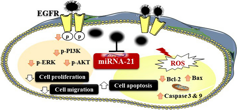
Results from different researches have suggested that myrrh possesses inhibitory properties against cell multiplication and brings about cell suicide of GC cells possibly achieved by downward control of COX-2 formulation in GC cells (Sun et al. 2020) (Fig. 3). Below is a schematic representation of relevant proteins showing concurrent upward control of Bax formulation, downward control of COX-2, and Bcl-2 formulation in cells during myrrh administration. In vitro studies have shown myrrh to induce apoptosis in a dose-dependent form in GC cells. GC cell migration rate could be possibly reduced by myrrh. Although, blockage of GC cell migration could increase with increase in administration time.
Fig. 3
Myrrh’s inhibition of proliferation of gastric cancer cells and induction of apoptosis (Sun et al. 2020)
Antiparasitic activityIn Egypt, the use of myrrh as an anti-parasitic agent saw its revolution in 1990 through evidence-based scientific research. The main focus of research in Egypt was the human trematode infection shrouded in stories of success and disagreement.
Myrrh as a schistosomicideMyrrh has been reported to have schistosomicidal activity on different phases of S. mansoni. The effect of the drug in infected mice was more pronounced at the 21st and 45th days of PI. The drug showed a promising prophylactic effect when administered 5 days before exposure (Massoud et al. 2004c). A unique myrrh formulation that contains volatile oil and myrrh resin was recorded at the beginning of the 2000s for its efficacy and safety in mice with S. mansoni infection. Extract of myrrh, administered at 250 mg/kg and 500 mg/kg, was described to have stimulated a noticeable reduction in worm burden, increasing hepatic displacement of worms and a radical reduction in immature egg percentages situated on the intestine wall (Badria et al. 2001). Mirazid was reported in mice model to be safe in the treatment of Schistosoma infection by S. mansoni, and it was also efficacious (Hamed and Hetta 2005). An improvement in liver enzyme activities and a significant worm load reduction by 81.10% and a reduction in ova count by 73.07% on daily administration of Mirazid, for 3-day fasting at 600 mg/kg (Hamed and Hetta 2005). In another study, myrrh was reported to have shown significant schistosomicidal activity at 500 mg/kg on administration every day for five consecutive days. The activity remained pronounced in groups that got the drug on the 21st and 45th days post infection (Massoud et al. 2004c). Furthermore, it was observed in the groups a significant reduction in granulomas restored jejunal mucosa continuity and led to paucity of eosinophils (Massoud et al. 2004c). However, results from other studies have reported otherwise the chances of myrrh application for the management of schistosomiasis. The most interesting report on inefficacy of myrrh on animals infected with S. mansoni was from a multi center research carried out by Botros et al. (2004). Mirazid, a commercial formulation, has been tested alongside myrrh resin derivatives at varying doses on various strains. In the Egyptian (CD) strain infected mice reduction in worm infection loads in mice was observed to be negligible. Solution of Mirazid, at high doses, was observed to be toxic to mice infected with Puerto Rican (Mill Hill) strain of S. mansoni but possessed modest to no worm reduction at lower doses. With Puerto Rican (NMRI) and Brazilian (LE) strains of S. mansoni mice and hamsters, no anti-schistosomal activity was observed. Furthermore, effects on tissue oogram and egg arrangement did not show significance (Botros et al. 2004).
Myrrh as a fasciolicideContrary to the inconsistent activity on schistosomiasis infection, myrrh has proven to possess a very potent fasciolicidal activity. This is evident from the significant data generated on this activity from experimental animals, infield studies, and clinical trials.
Myrrh showed high efficacy when tested on Fasciola in animal studies. It was reported in a study, that Mirazid completely eradicated Fasciola gigantica in rabbits with 20 mg/day dosage administered orally for six days consecutively. The immune response (IgG) was observed to be highest in infection treated rabbits compared to the infection untreated control (Mahmoud 2008). This lead to the conclusion, that at parasitology and immunology levels, Mirazid was safe and the most effective fasciolicidal drug (Mahmoud 2008).
Myrrh as a heterophycideHeterophytes are common parasite found on snails and fish serving as intermediary hosts, found along the Nile valley and lakes of Egypt reportedly subjected to myrrh. In animal and clinical studies, both showed promising results in terms of efficacy on the parasite. In an animal study, emulsion form of Mirazid showed significant activity on heterophyidiasis with very high reduction in load of worm, tegumental spines disfigurement, and attrition as seen under a scanning electron microscope. It was observed to be very active, leading to 100% worm load reduction at 500 mg/kg/day administered for consecutively for 3 days (Abdul-Ghani et al. 2009a).
Myrrh as a molluscicideAside from its reported termiticidal activity, myrrh extract has been reportedly tested on snail intermediate hosts of trematodes for activity. Based on the reports of it being safe and having activity on the parasite and its intermediate hosts, this provides it with the benefit of being effective in the control and treatment of diseases. It has been investigated the anti-molluscic potential on Egyptian snail species Bulinus truncates, Lymnaea cailliaudi, and Biomphalaria alexandrina. These snails’ species and their eggs were subjected to the drug at different conc. over a period of 24 and 48 h at 22–26 °C. The outcome of the experiment was B. alexandrina had an LD50 and LD90 (155 ppm and 195 ppm), higher than B. runcates (50 ppm and 95 ppm) and L. cailliaudi (50 ppm and 85 ppm) on 24-h exposure. A mortality rate of 100% for egg clutches of B. alexandrina and L. cailliaudiat was recorded at 100 ppm and 75 ppm respectively. However, in order to obtain similar results on 48 h of exposure, lower concentrations were required. Under laboratory conditions, reports on myrrh showed significant inhibitory effect on snail intermediate hosts, especially their eggs. On snail hosts of schistosomes, myrrh extract showed mulluscicidal activity. After 24 h of post exposure, molluscicidal activity on B. alexandrina and Biomphalaria alexandrina, at concentrations 20 and 10 ppm respectively, was reported (Massoud and Habib 2003). Prolonged exposure was observed by authors to have resulted to increase in infected snail’s number. Ovicidal activity of myrrh has been reported to be effective on a day-old egg mass of snail than on 5-day-old snail egg mass. Also, the eggs demonstrated more resistance to the drugs than adult snails.
Against respiratory infectionsMyrrh from Commiphora have been reported as having activity on sore throat and chest infection. It acts by subduing inflammatory responses associated with it (Khalil et al. 2020).
Extract of C. myrrh and its essential oil have been reported to be used as expectorants, essential for the management of respiratory diseases like chest infection and any ailment associated to it (Germano et al. 2017). Similarly, in myrrh the activity of aromatic gum resin has been reported infection in the chest (Su et al. 2015). The resin employs the anti-inflammation and cytotoxic mechanism on bacteria or fungi infection responsible for the chest ailment. Also, the C. myrrh extract and resin were reported to show analgesic and anti-inflammatory activity which further justifies their use as an essential herbal medicine for different chest pain (Su et al. 2011).
Nasal congestion effectsIn infections relating to cold and flu, myrrh or Commiphora myrrh extract reduces nasal congestion. Myrrh resin serves as an immune-stimulant in the season of cold and flu by accentuating the immunological system and acting as an expectorant in nasal congestion treatment (Kalra et al.
留言 (0)