
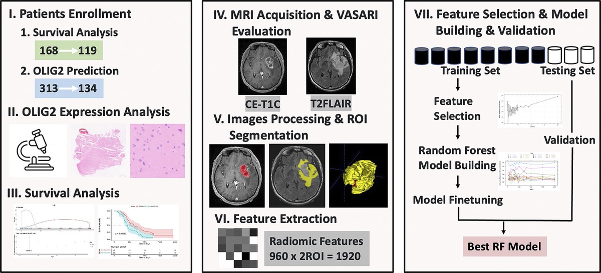
記住我

Sinonasal neoplasms are relatively uncommon compared with other head and neck tumors. Sinonasal malignancies comprise only 3% of all head and neck cancers and 1% of all malignancies.1,2 However, the sinonasal cavities are among the most histologically diverse areas of the body, with a large variety of neoplasms occurring in this region and many of these tumors having nonspecific presenting symptoms.2 The diagnosis often requires histologic examination of tissue specimens.
Imaging evaluation of sinonasal masses, including evaluation by computed tomography (CT) and magnetic resonance imaging (MRI), demonstrates the location, size, and extension of such masses. Such imaging techniques thus play an essential role in patient management and provide valuable guidance for successful biopsy or surgical treatment. Unfortunately, the appearance of sinonasal masses on conventional CT and MRI is not pathognomonic. Some benign lesions, like inverted papilloma, have ability to cause local destruction and extend into adjacent structures, mimicking malignancies.2–5 Hence, this may be a diagnostic challenge. One quantitative study defined the cutoff point of the apparent diffusion coefficient obtained from diffusion-weighted imaging in differentiating between benign and malignant sinonasal lesions. The study showed that the apparent diffusion coefficient value cutoff point had higher diagnostic performance than conventional MRI.6
However, CT is often the first examination performed because of its lower cost and higher availability than MRI. Computed tomography is also superior to MRI in visualization of osseous structures. Dual-energy CT or spectral CT is a new technology, and its potential lesion detection and characterization abilities extend beyond those of conventional single-energy CT by generating material-specific images based on the atomic number and unique mass attenuation coefficient of material at different x-ray energies. Therefore, we can generate dual-energy computed tomography (DECT)–based iodine-specific images and measure iodine density in a given region of interest (ROI) to detect subtle differences in attenuation. Dual-energy computed tomography also enables generation of virtual monoenergetic images.7–9 Previous studies have advocated a role for DECT in the characterization of diseases such as malignant pulmonary nodules and metastatic lymph nodes in oropharyngeal squamous cell carcinoma (SCC), pathological grading of clear cell renal cell carcinoma, and differentiation between orbital lymphoma and lymphoproliferative disease.10–13 However, no studies have evaluated the role of DECT for differentiation of inverted papilloma, which are the most common benign epithelial neoplasms in the sinonasal cavity, and sinonasal SCC/lymphoma, which are the first and second most common malignancies in the sinonasal tract.2
This study was performed to assess the value of quantitative measurements from DECT to differentiate inverted papilloma from sinonasal SCC/lymphoma.
MATERIALS AND METHODS Study PopulationAfter obtaining institutional review board approval for this study, we retrospectively reviewed DECT images of the sinus of patients who visited our hospital from January 2017 to August 2020. The inclusion criterion was a pathological diagnosis of inverted papilloma, SCC, or lymphoma with an enhancing sinonasal mass size of ≥10 mm. The exclusion criteria were incomplete DECT images and images that were performed posttreatment with radiation therapy or chemotherapy. In total, 28 patients were ultimately included (15 with inverted papilloma, 5 with SCC, and 8 with lymphoma). The pathologies were diagnosed after surgical resection in 9 patients with inverted papilloma, 1 with SCC, and 2 with lymphoma. The rest of the patients were diagnosed after biopsy.
Spectral CT ImagesAll patients underwent DECT (IQon Spectral CT; Philips Healthcare, Best, the Netherlands) of the sinus. The scanning parameters were 120 kVp, variable tube current, 1.0-mm section collimation, and 1-mm section interval. Typically, 50 to 60 mL of nonionic iodinated contrast material was power injected at a rate of 1.5 mL/s followed by a saline chaser. A postcontrast scan was performed 60 seconds after initiation of contrast administration.
Qualitative CT Imaging Features AnalysisAll spectral CT images were independently reviewed by 2 board-certified neuroradiologists with 3 and 8 years of experience in neuroimaging. Both reviewers were blinded to the patients' diagnoses and any other imaging studies. Disagreements were resolved by consensus between the 2 reviewers. The following imaging features were assessed: laterality (unilateral or bilateral), location (nasal cavity, maxillary sinus, ethmoid sinus, sphenoid sinus, or frontal sinus), enhancement pattern (homogeneous or heterogeneous), border (well-defined or ill-defined), necrosis, hemorrhage, calcification, bone destruction (no bone change/bone remodeling or bone destruction), pterygopalatine fossa (PPF) extension, adjacent invasion, and perineural spreading. The border was considered well-defined if two thirds or more of the lesion was sharply demarcated.
Quantitative Spectral CT AnalysisAll measurements were performed on an advanced workstation (IntelliSpace Portal; Philips Healthcare) by one in-training radiology resident. Three ROIs were drawn as circular areas of ≥10 mm2 within the relative homogeneous area of the most enhanced portion of the lesion, avoiding vessels and bone (Fig. 1). The measurements were performed twice at least 2 weeks apart to decrease memory bias. The average of the 2 measurements was calculated. The software automatically calculated the iodine density (mg/mL). A spectral attenuation curve was generated, representing the change in attenuation values in the ROI at different energy levels. Two representative data points were selected from the spectral curve, one at 40 keV and one at 70 keV, and the slope of a line between these 2 points represented the spectral attenuation curve slope (Fig. 2).
 FIGURE 1:
 Axial virtual monochromatic image at (A) 40 keV and (B) 70 keV in a patient with inverted papilloma seen as a heterogeneous enhancing mass involving the right nasal cavity and right maxillary sinus. Circular regions of interest were drawn in the relatively homogeneous area of the most enhanced portion of the lesion. The attenuation values were 153.4 HU and 75 HU in the regions of interest of 40 keV and 70 keV, respectively. (C) The mean iodine density was 1.35 mg/mL.
 FIGURE 2: Spectral attenuation curve in the same patient as shown in Figure 1. A spectral attenuation curve slope was calculated from an estimated line (yellow dotted line) drawn between the 2 representative points (white lines) at 40 keV and 70 keV. Figure 2 can be viewed online in color at www.jcat.org.
Spectral attenuation curve slope=HU40keV−HU70keV30
Statistical AnalysisStatistical analyses were performed with Stata/SE Version 16 (StataCorp, College Station, TX). Qualitative CT features were compared between the 2 groups using a χ2 test. The interobserver agreement between the reviewers was calculated with the κ statistic.
Mean values and SDs of the iodine density and spectral attenuation curve slope were compared between the 2 groups using Student's t test. Receiver operating characteristic (ROC) curves were generated for values with statistically significant differences to evaluate their diagnostic efficacy and calculate the cutoff values of the iodine density and spectral attenuation curve slope for differentiating inverted papilloma from sinonasal SCC/lymphoma, along with the corresponding sensitivity, specificity, positive predictive value (PPV), negative predictive value (NPV), and accuracy.
A P value of <0.05 was considered statistically significant. Interreader reproducibility was assessed using the intraclass correlation coefficient (ICC).
RESULTSThe patients' characteristics are summarized in Table 1. There were no significant differences between the inverted papilloma and malignant groups.
TABLE 1 - Demographic Data of All Patients Patients' Characteristics Inverted Papilloma SCC Lymphoma P Value (Inverted Papilloma vs. SCC/Lymphoma) Sex 0.978 Male 8 (53.3%) 2 (40.0%) 5 (62.5%) Female 7 (46.7%) 3 (60.0%) 3 (37.5%) Age, y 0.4646 Mean ± SD 58.13 ± 12.88 59.6 ± 11.67 63.75 ± 18.49 Median (range) 59 (31–76) 63 (46–75) 65 (31–87)The qualitative CT imaging features of inverted papilloma and sinonasal SCC/lymphoma are shown in Table 2. Bilateral lesions, sphenoid sinus involvement, presence of PPF extension, and adjacent invasion were significantly associated with sinonasal SCC/lymphoma. The adjacent structures that were invaded consisted of the nasopharynx, oropharynx, orbit, and intracranial structures. There was moderate agreement between the reviewers for bilateral lesions and the presence of PPF extension (κ = 0.58 and 0.45, respectively), excellent agreement for sphenoid sinus involvement (κ = 0.76), and almost perfect agreement for the presence of adjacent invasion (κ = 0.85). In the subgroup analysis, no imaging feature was significantly different between sinonasal SCC and lymphoma.
TABLE 2 - Frequency Distribution of Qualitative CT Imaging Features CT Imaging Features Inverted Papilloma SCC Lymphoma P Value (Inverted Papilloma vs. SCC/Lymphoma) Laterality 0.003 Unilateral 13 (86.7%) 0 (0.0%) 4 (50.0%) Bilateral 2 (13.3%) 5 (100%) 4 (50.0%) Location Nasal 15 (100%) 5 (100%) 8 (100%) N/A Maxillary sinus 12 (80.0%) 2 (40.0%) 5 (62.5%) 0.228 Ethmoid sinus 13 (86.7%) 4 (80.0%) 6 (75.0%) 0.639 Sphenoid sinus 0 (0.0%) 2 (40.0%) 4 (50.0%) 0.005 Frontal sinus 5 (33.3%) 2 (40.0%) 2 (25.0%) 1 Enhancement pattern 0.583 Homogeneous 1 (6.7%) 1 (20.0%) 1 (12.5%) Heterogeneous 14 (93.3%) 4 (80.0%) 7 (87.5%) Border 0.102 Well-defined 8 (53.3%) 0 (0.0%) 3 (37.5%) Ill-defined 7 (46.7%) 5 (100%) 5 (62.5%) Necrosis 0.153 No 14 (93.3%) 3 (60.0%) 6 (75.0%) Yes 1 (6.7%) 2 (40.0%) 2 (25.0%) Hemorrhage 0.464 No 15 (100%) 5 (100%) 7 (87.5%) Yes 0 (0.0%) 0 (0.0%) 1 (12.5%) Calcification N/A No 15 (100%) 5 (100%) 8 (100%) Yes 0 (0.0%) 0 (0.0%) 0 (0.0%) Bone destruction 0.114 No/remodeling 7 (46.7%) 1 (20.0%) 1 (12.5%) Yes 8 (53.3%) 4 (80.0%) 7 (87.5%) PPF extension 0.004 No 14 (93.3%) 1 (20.0%) 4 (50.0%) Yes 1 (6.7%) 4 (80.0%) 4 (50.0%) Adjacent invasion <0.001 No 13 (86.7%) 0 (0.0%) 2 (25.0%) Yes 2 (13.3%) 5 (100%) 6 (75.0%) Perineural spreading 0.464 No 15 (100%) 4 (80.0%) 8 (100%) Yes 0 (0.0%) 1 (20.0%) 0 (0.0%)N/A indicates not applicable.
The iodine density was significantly higher in sinonasal SCC/lymphoma (2.46 ± 0.22 mg/mL) than in inverted papilloma (1.42 ± 0.46 mg/mL; P = 0.001) (Fig. 3). In the subgroup analysis, iodine density was not significantly different between sinonasal SCC and lymphoma.
 FIGURE 3:
 Box plot showing the range of iodine density values within inverted papilloma and sinonasal SCC/lymphoma.
There was no statistically significant difference between the first and second iodine measurement sessions (P = 0.1747).
Spectral Attenuation Curve SlopeThe spectral attenuation curve slope was significantly higher in sinonasal SCC/lymphoma (4.35 ± 0.27 HU/keV) than in inverted papilloma (2.72 ± 0.88 HU/keV; P = 0.001) (Fig. 4). In the subgroup analysis, the spectral attenuation curve slope was not significantly different between sinonasal SCC and lymphoma.
 FIGURE 4:
 Box plot showing the range of spectral attenuation curve slopes within inverted papilloma and sinonasal SCC/lymphoma.
There was no statistically significant difference between the first and second measurements of the spectral attenuation at 40 keV and 70 keV (P = 0.0587 and 0.3879, respectively).
Diagnostic ImplicationReceiver operating characteristic curves were used to assess the diagnostic value of the iodine density and spectral attenuation curve slope (Fig. 5). A threshold iodine density of 1.74 mg/mL gave a sensitivity of 92.3%, specificity of 86.7%, NPV of 92.9%, PPV of 85.7%, and accuracy of 90.3%. The area under the ROC curve for iodine density was 0.903, with a 95% confidence interval (CI) of 0.778 to 1.000 (P = 0.001). A threshold spectral attenuation curve slope of 3.34 HU/keV gave a sensitivity of 92.3%, specificity of 86.7%, NPV of 92.9%, PPV of 85.7%, and accuracy of 90.8%. The area under the ROC curve for the spectral attenuation curve slope was 0.9077, with a 95% CI of 0.7856 to 1.000 (P = 0.001). Examples of the qualitative and quantitative analyses are shown in Figures 6 to 8.
 FIGURE 5:
 Receiver operating characteristic curves for (A) iodine density and (B) spectral attenuation curve slope in differentiating inverted papilloma and sinonasal SCC/lymphoma. AUC, area under the curve.
 FIGURE 6: Computed tomography images of a 59-year-old woman diagnosed with inverted papilloma (same patient as in Fig. 1). The images showed a heterogeneous enhancing mass involving the right nasal cavity, right ethmoid sinus, and right maxillary sinus. The mean iodine density was 1.47 mg/mL, and the mean spectral attenuation curve slope was 2.71 HU/keV.
 FIGURE 7:
 Computed tomography images of a 75-year-old woman diagnosed with sinonasal SCC. The images revealed a heterogeneous enhancing mass centered in the left nasal cavity and left ethmoid sinus with extension to the right nasal cavity, right ethmoid sinus, both frontal sinuses, bilateral inferior frontal lobes, extraconal space of left orbit, left sphenopalatine foramen, and left PPF. Associated bone destruction was noted. The mean iodine density was 2.21 mg/mL, and the mean spectral attenuation curve slope was 4.22 HU/keV.
 FIGURE 8:
 Computed tomography images of an 87-year-old man diagnosed with sinonasal lymphoma. Coronal (A) 70 keV, (B) axial 70 keV, and (C) axial 40 keV images demonstrated a homogeneous enhancing mass centered in the left nasal cavity, left ethmoid sinus, left sphenoid sinus, and left maxillary sinus. The mean iodine density was 1.83 mg/mL, and the mean spectral attenuation curve slope was 3.34 HU/keV.
Intraobserver reliability was excellent for repeated measurements of iodine density (ICC, 0.977; 95% CI, 0.960–0.994), attenuation at 40 keV (ICC, 0.965; 95% CI, 0.939–0.991), and attenuation at 70 keV (ICC, 0.945; 95% CI, 0.905–0.986).
DISCUSSIONA study by El-Gerby and El-Anwar6 showed that the MRI criteria suggestive of a malignant sinonasal lesion are a unilateral sinus lesion, bone involvement, tumor necrosis, soft tissue mass, lymphadenopathy, and involvement of surrounding structures. Some different to our study used CT imaging feature-based differentiation between inverted papilloma and sinonasal SCC/lymphoma, showing that bilateral lesions, sphenoid sinus involvement, presence of PPF extension, and adjacent invasion were significantly associated with sinonasal SCC/lymphoma.
Dual-energy computed tomography enables material differentiation beyond the measurement of attenuation.7–9 To the best of our knowledge, this study is the first quantitative study using DECT imaging to evaluate inverted papilloma and sinonasal SCC/lymphoma. The results revealed that sinonasal SCC/lymphoma had a significantly higher iodine density and spectral attenuation curve slope than inverted papilloma. Our ROC curve analysis showed that several quantitative parameters exhibited increased sensitivity, specificity, and accuracy for differentiation of inverted papilloma from sinonasal SCC/lymphoma. Using an optimal threshold iodine density of 1.74 mg/mL and spectral attenuation curve slope of 3.34 HU/keV was useful to differentiate inverted papilloma and sinonasal SCC/lymphoma with high diagnostic values. Angiogenesis is the process of formation of new vessels and is an important process in the pathogenesis of malignancy.14 The occurrence of angiogenesis is a possible explanation of the higher iodine density and spectral attenuation curve slope in sinonasal SCC/lymphoma (which has richer vasculature) than inverted papilloma. The subgroup analysis demonstrated no significant difference in either the qualitative or quantitative parameters between SCC and lymphoma.
In 2 inverted papillomas, the iodine density and spectral attenuation curve slope were higher than the threshold values. One of these (iodine density, 2.8 mg/mL; spectral attenuation curve slope, 5.29 HU/keV) was pathologically diagnosed as inverted papilloma with a focal area of moderate dysplasia. Inverted papillomas are benign epithelial neoplasms but can transform to SCC (1.9%–27.0%).4 Some studies have shown that a focal loss of convoluted cerebriform pattern and low apparent diffusion coefficient values on MRI as well as increased SUVmax on positron emission tomography/computed tomography may help identifying inverted papilloma with SCC although not as specific.15 However, there is no evidence to support the use of CT to determine the malignant transformation. In a study of the clinicopathologic and immunophenotypic features of carcinoma ex-Schneiderian papilloma, Nudell et al.16 found that many cases of carcinoma ex-Schneiderian papilloma developed progressively worsening dysplasia before converting to SCC. A high iodine density or high spectral attenuation curve slope of an inverted papilloma may predict the risk of malignant transformation. Further research in this area will be needed to provide reliable evidence.
Quantitative analysis has shown great diagnostic performance for the differentiation of inverted papilloma and sinonasal SCC/lymphoma. Our measurement method is feasible and reproducible and provides excellent intraobserver agreement based on ICCs.
A limitation of our study was the small number of patients with sinonasal SCC. Another limitation was the quantitative analysis of spectral CT based on ROIs placed at a focal area of the tumor; that is, the ROIs did not cover the whole lesion. However, the sample bias was reduced by sampling multiple ROIs. Moreover, the excellent ICCs in this study emphasize the reproducibility of this measurement method.
To conclude, the quantitative results of our study indicated significant differences in the DECT-derived iodine density and spectral attenuation curve slope between inverted papilloma and sinonasal SCC/lymphoma. Sinonasal SCC/lymphoma had a significantly higher iodine density and spectral attenuation curve slope than inverted papilloma. Using optimal thresholds of quantitative measurements provides great diagnostic efficacy.
ACKNOWLEDGMENTThe authors thank Angela Morben, DVM, ELS, from Edanz (https://www.edanz.com/ac) for editing a draft of this manuscript.
REFERENCES 1. Koeller KK. Radiologic features of sinonasal tumors. Head Neck Pathol. 2016;10:1–12. 2. Kawaguchi M, Kato H, Tomita H, et al. Imaging characteristics of malignant sinonasal tumors. J Clin Med. 2017;6:116. 3. Kanwar S, Mital M, Gupta PK, et al. Evaluation of paranasal sinus diseases by computed tomography and its histopathological correlation. J Oral Maxillofac Radiol. 2017;5:46–52. 4. Tatekawa H, Shimono T, Ohsawa M, et al. Imaging features of benign mass lesions in the nasal cavity and paranasal sinuses according to the 2017 WHO classification. Jpn J Radiol. 2018;36:361–381. 5. Eide JG, Welch KC, Adappa ND, et al. Sinonasal inverted papilloma and squamous cell carcinoma: contemporary management and patient outcomes. Cancer. 2022;14:2195. 6. El-Gerby KM, El-Anwar MW. Differentiating benign from malignant sinonasal lesions: feasibility of diffusion weighted MRI. Int Arch Otorhinolaryngol. 2017;21:358–365. 7. McCollough CH, Leng S, Yu L, et al. Dual- and multi-energy CT: principles, technical approaches, and clinical applications. Radiology. 2015;276:637–653. 8. Postma AA, Das M, Stadler A, et al. Dual-energy CT: what the neuroradiologist should know. Curr Radiol Rep. 2015;3:16. 9. Kim H, Park CM, Kang CK, et al. Effect of CT acquisition parameters on iodine density measurement at dual-layer spectral CT. AJR Am J Roentgenol. 2018;211:748–754. 10. Zhang Y, Cheng J, Hua X, et al. Can spectral CT imaging improve the differentiation between malignant and benign solitary pulmonary nodules?PLoS One. 2016;11:e0147537. 11. Foust AM, Ali RM, Nguyen XV, et al. Dual-energy CT-derived iodine content and spectral attenuation analysis of metastatic versus nonmetastatic lymph nodes in squamous cell carcinoma of the oropharynx. Tomography. 2018;4:66–71. 12. Wei J, Zhao J, Zhang X, et al. Analysis of dual energy spectral CT and pathological grading of clear cell renal cell carcinoma (ccRCC). PLoS One. 2018;13:e0195699. 13. Tunlayadechanont P, Panyaping T, Kaewkerd B. Role of quantitative spectral CT analysis for differentiation of orbital lymphoma and other orbital lymphoproliferative disease. Eur J Radiol. 2020;133:109372. 14. Gavalas NG, Liontos M, Trachana S, et al. Angiogenesis-related pathways in the pathogenesis of ovarian cancer. Int J Mol Sci. 2013;14:15885–15909. 15. Long C, Jabarin B, Harvey A, et al. Clinical evidence based review and systematic scientific review in the identification of malignant transformation of inverted papilloma. J Otolaryngol Head Neck Surg. 2020;49:25. 16. Nudell J, Chiosea S, Thompson LD. Carcinoma ex-Schneiderian papilloma (malignant transformation): a clinicopathologic and immunophenotypic study of 20 cases combined with a comprehensive review of the literature. Head Neck Pathol. 2014;8:269–286. Keywords:dual-energy CT; inverted papilloma; sinonasal squamous cell carcinoma; lymphoma
Copyright © 2022 The Author(s). Published by Wolters Kluwer Health, Inc.
留言 (0)