Our report describes the case of a patient with atypical anti-GBM disease with proteinuria, hematuria, and acute kidney injury. Pathologically, crescentic glomerulonephritis was detected in kidney tissues. Because anti-GBM antibodies binding to alpha3 (IV) NC1 were not detected by FEIA, IIF was used to identify anti-GBM antibodies. Finally, combination therapy with high-dose steroids and plasma exchange was effective for atypical anti-GBM disease with acute kidney injury, resulting in improvements in proteinuria, hematuria, and renal function.
Anti-GBM disease is a type of nephritis that histologically presents as crescentic necrotizing glomerulonephritis, with linear deposits of IgG against the GBM and serological positivity for anti-GBM antibodies. It was first reported by Lerner et al in 1967 [14]. Classic anti-GBM disease is associated with rapidly progressive glomerulonephritis and often pulmonary hemorrhage, shortened life expectancy, and poor renal prognosis [3,4,5]. Atypical anti-GBM disease presents with better renal outcomes and milder symptoms compared with classic anti-GBM disease, and circulating antibodies to α3NC1 are not detected by EIA in atypical anti-GBM disease.
Anti-GBM antibodies recognize amino acid residues at the N-terminal 17–31 (EA) and C-terminal 127–141 (EB) of the NC1 domain of the type IV collagen α3 chain as antigen epitopes [15, 16]. In addition to these epitopes, the NC1 domain EA region of the α5 chain has also been reported as an antigenic epitope. These antigen epitopes reside in a hexamer composed of type IV collagen α345 chains and are localized within the basement membrane under normal conditions. Infectious diseases (e.g., influenza), toxic substance inhalation (organic solvents and carbon tetrachloride), and smoking damage the basement membrane of the lungs and kidneys, which exposes antigen epitopes on the α3 and α5 chains and produces anti-GBM antibodies that react to them [17].
The pathological feature of anti-GBM is linear staining of IgG deposited in the GBM, usually accompanied by autoantibodies against structural collagen IV elements of the GBM. Most of these antibodies are directed against α3NC1 and react with epitopes (17–31 = EA, 127–141 = EB) normally masked by intrachain methionine cross-links [18, 19]. However, antibodies that react with other collagen IV epitopes (e.g., α5NC1 and α4NC1) have also been identified. Furthermore, antibodies that specifically target the α345NC1 hexamer but not the α3NC1 monomer have been reported to be associated with mild nonprogressive glomerulonephritis [20].
Anti-GBM antibodies are detected by IIF, Western blot, and the EIA method, which includes ELISA, CLEIA, and FEIA. The EIA results can be obtained quickly using a fully automated assay. The EIA method has high sensitivity and specificity [21,22,23]. However, there are cases in which anti-GBM antibodies cannot be detected by EIA; therefore, it is crucial to use IIF or other methods to recognize the presence of anti-GBM disease.
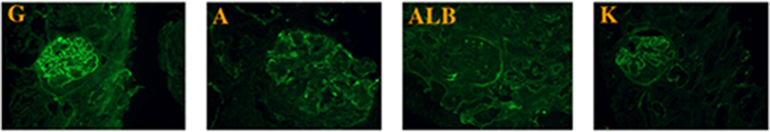
There are several potential reasons why antibodies are not detected by the commercial EIA method. First, the titers of anti-GBM antibodies may be too low. In some reports, low titers of circulating anti-GBM antibodies have been associated with mild proteinuria, hematuria, and renal dysfunction. Low-affinity antibodies to the α3NC1 peptide have also been correlated with reduced crescentic glomerulonephritis and better prognosis. Antibodies with a low affinity can only be detected by sensitive assays, such as Western blot or biosensor experiments, rather than by routine methods [21]. Second, the autoantibodies in some patients may be directed against antigens located on GBM components other than α3NC1. Therefore, the antibodies cannot be detected using routine assays. In the present case, serum anti-GBM antibodies were detected by IIF using normal kidney tissue. Third, similar to other autoimmune diseases, antibody production ceases during the reestablishment of immune homeostasis, and circulating antibodies are destroyed by the liver to a greater extent than tissue antibodies [19]. In these settings, minimal or no antibodies would remain in the circulation, and antibodies would only be present in the GBM.
Classic anti-GBM patients, especially those with initial serum creatinine levels below 5 mg/dL and/or pulmonary hemorrhage, are usually treated with high-dose steroids, cyclophosphamide, and plasmapheresis [13]. However, it remains unclear whether the benefits of these aggressive regimens outweigh the risks in patients with atypical anti-GBM who are clinically and pathologically mild and often without pulmonary hemorrhage. The patient in this case had no pulmonary hemorrhage, but her renal function was declining; therefore, aggressive treatment was administered. Aggressive treatment, including high-dose steroids and plasma exchange, resolved proteinuria and hematuria and improved renal function. Further studies are needed to determine the optimal treatment.
In conclusion, we reported the case of a patient with atypical anti-GBM. If anti-GBM antibodies cannot be detected by EIA, such as ELISA, CLEIA, or FEIA, determining the presence of anti-GBM disease using IIF or other methods to initiate optimal treatment in a timely manner may be useful.


留言 (0)