What Is the Rate of Displacement of Occult Posterior Malleolus Fractures in Nailed Tibial Shaft Fractures?
Objective:
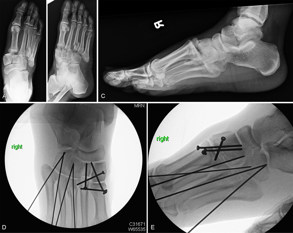
To describe the radiographic consequences of posterior malleolus fractures (PMF) present with tibial shaft fractures fixed with intramedullary nails.
Design:
Retrospective cohort study.
Setting:
Level 1 trauma center.
Patients/Participants:
Seven hundred thirty-three patients with tibial shaft fractures.
Intervention:
Intramedullary nail fixation and prophylactic articular fixation.
Main Outcome Measure:
Displacement of PMF with intramedullary nail insertion.
Results:
Seven hundred thirty-three patients were identified with tibial shaft fractures treated with intramedullary nail fixation at a Level 1 trauma center without a uniform preoperative computed tomography protocol. One hundred thirty-three patients had an identifiable PMF appreciated on preoperative imaging. Of the 600 remaining patients without a known PMF, 29 had PMF identified after nail insertion: 24 patients with nondisplaced fractures that all healed radiographically at final follow-up, 3 patients had fractures <30% of the articular surface displaced 1–2 mm, and 2 patients had fractures >30% of the joint surface that displaced 1–2 mm.
Conclusions:
The incidence of radiographically observable PMF associated with tibial shaft fractures is high, even without a preoperative computed tomography screening protocol in place. In patients without an appreciable PMF on injury films, less than 1% (2/600) had displacement of a large, clinically significant PMF with nail placement.
Level of Evidence:
Prognostic Level IV. See Instructions for Authors for a complete description of levels of evidence.


留言 (0)