The EAC is a pathway for transmitting sound to the tympanic membrane [2], but its morphology varies. The curved overhang of the EAC can obstruct the field of vision, affect diagnosis [1], treatment and render operation on the middle ear [3] difficult. Especially when performing middle ear surgery, sometimes it may be necessary to shave the EAC bone to perform the operation. Recent endoscopes have a wide field of view, which could reduce the need for EAC excision [4, 5], but they offer limited operability. When performing procedures or surgical operations via the EAC, it is crucial to understand the condition of vital structures in the middle ear to anticipate the difficulty of surgical operations and prevent secondary damage.
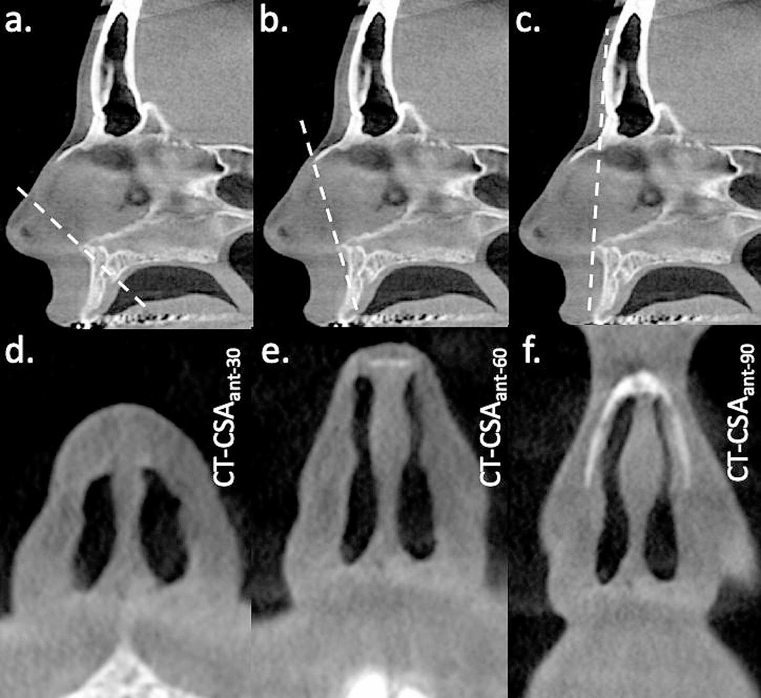
In this study, 3D reconstruction processes MPR (multiplanar reconstruction) and MIP, were used to evaluate the morphology of the EAC. MPR is a process for visualizing an arbitrary cross-sectional image, commonly used in temporal bone CT [6], whereas MIP is a method of projecting the relative maximum value in the region by freely setting the visual axis from the reader’s line of sight to any point in the target structure and determining the arbitrary thickness [7]. These two processes allow the creation of a 3D image since it is possible to visualize multiple slices at the same time. In contrast, the temporal bone CT has limited usefulness because visualization is limited by bone tissue [8]. Regarding the usefulness of MIP in the temporal bone region, evaluation of the auditory ossicles by CT [6, 8] and evaluation of cochlear basal turn by magnetic resonance imaging [9] have been reported; however, only a relatively small number of cases have been reported. In this study, by setting the tympanic umbo as the target point with MIP, moving the visual axis, and confirming the position where the tympanic membrane can be confirmed best, the visual axis that can respond to the curved EAC was determined. By creating MPR slices that pass through the short and long axes of the bony tympanic ring based on this image, it was possible to create slices showing the short and long axes of the bony tympanic ring that also correspond to the curvature of the EAC. Kavita et al. [3] used axial and coronal slices to evaluate EAC curvature in pediatric cases. However, in order to evaluate the morphology of the adult EAC that was curved in a complicated direction due to growth, it was difficult to use axial and coronal slices as horizontal and vertical slices along the long axis of the EAC. MIP is a reconstruction process that can be performed in a relatively short time [6, 8]. Since the visual axis of the reader’s line of sight for the target structure can be freely set in MIP, it is useful for evaluation of the complicatedly curved EAC morphology.
Furthermore, in this study, we investigated the individual anatomical differences of the tympanic sinus, the vertical portion of the facial nerve, and the jugular bulb as important structures during middle ear surgery. The tympanic sinus is the space behind the tympanum [10], a difficult-to-visualize area considered to be a clinically important structure because lesions tend to remain in this space [11].
The facial nerve runs in a complicated manner in the temporal bone, and the closest part to the tympanic ring is the vertical portion [12]. There is no good landmark to estimate the position of the vertical portion of the facial nerve when performing an EAC procedure, and due circumspection is required when performing an EAC excision [13]. The jugular bulb is sometimes present as a high jugular bulb (HJB) near the EAC. It has received significant attention as it may cause massive bleeding during procedures such as tympanostomy [14]. The EAC and these vital structures in the middle ear have large individual anatomical variations, and many studies have been conducted so far. However, it has been reported that it is associated with changes in mastoid cell development mainly due to chronic inflammation. In this study, a slight correlation could be found between the morphology of the bony tympanic ring with a short antero-posterior length and the severe overhang of the inferior wall of the EAC, the shallow tympanic sinus, and the lateral process of the vertical portion of the facial nerve.
For EAC curvature, visual field limitation due to overhangs of the anterior, posterior, and inferior walls of the EAC is clinically important [3]. When the anterior canal overhang (ACOH) is high, the operability of middle ear surgery is often difficult, and ACOH has been discussed for some time; however, there are only a few reports on inferior canal overhang (ICOH) and posterior canal overhang (PCOH). Dedhia et al. [3] first reported that overhang was the most severe in ICOH, and cases with large ICOH overhang were predominantly cases with a history of tympanic tube placement. This finding suggests that the higher the ICOH angle, the more likely it is for eustachian tube dysfunction and otitis media to occur. Furthermore, Park et al. [15] reported that the antero-posterior length of the tympanic membrane was significantly shorter in the COM group than in the normal temporal bone group. However, there is no difference in the superior-inferior length; the antero-posterior direction is more important for the growth of the tympanic membrane after birth than the superior-inferior direction. As such, this suggests that the cavity of the bony EAC may become narrow when its growth is inhibited due to inflammation.
Recently, there has been an increase in reports stating that the growth of mastoid cells is suppressed by chronic inflammation of the middle ear [16,17,18]. However, regarding changes in vital structures of the middle ear, the tympanic sinus becomes shallow due to chronic inflammation [11, 19] and the vertical portion of the facial nerve runs more outward [20]. Since the tympanic sinus progresses toward the less resistant region during development of mastoid cells, it is thought that suppression of growth of mastoid cells due to chronic inflammation changes the depth of the tympanic sinus [11]. Moreover, the facial nerve has a larger anatomical individual variation in the peripheral vertical portion than in the proximal portion such as the labyrinthine and tympanic segments [13]. Compared to adults, it runs more outward in children, and its position changes as one grows [21]. In cases where growth of mastoid cells is suppressed, the vertical portion of the facial nerve runs more anteriorly and laterally. The vertical portion of the facial nerve also suppresses the medio-posterior movement with growth by suppressing the mastoid growth [20].
From the results obtained in this study, anatomical morphologies such as the EAC, tympanic sinus, and the vertical portion of the facial nerve that change after birth, may change in conjunction to some extent. Similar to suppression of the growth of mastoid cells by chronic inflammation, morphological changes may occur mainly due to chronic inflammation. Therefore, if there are findings such as a short antero-posterior length of the bony tympanic ring or a severe overhang of the inferior wall of the EAC during treatment or surgery via the EAC, it can be predicted that the tympanic sinus is shallow, and the vertical part of the facial nerve is likely to run outward. Temporal CT is often performed prior to the middle ear surgery; however, the fact that these structures can be predicted from the EAC morphology is meaningful in assuming the difficulty of preventing secondary damage and removing cholesteatoma in surgery.
From this study, a favorable correlation between the height of HJB and the morphology of the EAC could not be obtained. There have been reports that the height of the jugular bulb is also affected by the growth of mastoid cells, and that better the growth, the higher the apex of the jugular bulb [22, 23]. Therefore, it was speculated that there may be a correlation between the EAC morphology and the height of the jugular bulb. However, in this study, the height of non-HJB cases classified as class 0 could not be attributed to why the correlation with the height of HJB and the class classification could not be obtained, and it is possible that only the height of the case defined as HJB was measured. Although future studies are needed, the relationship between clinically significant HJB, and its height and EAC morphology is low.
There are some limitations in this study. First: since the temporal bone CT used in this anatomical examination used a slice with a width of 0.5 mm, the reliability may be low for measurements of 0.5 mm or less. Second: in this study, as in previous reports, ears with COM tended to have shorter bony tympanic rings, severe overhangs in the lower wall of the EAC, and shallower tympanic sinus, and the vertical portion of facial nerves tended to run laterally compared to normal ears. This study further suggested that there may be a correlation between changes in EAC morphology and changes in middle ear structures. However, it was not possible to elucidate how these changes affect the duration and extent of chronic inflammation in this study. Thus a prospective study that includes the duration of chronic inflammation is needed in the future.


留言 (0)