TCGA data acquisition and reprocessing
Expression profiles and somatic mutation data of endometrial cancers were obtained using cBioPortal (https://www.cbioportal.org/). Expression profiles of normal endometrial tissues were obtained using the GDC Data Portal (https://portal.gdc.cancer.gov/projects/TCGA-UCEC). Normalization and differential expression analyses of transcriptome data were conducted using the edgeR package.
Ethics statement and tissue samples collection
This study was approved by the Human Investigation Ethics Committee of Shanghai First Maternity and Infant Hospital, Tong Ji University School of Medicine. EC and normal endometrial tissues were collected after written informed consent from the patients. Paraffin-embedded tissue samples for immunohistochemistry were obtained from patients who underwent surgical treatment from 2019 to 2021 (Additional file 5: Table S1). Two independent pathologists verified the histological diagnosis of all tissue samples. None of the patients had undergone hormone therapy, radiotherapy, or chemotherapy prior to surgery.
Immunohistochemistry
All tissue sections (4 mm thick) were prepared in paraffin-embedded specimens. Staining was performed using primary antibodies as follows: rabbit polyclonal antibody against JAK1 (1:100; CST, Danvers, MA, USA). Two independent pathologists who were blinded to the clinical and pathological data evaluated the specimens. Sections were evaluated according to semi-quantitative immunoreactivity scores. The percentage of positive staining was scored as follows: 0 = 0–5%, 1 = 6%-25%, 2 = 26–50%, 3 = 51–75% and 4 > 75%. Staining intensity was scored as follows: 0 = none, 1 = weak, 2 = moderate, and 3 = strong. For each specimen, the final score (Immunohistochemistry scores, IS) was the multiply of the values of the two parameters.
Cell culture and hypoxic conditions
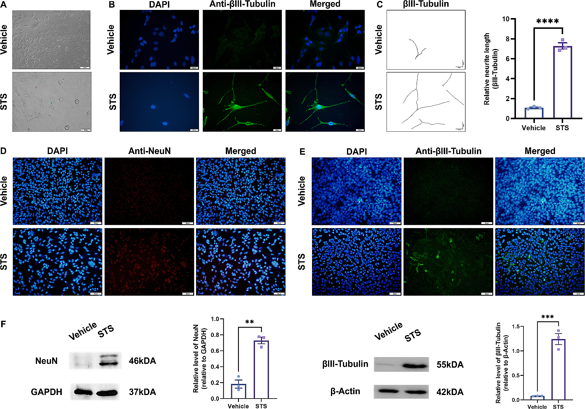
293 T, KLE, SPEC-2, AN3CA, Ishikawa, HEC-1A, and HEC-1B were obtained from the Chinese Academy of Sciences Committee Type Culture Collection cell bank (Shanghai, China). These cell lines were grown in Dulbecco’s modified Eagle’s medium (DMEM)/F12 (Gibco, Auckland, NZ) supplemented with 10% fetal bovine serum (Gibco) in a humidified atmosphere of 5% CO2 at 37 °C. Ruxolitinib was prepared for use in cell culture. For hypoxia experiments, cells were grown in an in vitro hypoxic (2% O2) container system (BD Diagnostics), and protein and RNA were collected immediately when cells were removed from the hypoxic container [23].
Transient transfection
shRNAs and siRNAs were purchased from GeneCHEM (Shanghai, China). The sequences are listed in Additional file 5: Tables S2 and S3. The JAK1 expression plasmid, PCDH, was purchased from GenePharma Biotech (Shanghai, China). The Myc-tagged JAK1 and Flag-tagged HIF-1/2α expression plasmids were purchased from WZ Biosciences Inc (Shanghai, China). Transient transfection was performed using LipofectamineTM 2000 (Invitrogen) according to the manufacturer’s protocol.
RT-qPCR
Total RNA was extracted from cell lines using TRIzol (Invitrogen), and cDNA was prepared using a reverse transcriptase kit (TaKaRa) according to the manufacturer’s instructions. cDNA was analyzed by real-time PCR using SYBR Premix Ex Taq (TaKaRa) in an Eppendorf Mastercycler ep realplex. The housekeeping gene, GAPDH, was used as an internal control. Data were calculated using the 2−△△Ct formula. The primer sequences used are listed in Additional file 5: Table S4. The experiments were repeated at least three times.
Western blotting
Proteins were extracted using the RIPA kit (Beyotime, Shanghai, China) containing a 1% dilution of the protease inhibitor PMSF (Beyotime). Protein concentrations were determined using a BCA Protein Assay kit (Beyotime). Equal amounts of protein were loaded into each lane of an SDS-PAGE gel for protein separation and transferred to polyvinylidene fluoride (PVDF) membranes (Millipore). Membranes were blocked and then incubated with rabbit polyclonal antibody against JAK1 (1:1000; CST), rabbit polyclonal antibody against HIF-1α (1:1000, CST), rabbit polyclonal antibody against HIF-2α (1:1000, CST), and rabbit polyclonal antibody against GAPDH (1:5000, CST) individually at 4℃ overnight. HRP-conjugated Goat anti-rabbit antibodies (1:2000, Proteintech) were used to detect the bound primary antibodies.
RNA-sequencing and data analysis
RNA extracted from KLE cells and KLE cells transfected with JAK1 shRNA was used for RNA-sequencing (APExBIO). Differentially expressed genes ((DEGs) between KLE cells and KLE cells transfected with JAK1 shRNA were determined using DESeq2 and the Wald hypothesis test. Genes were considered significant and reported according to the following criteria: FDR < 0.05 and |log2FoldChange|> 1. Heat maps and hierarchical clustering were performed with R software.
To further explore the potential functions of JAK1 in the pathology of EC, DEGs were analyzed using Gene Ontology (GO, http://www.geneontology.org/) and the Kyoto Gene and Genomic Encyclopedia (KEGG, https://www.kegg.jp/) pathway enrichment analysis. Differences were considered statistically significant at P < 0.05.
Cell proliferation
For Cell proliferation assays, cells were seeded into 96-well plates at 1 × 10³ cells/well and cultured for 1–5 days. Cell proliferation assays were performed using the CCK-8 Kit (Dojindo). Absorbance was measured at 450 nm using a Multimode Plate Reader (Molecular Devices, USA).
Colony formation assays
For colony formation assays, cells were seeded into 6-well plates. When identifiable cell clones had formed, the colonies were fixed with methanol and stained with 0.5% crystal violet. All experiments were repeated at least three times.
Cell migration assays
Cells were suspended in 200 μl serum-free medium and plated at a density of 6 × 10⁴ cells/well in 6.5 mm transwell chambers equipped with 8.0 μm pore-size polycarbonate membranes. The complete medium (600 μl) was added to the lower chamber. After incubation for 48 h, cells were fixed in 4% paraformaldehyde and stained with crystal violet. The cells that migrated to the basal side of the membrane were counted using a microscope.
Co-immunoprecipitation assay (Co-IP)
Co-IP assays were performed using Pierce Classic Magnetic IP/Co-IP Kit (Thermo Fisher) and Flag Tag IP/Co-IP Kit (Biolinkedin, China) according to the manufacturer’s instructions. Co-immunoprecipitated proteins were analyzed by western blotting as described above. Anti-Flag and anti-Myc antibodies were purchased from Sigma-Aldrich.
Statistical analysis
All data were statistically analyzed using Graphpad prism 7.0. Measured data were assessed using unpaired Student’s t-test or one-way ANOVA for multiple comparisons, and the χ2 test for 2 × 2 tables was used to compare the categorical data. *p < 0.05 **p < 0.01, ***p < 0.001, ****p < 0.0001.


留言 (0)