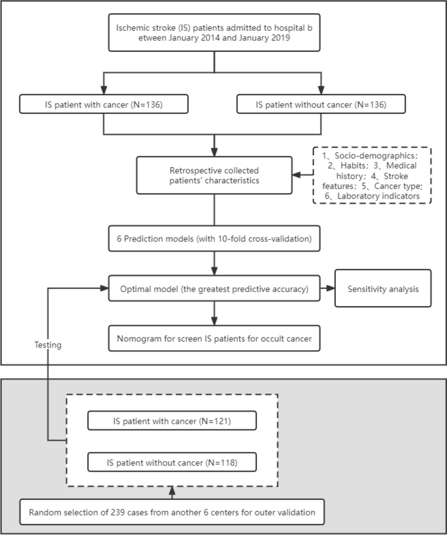
Currently, the paradigm shift in cancer treatment from traditional reactive medicine to PPPM remains a hot topic. Using the PPPM approach, such as noninvasive imaging methods, to early and accurately predict the prognosis after treatment of each individual is a major facilitator of this shift, which is critical for the early adjustment of treatment protocols. This two-centre study was the first based on RS-EPI DWI to extract radiomic features from baseline data and integrate ML algorithms as a method to investigate its utility in predicting DFS of RC patients with direct surgery, culminating successfully in the development and validation of an optimal model. Our results revealed that the constructed radiomics model performed well in predicting the prognosis and stratifying patients according to the risk of ROM. Compared to the use of clinicopathological information alone, the integrated application of radiomic and clinicopathological information significantly improved the predictive capability of the model. Therefore, the proposed predictive model may support the implementation of this significant shift in RC treatment.
Radiomics with the ML algorithm might provide essential additional information
Using the framework of PPPM, radiomics methods with ML algorithms might be a more effective approach than conventional morphology imaging. Due to tumour heterogeneity and differences in the microenvironment, variability in the outcome of treatment response and ROM for patients with RC consistently exists, which has been a great challenge for choosing the optimal protocol that avoids both under- and overtreatment. Researchers have increasingly focused on identifying image features as predictors for the long-term prognosis, which provides an essential foundation for the development of individualized medicine [20]. However, morphological manifestations indicating tumour progression are often extremely subtle and relatively lag in occurrence, indicating that they are difficult to detect on initial images through a traditional visual assessment [21]. Radiomics methodology enables the quantification of heterogeneity, which is inaccessible by the human eye, via high-throughput extraction of features hidden behind the radiology image and enables the construction of models for decision support.
One reason for the suboptimal adaptation of standard cancer treatment protocols may be the insufficient availability and utilization of information about the characteristics of the tumour itself. As an increasing number of features are developed, traditional statistical methods, such as logistic regression and multiple linear regression models, have become increasingly overwhelmed with such large and complex data. Among numerous other studies, ML algorithms have exhibited excellent performance in discovering potential connections from intricate data [12, 22, 23], facilitating the selection of the most suitable fraction from this vast amount of radiomic features beyond human understanding. Therefore, the accurate predictive ability of the model, a crucial aspect highlighted in the PPPM, might be guaranteed by the powerful data processing capacity of ML algorithms.
The application of RS-EPI DWI radiomics showed great predictive accuracy
The application of RS-EPI DWI to predict aggressive features, such as the status of EGFR and tumour differentiation, in RC has been reported in a limited number of previous studies [24,25,26]. Wen et al. further applied an ML method based on T2WI and RS-EPI DWI to predict the T stage of RC, which exhibited fairly high accuracy with AUCs of 0.893 and 0.810 in the training and testing cohorts, respectively. These findings revealed the great prognostic potential of this technique. However, to the best of our knowledge, this study was the first to apply radiomic features derived from RS-EPI DWI in predicting the ROM risk of patients with RC.
RS-EPI DWI provides remarkable advantages in image quality, geometric distortion, and discrimination of tissue variability, as we previously reported [27], generating a sharper visual boundary between the lesion and the surrounding normal tissue to improve the accuracy of manual lesion delineation and reducing the effect of image noise on high-dimensional features. Thus, the extracted features might be more representative of the actual tumour microenvironment. In a study predicting DFS, the radiomics model based on SS-EPI DWI was modestly evaluated by calculating the C-index in both the training (0.627, 95% CI%: 0.529–0.726) and test cohorts (0.658, 95% CI%: 0.536–0.779), while the performance of the model based on RS-EPI DWI in our study was substantially improved (C-index = 0.78 ± 0.04, 95% CI: 0.69–0.87), indicating that the technical superiority may indeed contribute to model optimization and was reflected in the forecasting results to some extent.
The proposed models effectively stratify the ROM risk of RC patients and thus provide a reference for the application of PPPM
In previous studies a variety of clinicopathological features are intimately associated with the prognosis of RC patients [7, 8, 28, 29]. For example, Ceyhan et al. [28] suggested that PNI is useful as a prognostic factor for patients with RC. A nomogram model based on multiple clinicopathological factors, including pathological stage and adjuvant therapy, was proven to be effective in predicting ROM [29]. These findings were highly consistent with the key features of the clinical model selected by ML algorithms in our study, which supported the validity of our model construction approach.
Furthermore, some studies have proven the incremental value of integrating imaging and clinicopathological information for the performance of tumour assessment and prognostic prediction in patients with glioblastoma multiforme, advanced nasopharyngeal carcinoma, breast cancer, lung cancer, prostate cancer, and RC [30, 31]. The same was true for the results of our study. In our study, no significant differences were observed between the radiomics model and clinical model, as evaluated using the AUC (p = 0.118) or the C-index (p = 0.062). However, after combining the two features, a significant improvement was observed in the overall performance of the merged model compared to the clinical model (AUC = 0.87 [95% CI: 0.80–0.93] vs. 0.71 [95% CI: 0.59–0.81], p = 0.009; C-index = 0.829 [95% CI: 0.764–0.895] vs. 0.676 [95% CI: 0.560–0.792], p = 0.002). Although the predictive ability inevitably decreased slightly as the interval between the baseline and prediction time points increased, it still maintained a comparatively high level of predictability at any point of 1, 3, and 5 years (AUC = 0.887 [95% CI: 0.816–0.958] vs. 0.704 [95% CI: 0.541–0.868], 0.813 [95% CI: 0.713–0.913] vs. 0.690 [95% CI: 0.550–0.830], 0.794 [95% CI: 0.655–0.932] vs. 0.711 [95% CI: 0.553–0.868]; C-index = 0.863 vs. 0.693, 0.821 vs. 0.672, 0.826 vs. 0.705, respectively). Regarding the clinical utility of the model, the results of DCA also revealed that the merged model potentially provided a significant increase in benefits compared to the clinical model, which further reflected the improved accuracy of the merged model. After external validation, the combined model continued to achieve high accuracy (AUC: 1 year = 0.819 [95% CI: 0.673–0.965], 3 years = 0.795 [95% CI: 0.599–0.991], and 5 years = 0.783 [95% CI: 0.580–0.987]). However, limited by the small number of samples and endpoint events (9/37), some of the baseline characteristics differed between the two cohorts. The general applicability of the model may require further validation in a larger dataset of patients from more centres.
In addition, the results from the K-M curve indicated that the merged model provided a favourable stratification of the risk of ROM, with HR values of 12.189 (95% CI: 4.976–29.853, p < 0.001) and 6.427 (95% CI: 0.515–80.245, p = 0.002), which had significant implications for the arrangement of pre- and postoperative sequential treatment protocols. Adjustments in therapeutic regimens may require extreme caution, particularly for those patients whose results of risk stratification according to standard guidelines are inconsistent with our model. Therefore, the implementation of accurate ROM risk stratification and targeted pre-emptive interventions before poor prognostic outcomes occur for RC patients could be achieved using our proposed model, which represents the achievement of a momentous shift from delayed intervention to the PPPM treatment paradigm.
Study strengths and limitations
We have developed certain innovations in the modelling approach to optimize the predictive power of the model. For the merged model, we adopted the same approach as for the radiomics model and clinical model, filtering the useful prognostic features from the pool that fused all clinicopathological factors and radiomic features directly, rather than by fusing the two models using a linear method such as a weighted average fusion strategy [32]. Our approach defined the clinicopathological and imaging information as the same dimension, thus reducing missing information in the data downscaling process and eliminating the effect on the performance due to the differences in the modelling methods. Differences in the selected features may imply the surrogate availability of certain clinicopathological factors and radiomic features for each other, consistent with our objectives in developing the radiomics profile to some extent.
Several limitations still exist in our study. First, since the RS-EPI DWI sequence is a new technique that has only been applied in the clinic for a short period, our sample size is relatively small, which partially contributed to the differences in baseline characteristics between the two cohorts. Although we have endeavoured to maximize the utilization of samples and prevent overfitting of the model using the LOOCV method, a larger sample is still required to further investigate the model for universal applicability. We are currently developing an advanced self-adaptive model that can automatically adjust model parameters as more criteria-compliant cases from different centres are continuously incorporated, thus constantly improving the versatility of the model. Second, the retrospective nature of patient enrolment inevitably leads to selection bias, which partially contributed to the differences in baseline characteristics between the two cohorts. Thus, prospective research will still be needed in the future. Third, regarding the factor of postoperative adjuvant therapy, we only focused on whether the patient had received it and did not explore the detailed protocol, which has been reported to be a valid DFS risk factor. We will further explore the aforementioned problem in our future studies. In addition to the protocol of manually contouring the lesions, which varied according to the clinical experience of the reviewers and image quality, the compatibility of image pathology remains a considerable obstacle to be overcome, although the image quality of RS-EPI DWI applied in our study was significantly improved compared to conventional DWI. In future research, we will further introduce premium methods such as deep learning algorithms to make improvements that might solve the aforementioned problems. Finally, the low interpretability of radiomics results continues to be the major obstacle to its progression.


留言 (0)