
記住我

It is estimated that 10% to 40% of patients with cancer will develop 1 or more brain metastases.1–3 These originate most frequently from lung cancer, followed by breast cancer and malignant melanoma.4 Patients with brain metastases have a poor prognosis with a median survival of 6 months and a 2-year survival of 8.1%.5,6 Stereotactic radiosurgery (SRS) is frequently the preferred treatment for patients with brain metastases, but whole-brain radiation therapy, neurosurgical resection, and/or systemic treatments such as chemotherapy, immunotherapies, and targeted therapy can also be considered.7,8 Late diagnosis may limit treatment options,9 and early identification and accurate localization of brain metastases are therefore important for treatment planning and preventing further deterioration of the patient.10 Precise tumor delineation and tools to differentiate brain metastases from treatment-related changes, such as radiation necrosis, are also of high importance.
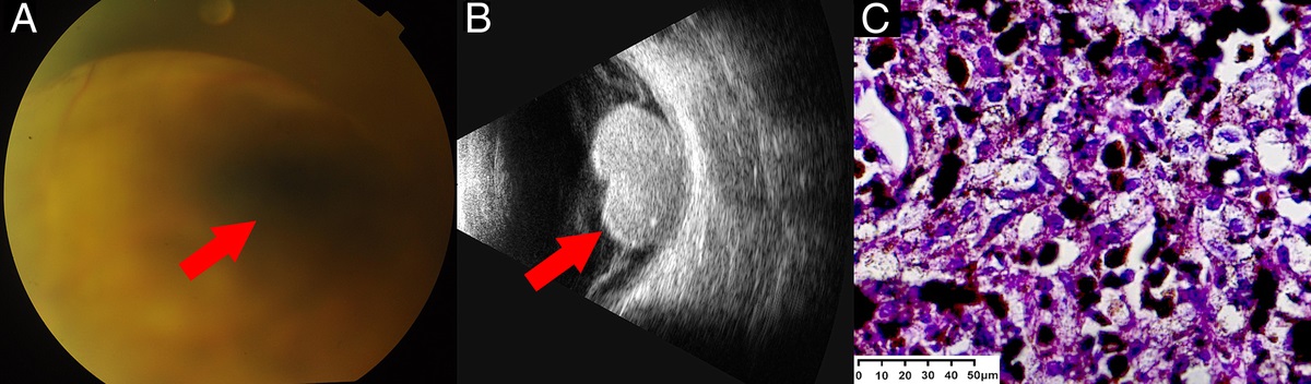
Contrast-enhanced MRI (ce-MRI) is currently the recommended imaging modality for the detection of brain metastases.11 MRI provides high-resolution anatomical images with excellent soft tissue contrast, and contrast agents increase visibility of pathology when the blood-brain barrier (BBB) is disrupted. Brain metastases are considered to be well delineated on ce-MRI, and the sensitivity for detection is high also for small metastases. However, contrast enhancement is a marker for BBB disruption, which may be caused by radiotherapy, infection, inflammation, ischemia, and other afflictions.11 Contrast enhancement is therefore not specific for malignancy, and ce-MRI has limitations for instance in separating recurrence of brain metastases from treatment-related changes, a challenge regularly occurring in clinical practice. The accuracy may be improved with perfusion-weighted MRI, but the literature is still limited, and alternative tools for diagnosing recurrent brain metastases after treatment are warranted.12
Amino acid (AA) PET may add complementary information to MRI in the management of patients with brain metastases. Brain malignancies accumulate AAs through overexpression of AA transporter systems, which is a result of alterations in tumor vasculature and proliferation.13 Amino acid PET tracers do not accumulate as much in normal brain parenchyma as the most commonly used PET tracer, the glucose analog 18F-FDG. This results in improved tumor-to-normal brain contrast for AA tracers over 18F-FDG. The transport of AAs across the BBB is facilitated by specific AA transporters, which allows for uptake in neoplastic tissue even with an intact BBB for common AA tracers, in contrast to MRI.14
Amino acid PET tracers (i.e., 11C-methyl-l-methionine [11C-MET], O-[2-18F-Fluoroethyl]-l-tyrosine [18F-FET], and l-3,4-dihydroxy-6-18F-fluorophenylalanine [18F-FDOPA]) are recommended by international guidelines to complement MRI in the clinical management of patients with gliomas.13,15,16 For patients with brain metastases, the Response Assessment in Neuro-Oncology (RANO) working group has concluded that ce-MRI is the imaging modality of choice for detecting brain metastases due to the high spatial resolution.11 However, the RANO group has also stated that AA PET is useful for differentiating brain metastasis recurrence from radiation-induced changes, but that the literature is still limited.1118F-FET has shown to differentiate recurrence of brain metastases from radiation necrosis with high accuracy, especially when supplemented with dynamic PET acquisitions.17–19
The artificial AA anti-1-amino-3-18F-fluorocyclobutane-1-carboxylic acid (18F-FACBC),20 also known as fluciclovine (18F) or Axumin (Blue Earth Diagnostics Ltd, United Kingdom), was initially developed for the assessment of brain tumors but is most commonly used for prostate cancer.21 Recent studies have demonstrated increased 18F-FACBC uptake in gliomas compared with normal brain parenchyma, especially for high-grade gliomas.22–28 Several glioma studies have reported 18F-FACBC uptake in areas without contrast enhancement, in addition to generally larger tumor volumes with 18F-FACBC than ce-MRI.22,24,27,29 In addition, in studies exploring glioma, 18F-FACBC provides greater visual contrast than 11C-MET due to lower uptake in healthy brain tissue.25,26,29 This high contrast has likely also allowed for the detection of small satellite gliomas not visualized with MRI.23
18F-FACBC PET has only been evaluated in a limited number of patients with brain metastases.30,31 The results are promising with relatively high tumor-to-background ratios (TBRs) compared with 18F-FET and 11C-MET. This may increase the possibility to detect small brain metastases. 18F-FACBC PET has also shown potential to differentiate recurrence from radiation necrosis.30 The potential of 18F-FACBC in the management of brain metastases should therefore be further explored. The aims of this study were to assess the diagnostic value of static and dynamic 18F-FACBC PET in patients with brain metastases and to compare tumor volumes between 18F-FACBC PET and ce-MRI.
PATIENTS AND METHODS SubjectsBetween January 2020 and July 2021, 18 patients (7 female) were recruited to a multicenter study at St. Olavs Hospital, Trondheim University Hospital, and the University Hospital of Northern Norway for simultaneous brain 18F-FACBC PET/MRI examinations. Patients were eligible for inclusion if they had a known or confirmed cancer and MRI findings consistent with a new metastatic brain lesion and/or suspected recurrence if previously treated with surgery or SRS in this location.
The average age of the subjects was 64.5 years (range, 51–77 years), and their primary cancers were lung (n = 9), gastrointestinal (GI) (n = 5), malignant melanoma (n = 2), breast (n = 1), and thyroid (n = 1) (Tables 1 and 2). The patients had a median graded prognostic assessment of 1.5 (range, 0–3).32 Four patients (4 lesions) had prior intracerebral resection, and 4 patients (6 lesions) had prior SRS in the same location as the lesion of interest. One patient received chemotherapy (FOLFOX + SOX), and 1 patient had immunotherapy treatment (nivolumab, Opdivo; Bristol-Myers Squibb) during the last month before the PET/MRI examination. Thirteen patients had ongoing glucocorticosteroid treatment at the time of the examination. The study was approved by the Regional Ethics Committee (REC, reference number: 2018/2243). All patients gave written informed consent to participate in the study.
TABLE 1 - Patient Information and Results From Evaluations of Brain Lesions Not Treated With Stereotactic Radiosurgery (No-SRS Group) Before PET/MRI Patient ID Age Primary Cancer GPA Lesion Number MRI Positive* PET Detected† Dmax MR, (mm) VMRI, (mL) VPET, (mL) DSC SUVmax SUVmean TBR TAC Slope 1‡ 54 Breast 2.5 1 Yes Yes 19 1.68 1.7 0.76 3.2 0.3 12.3 III 2 61 Esophagus (GI) 0 1§ Yes Yes 55 5.00 9.3 0.55 3.9 0.4 9.0 III 2 Yes No 5 0.03 — — 1.0 0.4 2.2 — 3 Yes No 5 0.02 — — 1.2 0.4 2.7 — 4 Yes No 5 0.02 — — 0.5 0.4 1.2 — 5 Yes No 4 0.02 — — 0.7 0.4 1.6 — 3‡∥ 63 Coli (GI) 1 1 Yes Yes 22 2.60 2.1 0.77 7.3 0.6 12.8 III 4 66 Lung 1.5 1 Yes Yes 10 0.32 1.0 0.50 1.8 0.3 5.4 — 5‡ 66 Lung 0 1 Yes Yes 18 1.53 2.2 0.69 3.5 0.4 8.6 III 2 Yes Yes 9 0.14 0.6 0.35 1.9 0.4 4.7 — 3 Yes No 4 0.01 — — 0.5 0.4 1.3 — 4 Yes No 5 0.01 — — 0.5 0.4 1.2 — 6‡ 74 Melanoma 3 1 Yes Yes 30 5.38 3.7 0.77 7.4 0.3 21.9 III 2 Yes Yes 17 0.91 0.9 0.63 3.6 0.3 10.8 III 8¶ 73 Melanoma 3 1§ Yes Yes 13 0.36 0.5 0.58 6.9 0.3 19.8 II 2 Yes Yes 7 0.10 0.5 0.30 1.7 0.3 4.7 — 3 Yes No 3 0.01 — — 0.4 0.3 1.0 — 9‡ 51 Coli (GI) 0 1 Yes Yes 24 4.18 6.6 0.76 2.3 0.4 5.8 II 12‡ 56 Esophagus (GI) 0 1 Yes Yes 49 27.02 29.6 0.90 5.2 0.4 13.2 — 13‡ 69 Thyroid NA 1 Yes Yes 8 0.14 0.9 0.24 1.2 0.3 3.9 — 2 Yes No 6 0.04 — — 0.4 0.3 1.3 — 3 Yes No 5 0.02 — — 0.3 0.3 1.1 — 4 Yes No 8 0.09 — — 0.7 0.3 2.2 — 14‡ 58 Lung 3 1 Yes Yes 26 4.47 3.6 0.86 8.4 0.3 25.9 III 15‡ 67 Rectal (GI) 0 1 Yes Yes 33 7.83 10.7 0.83 3.8 0.4 10.6 II 16 74 Lung# 1.5 1 Yes Yes 51 25.60 30.9 0.88 4.3 0.3 16.4 II 17‡ 63 Lung# 0.5 1 Yes Yes 38 12.85 10.8 0.76 6.5 0.3 21.7 III 2 Yes Yes 47 18.85 18.8 0.89 3.2 0.3 10.7 II 18‡ 63 Lung# 3 1 Yes Yes 38 11.30 11.7 0.78 2.8 0.3 9.2 II*“Yes” if new contrast-enhancing lesion.
†“Yes” if visually detected on PET, “no” otherwise.
‡Ongoing steroid treatment.
§Prior resection at same location.
∥Chemotherapy last month.
¶Immunotherapy last month.
#Most likely lung cancer.
Dmax MR, maximum lesion diameter on ce-MRI; VMRI, tumor volume defined on ce-MRI; VPET, tumor volume defined on 18F-FACBC PET; NA, not available, GPA not defined for thyroid cancer.
*“Yes” if lesion showed contrast enhancement and progressive enlargement compared with previous MRIs, “no” if lesion was stable or decreased in size compared with previous MRIs.
†“Yes” if visually detected on PET.
‡Based on follow-up MRI.
§Prior SRS at same location.
∥Prior resection at same location.
¶Ongoing steroid treatment.
GPA, graded prognostic assessment; TP, time period, Dmax MR, maximum lesion diameter on ce-MRI; VMRI, tumor volume defined on ce-MRI; VPET, tumor volume defined on 18F-FACBC PET.
The patients underwent a simultaneous brain PET/MRI examination (Siemens Biograph mMR; Erlangen, Germany) after at least 4 hours of fasting. 18F-FACBC (3.0 ± 0.2 MBq/kg; average activity, 234 ± 50 MBq) was manually injected at t = 0 of the PET acquisition (list mode; 0–35 minutes post injection). Two patients (ID 10 and 11) had a shorter acquisition (30 minutes) due to delays and expiration of the tracer. MRI sequences acquired were ultrashort echo time (UTE) for PET attenuation correction, 3D fluid-attenuated inversion recovery, and contrast-enhanced 3D T1 magnetization-prepared rapid gradient echo. One patient (ID 12) was examined on a PET/CT system (Siemens Biograph Vision 600) due to technical problems with the PET/MRI system (only static acquisition 20–35 minutes post injection). This patient had a separate MRI examination later the same day.
PET ReconstructionStatic and dynamic PET image reconstructions were performed using iterative OSEM reconstruction (PET/MRI reconstruction: 3 iterations, 21 subsets, 344 image matrix, 4 mm Gaussian filter; PET/CT reconstruction: 8 iterations, 5 subsets, 440 image matrix, 4 mm Gaussian filter, TOF), point spread function modeling, and relative scatter correction. MR-based attenuation correction was performed with a deep learning–based method (DeepUTE) using the UTE MR sequence as input for making MR-based attenuation correction maps.33 Static PET reconstructions were generated from the last 15 minutes of the list mode data, whereas the dynamic reconstructions were performed with frames of 12 × 5 seconds, 6 × 10 seconds, 6 × 30 seconds, 5 × 60 seconds, and 5 × 300 seconds, based on recommendations for dynamic 18F-FET PET imaging of gliomas.16
Image EvaluationThe PET and MR images were jointly investigated by an experienced nuclear medicine physician (years of experience: H.J., 5 years/T.V.B., 20 years) and an experienced neuroradiologist (years of experience: E.M.B., 7 years/J.A.T., 8 years). As both recurrence and radiation necrosis show contrast enhancement, the MRI interpretation differed depending on whether the lesions were treated with SRS before PET/MRI or not, and the lesions were divided in 2 groups. Lesions without previous SRS in the same location (no-SRS group) were categorized as “MRI positive” if they showed contrast enhancement and were not present on previous MRIs or if no previous MRIs were available, thus radiologically evident a metastasis. Contrast-enhancing lesions in operation cavities were also defined as “MRI positive.” Lesions with previous SRS in the same location (SRS group) were categorized as “MRI positive” if showing contrast enhancement and progressive enlargement according to the RANO criteria for brain metastasis34 (>20% enlargement in longest diameter) compared with previous MRI scans. On the contrary, if the lesions had been stable or decreased in size compared with previous MRI scans and thus indicative of radiation necrosis, they were categorized as “MRI negative.” A follow-up status of the lesions in the SRS group was assessed based on follow-up MRIs after the PET/MRI, to serve as a surrogate marker for separating true recurrence from radiation necrosis in the lack of histopathological verification. PET findings with 18F-FACBC uptake distinctly higher than surrounding tissue were defined as detected on PET.
Image AnalysisImage analysis was performed with the software PMOD (version 4.203; PMOD Technologies LLC, Zürich, Switzerland). A volume of interest (VOI) was drawn on the static PET images to encompass the lesion of interest to extract the SUVmax of the lesion. For lesions not detected on PET, SUVmax was measured in the volume defined on ce-MRI. To measure reference tissue, a crescent-shaped VOI was placed in normal brain parenchyma (in the contralateral hemisphere if only 1 lesion and in an area not affected by the disease if lesions in both hemispheres) as described by Unterrainer et al35 (Fig. 1). The SUVmean in the normal brain VOI was measured. TBR was defined for all brain lesions of interest in the static PET images as SUVmax divided by SUVmean. SUVmax and TBR were compared between lesions of different tumor origins.
 FIGURE 1:
 Background VOI in normal brain parenchyma consisting of 6 consecutive crescent-shaped ROIs fused together, avoiding inclusion of ventricles and veins.
PET and MRI tumor volumes, VPET and VMRI, were defined subsequent to rigid registration of the static PET image to the T1 MR image to ensure best possible alignment and equal voxel dimensions. No guidelines exist for tumor delineation with 18F-FACBC PET. A threshold-based method of 41% of SUVmax was found to visually perform best for all lesion sizes and was applied in this study. Manual modifications were made to include necrotic areas inside tumors and to exclude nontumor tissue, such as veins. Contrast-enhanced MRI volumes were manually delineated in accordance with an experienced neuroradiologist, and necrotic areas inside tumors were included. The maximum diameter of the lesions (in any direction) on ce-MRI (Dmax MR) was automatically calculated in PMOD. The spatial similarity of PET and MRI tumor volumes was evaluated using the Dice similarity coefficient (DSC).36
DSC=2VMRI∩VPETVMRI+VPET
The VOIs drawn on static PET were transferred to the dynamic PET images to generate time-activity curves (TACs) for SUV
留言 (0)