18F-FDG Muscular Uptake in Statin-Associated Symptoms Without Myositis: How Long to Stop Treatment for Image Quality Improvement?
Statin-associated muscle symptoms (SAMSs) are a frequent adverse effect of statin treatment (7%–29%) and the main reason for nonadherence.
1 SAMS can occur with normal or slightly elevated serum creatine kinase (CK). The etiology is complex with multifactorial mechanisms and appears more frequently in women.
2 Myopathy is a rarer complication with muscular inflammation and increased CK. Diffuse 18F-FDG muscular uptake on PET was reported in statin-related rhabdomyolysis
3,4 and other myopathies.
5–7 In men, but not in women, the risk of statin-related myopathy is dose-dependent.
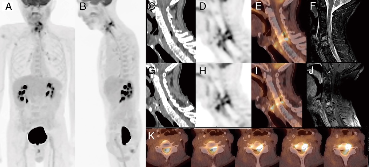
2 In this case, a 80-year-old man was referred for 18F-FDG PET in hemopathy follow-up. A long-term statin treatment was prescribed for cardiovascular prevention. On examination, the patient described mild chronic myalgia for more than 6 months. CK and C-reactive protein were normal. Clinically, the patient was classified statin-related myotoxicity (SRM) 1 on the 7-point SRM scale. Acquisitions were all obtained on PET/CT (Biograph mCT Flow; Siemens) 60 minutes after 3.5 MBq/kg 18F-FDG injection. Table flow acquisition was 1.3 mm/s. The patient was fasting without any treatment for at least 6 hours, and glycemia was controlled and normal (5.4–7.0 mmol/L). Reference PET acquisition (A) showed an abnormal diffuse 18F-FDG muscular uptake on MIP, more pronounced on shoulders, arms, hips, legs, and feet. This aspect remained similar on the second examination without treatment for 6 hours (B). On the follow-up with a statin discontinuation for 3 days (C) and 7 days (D), an image quality improvement was observed with less muscular uptake and a better 18F-FDG bioavailability, especially on brain. The 1-week statin-free medication showed a better overall image quality except arm movements during the acquisition. Cervical hypermetabolisms were related to uptakes in contracting skeletal muscles. On pelvic transaxial fused PET/CT slices (below MIP images), there was an hypermetabolism of gluteal muscles, which was gradually reduced after 7 days of statin discontinuation. The 2 last acquisitions also revealed osseous and pulmonary focal uptakes related to hemopathy. Diffuse muscular uptake has been reported to alter the tumoral contrast and PET lesion detectability.
8 This case illustrates the possibility of diffuse muscular FDG uptake in SAMS without biological myositis and the need for a minimal 1-week statin discontinuation to improve image quality. This treatment interruption could be planned as the first biological modifications described in the literature were observed from a 5-day statin interruption
9 with only impact on long-term cardiovascular risks after a 3-month discontinuation.
10–12 Thus, this short statin discontinuation could be conceivable without impact on the patient cardiovascular risk. A study on a larger cohort would allow a better assessment of an ideal discontinuation time.


留言 (0)