
記住我


KEY PERSPECTIVE
What is novel?
Primary prevention efforts are needed to address the high burden of atherosclerotic cardiovascular disease (ASCVD). A quantitative assessment is the foundation for assessing ASCVD risk in primary prevention. Life's Essential 8 incorporates eight different metrics that capture overall cardiovascular health.What are the clinical and/or research implications?
All individuals should be counseled on a heart healthy lifestyle and have a cardiovascular health assessment. Statin therapy can be considered among those with a 10-yr ASCVD risk of ≥5% in primary prevention, especially with the presence of risk-enhancing factors. When the decision regarding statin therapy is unclear, coronary artery calcium score can help refine risk.Over the last several decades, the global prevalence and mortality from atherosclerotic cardiovascular disease (ASCVD) have increased, with 23.6 million annual CVD deaths predicted to occur by 2030.1,2 In the United States, and other high-income nations, mortality from ASCVD has declined because of advances in treatment and preventive measures; however, ASCVD remains the leading cause of death.2,3 In addition, age-standardized ASCVD mortality rates have recently risen in certain regions of the United States and other high-income countries.2 This high burden of ASCVD has a significant economic impact, with ASCVD-related costs projected to rise by $183 billion in the United States from 2015 to 2035.4 These figures highlight the large opportunity for appropriate risk assessment and prevention of ASCVD.
Identifying risk factors associated with ASCVD has been paramount to informing public health policies and interventions to reduce ASCVD risk.5 In fact, a study found that 44% of the decline in mortality due to coronary heart disease in the United States from 1980 to 2000 was due to optimization of several CVD risk factors, including hypercholesterolemia, hypertension, tobacco use, and physical inactivity.6 Assessing ASCVD risk based on these CVD risk factors helps guide treatment, primarily aimed at reducing low-density lipoprotein-cholesterol (LDL-C) in primary prevention, as cumulative exposure to LDL-C is the main driver for ASCVD.3,7–9 The purpose of this review is to discuss the principals of ASCVD risk assessment and the management of ASCVD risk factors in primary prevention.
REVIEW OF RELEVANT LITERATURE AND DISCUSSION POOLED COHORT EQUATIONSAlthough several tools exist to assess ASCVD risk all over the world, they all focus on quantitative risk assessment, helping match individual ASCVD risk with intensity of treatment.10,11 Research has shown that estimating ASCVD risk leads to greater initiation of lipid-lowering therapy and has a low likelihood of causing harm.10 In addition, calculating ASCVD risk has been shown to enhance shared decision making and improve patient satisfaction.12 Furthermore, recent data from the Million Hearts CVD Risk Reduction Model illustrate a decrease in myocardial infarctions and strokes with the use of ASCVD risk assessment in clinical practice.13 This review will focus on application of the US-derived sex- and race-specific pooled cohort equations (PCE) to quantitatively assess ASCVD risk in primary prevention.
The PCE were developed in 2013 from four large and diverse US cohorts, including the Cardiovascular Health Study, Coronary Artery Risk Development in Young Adults, Atherosclerosis Risk in Communities, and Framingham Original and Offspring Study to predict absolute 10-yr ASCVD risk, defined as fatal and nonfatal myocardial infarction or stroke.8,14 All of these cohorts had a follow-up period of >11 yr with adjudicated ASCVD outcome data.14 The PCE have since been well validated in other cohorts and US populations, performing well in both men and women.15,16 That said, the PCE were developed using only White and African American participants, and thus, while they can be used in all populations, studies have shown over and underestimation of ASCVD risk in certain Asian and Hispanic racial/ethnic subgroups.14,17,18 In addition, the PCE were intended for use in adults with an LDL-C ≥70 mg/dL and <190 mg/dL, aged 40-75 yr.8 However, in patients younger than 40 yr, the PCE can be used to calculate lifetime risk, guiding intensity of lifestyle preventive measures.8 The ASCVD risk estimation is controversial in adults older than 75 yr, as there are limited data for statin therapy in primary prevention in this population.19
The parameters included in the PCE are age, sex, race, systolic blood pressure (BP), BP medication use, total cholesterol, high-density lipoprotein-cholesterol (HDL-C), current tobacco use, and history of diabetes.14 The PCE calculate the estimated absolute 10-yr ASCVD risk and categorize individuals as low risk (<5%), borderline risk (5 to <7.5%), intermediate risk (7.5 to <20%), and high risk (≥20%).8 The PCE risk assessment online tool can be found here: https://tools.acc.org/ascvd-risk-estimator-plus/#!/calculate/estimate/.20
The ASCVD risk category serves as the foundation for guiding the type and intensity of primary prevention interventions.3,8 All individuals, regardless of ASCVD risk, should be counseled on dietary and physical activity (PA) recommendations to promote a heart healthy lifestyle.3,8 Those at low risk (<5%) can be monitored with heart healthy lifestyle recommendations alone, as there has not been found to be a clear benefit for statin therapy in these individuals.3,8 Individuals with borderline risk (5 to <7.5%) can be considered for moderate-intensity stain therapy if specific risk enhancers are present.3,8 Those with intermediate risk (7.5 to <20%) should be considered for moderate-intensity statin therapy, especially if risk enhancers are present.3,8 This recommendation is further supported by data from a large randomized controlled trial that found a reduction in ASCVD events with moderate-intensity statin therapy in intermediate ASCVD risk individuals.8,10,21 Finally, those with high risk (≥20%) should be strongly considered for high-intensity statin therapy.8,10 After ASCVD risk is calculated, a patient-centered discussion should take place regarding present CVD risk factors and degree of control, interpretation of ASCVD risk, benefits and harms of pharmacotherapy, and patient wishes regarding preventive therapy for myocardial infarction and stroke.8,10 Part of this conversation should also include a discussion about risk-enhancing factors and how they influence ASCVD risk.
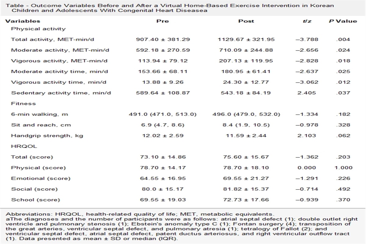
The 2018 Multi-Society Guidelines on Management of Blood Cholesterol considers several risk-enhancing factors that can help refine ASCVD risk assessment in those with borderline (5 to <7.5%) or intermediate (7.5 to <20%) ASCVD risk.8 Risk enhancers are displayed in the Table, adapted from Grundy et al, and provide a more individualized risk assessment, especially where PCE have been shown to underestimate risk, including those with chronic inflammatory conditions or South Asian ancestory.3,8,22,23 There are four biomarkers that are considered risk enhancers: lipoprotein(a) [Lp(a)], apolipoprotein B (apoB), high-sensitivity C-reactive protein (hsCRP), and ankle brachial index (ABI).8 Lipoprotein(a) is an LDL-like molecule covalently linked to an apolipoprotein(a) component with proatherogenic, proinflammatory, and antifibrinolytic properties.24 Multiple lines of evidence have shown a linear association between Lp(a) and CVD, with considerable risk at levels ≥50 mg/dL, which is the threshold used to qualify Lp(a) as a risk enhancer.8,24 Another emerging biomarker is ApoB, which encompasses all atherogenic lipids, including Lp(a), and is associated with ASCVD when elevated (≥130 mg/dL), even when LDL-C is controlled.25 Next, hsCRP is an excellent marker of chronic inflammation, associated with ASCVD at levels ≥2 mg/L.26 In fact, anti-inflammatory treatment leading to a reduction of hsCRP was found to have a cardiovascular benefit, even when LDL-C remained the same.26 Recently, a significant interaction was found between Lp(a) and hsCRP, indicating that ASCVD risk due to Lp(a) may be present only when hsCRP is elevated.27 Finally, ABI is a marker of peripheral artery disease and is calculated for each lower limb by dividing the ankle systolic BP by the highest systolic BP from both arms.8,28 An ABI ≤0.9 is associated with ASCVD risk and qualifies as a risk enhancer.8,28 Risk-enhancing factors have been studied in various large cohorts, and while individually associated with ASCVD, they have little to no improvement in net reclassification when incorporated in the PCE.29,30 However, a recent analysis found significant net reclassification in those with ≥3 risk-enhancing factors and intermediate ASCVD risk.31 Thus, the presence of risk-enhancing factors supports further discussion for consideration of statin therapy in those with borderline and intermediate risk.8
Table - Individual Risk-Enhancing Factorsa Risk Enhancers Example or Description Qualifying as Risk Enhancer Race/ethnicity with elevated ASCVD risk South Asian ancestry (India, Pakistan, Bangladesh, Bhutan, Maldives, Nepal, Sri Lanka)72 Family history of premature ASCVD Males <55 yr; females <65 yr Medical conditions Chronic kidney disease Stages 2-4 Chronic inflammatory diseases Rheumatoid arthritis, systemic lupus erythematous, psoriasis, human immunodeficiency virus Metabolic syndrome At least three of the following: abdominal obesity, hypertriglyceridemia, hypertension, hyperglycemia, and low HDL-C Premature menopause <40 yr Pregnancy conditions Preeclampsia, gestational diabetes mellitus22 Primary hypercholesteremia LDL-C: 160-189 mg/dL; or non-HDL-C: 190-219 mg/dL Primary hypertriglyceridemia Nonfasting ≥175 mg/dL Biomarkers Elevated Lp(a) ≥50 mg/dL Elevated apoB ≥130 mg/dL Elevated hsCRP ≥2.0 mg/L Abnormal ABI <0.9Abbreviations: ABI, ankle brachial index; apoB, apolipoprotein B; ASCVD, atherosclerotic cardiovascular disease; HDL-C, high-density lipoprotein-cholesterol; hsCRP, high-sensitivity C-reactive protein; LDL-C, low-density lipoprotein-cholesterol; Lp(a), lipoprotein(a).
aAdapted from Grundy et al.8Statin allocation in primary prevention is based on ASCVD risk assessment; however, there are other important indications for initiating statin therapy that do not rely on calculating ASCVD risk, including secondary prevention for ASCVD, severe hypercholesterolemia, and diabetes.8 Secondary prevention refers to individuals with previous atherosclerotic disease, including acute coronary syndrome, myocardial infraction, cerebrovascular accident, and peripheral arterial disease.32 In these individuals, high-intensity statin therapy should be started with an aim of reducing LDL-C by ≥50%.32 Other individuals who should receive statin therapy without calculating ASCVD risk are those with severe hypercholesterolemia.8 These are individuals with an LDL-C ≥190 mg/dL and thus have higher lifetime risk for ASCVD for which high-intensity statin therapy should be started.33 Finally, individuals with diabetes aged 40-75 yr all have at least intermediate risk for ASCVD and should therefore be started on statin therapy; however, calculating ASCVD risk helps refine treatment dosing.8,34 In addition, there are diabetes-specific risk enhancers that support use of high-intensity statin therapy, including long-duration of diabetes, albuminuria, chronic kidney disease, retinopathy, neuropathy, and an ABI <0.9.8 For individuals with no history of ASCVD, severe hypercholesterolemia, or diabetes, performing an ASCVD risk assessment is foundational to guiding management in primary prevention.3,8
IMAGINGAfter quantitative ASCVD risk assessment and consideration of risk-enhancing factors, if uncertainty regarding initiating lipid-lowering therapy still exists, coronary artery calcium (CAC) may be considered for those with borderline or intermediate ASCVD risk.3,8 The ability for CAC to reclassify individuals is much stronger than any one particular risk-enhancing factor.3,8 Studies have consistently documented that individuals with a CAC of 0 have very low 10-yr ASCVD risk and are unlikely to benefit from initiation of statin therapy.35–38 In the Multi-Ethnic Study of Atherosclerosis, a primary prevention cohort, Nasir et al36 found that a CAC score of 0 reclassified risk in almost half of individuals with borderline or intermediate ASCVD risk (5 to <20%). Here, a CAC of 0 can serve as justification to delay initiation of statin therapy, minimizing costs, and polypharmacy.8,39 However, individuals with a CAC of 0 make up more than a quarter of total CVD events.35 These individuals typically have a greater burden of known cardiovascular risk factors, including tobacco use and family history of premature ASCVD.8,35 In fact, current guidelines recommend against CAC testing in those with tobacco use, family history of premature ASCVD, and/or diabetes mellitus.8,40 Thus, while a CAC score of 0 can be helpful in down-risking individuals, it should be interpreted within the overall clinical context to help refine risk and further the patient-centered conversation.3,8 For individuals who do not have a CAC score of 0, a score of ≥1 favors consideration for statin therapy.8,40
RISK FACTORSAlthough PCE allow for a quantitative risk assessment, there are several CVD risk factors that serve as metrics for assessing overall cardiovascular health.41 The initial construct by the American Heart Association, known as Life's Simple 7, included diet, PA, smoking, body mass index (BMI), fasting glucose, total cholesterol, and BP.41 After over a decade of research using Life's Simple 7, several metrics were redefined and a new metric, sleep, was included, forming the updated Life's Essential 8.41 Each metric is scored from 0 to 100 and an unweighted average of all the metrics creates the composite score, also from 0 to 100.41 Identifying and managing each of these individual metrics are imperative to lower ASCVD risk in primary prevention. Therefore, it is important to provide lifestyle modification counseling to individuals to control CVD risk factors.42,43
DietThere is consistent evidence that following a heart healthy diet promotes quality cardiovascular health.44,45 Most of the data regarding dietary patterns in research are from dietary records, food recalls, and food frequency questionnaires.46 As nutrients are not consumed independently, current research has focused on dietary patterns, rather than individual food components.47,48 For example, a study from the Progression of Early Subclinical Atherosclerosis found that a social-business eating pattern (high in processed foods, red meat, alcohol, and sugary beverages) was associated with increased CVD risk.48 Conversely, another dietary pattern, the Mediterranean diet, has been shown to reduce CVD risk through its anti-inflammatory properties and positive impact on the gut microbiome.44 The largest study of the Mediterranean diet was from a multicenter trial in Spain that randomized 7447 individuals to a Mediterranean diet with extra virgin olive oil, Mediterranean diet with mixed nuts, or a control diet.46 The authors found that after a mean follow-up of 4.8 yr, individuals in the Mediterranean diet with extra virgin olive oil and the Mediterranean diet with mixed nuts had lower CVD risk compared with the control group, hazard ratio: 0.69 and 0.72, respectively.49 Similarly, the Dietary Approaches to Stop Hypertension, a diet low in total and saturated fat but rich in fruits, vegetables, and low-fat dairy, was found to reduce BP and inflammatory markers.44,50 Based on the aforementioned evidence, the American Heart Association has several dietary recommendations for primary prevention, encouraging a plant-based or Mediterranean diet, high in fruits, vegetables, and legumes.3 The American Heart Association also recommends avoiding trans-fats, red meats, processed meats, sugar sweetened or artificially sweetened beverages, and a high salt diet.3
Physical ActivityThe cardioprotective effects of PA are well understood, and, thus, the American Heart Association recommends that adults obtain 150 min/wk of moderate-intensity aerobic exercise.3 Alternatively, individuals can also complete 75 min/wk of vigorous-intensity aerobic exercise.3 Furthermore, it is also recommended that adults engage in resistance training ≥2 times/wk, as it has been shown to lower BP and improve physical strength and performance in elderly individuals.51–53 Yet, approximately 50% of individuals in the United States, and 33% globally, do not achieve minimum PA recommendations.53,54 Even more, a cross sectional study using accelerometers found that <5% of adults in the United States followed the recommended 30 min/d of PA.55 This lack of PA results in enormous costs, approximately 11% of the health care spending budget in the United States.53 Thus, there is a dire need to improve PA at a population level.53
Exercise prescription and counseling are beneficial interventions at increasing PA and should be offered to patients.42,56 Physical activity prescriptions are typically tailored on the basis of comorbidities and current level of PA.53 In addition to promoting moderate-intensity PA, counseling should also include measures to minimize sedentary activity, as higher levels of sedentary activity are associated with elevated CVD risk.53 During the office visit, it is important to document PA details in the electronic health record to allow for monitoring and clear communication with other providers.53 A PA tool, such as the Exercise Vital Sign, should be used to standardize documentation and assessment of PA.57 The Exercise Vital Sign contains two questions: how many d/wk do you perform moderate-intensity exercise and how many minutes do you perform this activity?57 The scores of the two questions are multiplied to create a composite score that can be used for documentation and can also be added as a vital sign.57 If available, documentation of information provided by wearable technology can also provide valuable objective PA data.53
Tobacco UseGlobally, tobacco use is the leading and most preventable cause of death.58 In the United States, approximately 480 000 individuals pass away yearly, and >8 million individuals are disabled from tobacco use.45 In addition, in the United States, 20% of the CVD mortality is due to tobacco use.59 Research has found no safe level of tobacco exposure, both primary and secondary.3 Several studies have attempted to quantify the time post-smoking cessation where CVD risk approaches that of a never smoker, with varying estimates.59 Recently, Duncan et al59 investigated the relationship between time post-smoking cessation and CVD risk and found that CVD risk approaches the risk of a never smoker 10-15 yr after quitting smoking. However, it should also be noted that the authors found significantly lower CVD risk in those who quit smoking for 5 yr.59 Interestingly, the PCE incorporate smoking; however, risk associated with former smoking after cessation for ≥5 yr is considered the same as a never smoker.59 Thus, the PCE can underestimate risk in former smokers, who are increasing in prevalence in the United States.3
Given the considerable risk associated with tobacco use, it is recommended that clinical providers assess smoking status in all adults.3 Smoking cessation rates are higher in those who are provided tailored behavioral interventions, assessing readiness and benefits to quitting.3 In addition to behavioral interventions, pharmacotherapy can be used to aid in cessation.3 These therapies include over-the-counter nicotine replacement therapy, as well as bupropion and varenicline, which are both oral prescription medications.3 Of note, Electronic Nicotine Delivery Systems should not be used in smoking cessation as these devices result in inhalation of toxic gases that increase oxidative stress.3
Body Mass IndexCurrently, there is a global obesity pandemic impacting an approximate 603 million adults.3,60,61 In fact, between 1975 and 2016, the global obesity prevalence tripled.61 Obesity promotes atherosclerosis through inflammatory processes and development of CVD risk factors, including diabetes and hypertension.3 The BMI is used to categorize overweight (25 to <30kg/m2) and obesity (≥30kg/m2) status.3,62 It is important to recognize that while BMI correlates with body fat composition, there is variation across race/ethnicities, for which different thresholds can be used.3 The distribution of adiposity impacts ASCVD risk, with central adiposity associated with the highest risk, assessed by waist circumference.3 Thus, for every level of BMI, higher waist circumference confers greater ASCVD risk.3 Although obesity is due to energy-balance dysregulation, there are complex environmental and genetic factors at play; thus, management is often multipronged and multidisciplinary.62 Treatment should be individualized, incorporating management of comorbidities, lifestyle interventions, pharmacotherapy, and weight loss surgery.62
DiabetesType 2 diabetes mellitus is diagnosed with hemoglobin A1c≥6.5% and makes up 90-95% of all cases of diabetes mellitus.3,63 The global prevalence of type 2 diabetes mellitus is 9.3% and individuals with type 2 diabetes mellitus have twice as much ASCVD risk compared with those without.3,63 All individuals with type 2 diabetes mellitus should be started on statin therapy and provided lifestyle recommendations to reduce ASCVD risk.3 The major microvascular and macrovascular complications from type 2 diabetes mellitus arise from chronic exposure to elevated levels of glucose.63 Thus, treatment should also focus on pharmacotherapy to achieve glycemic targets, often hemoglobin A1c below 7.0%; however, thresholds should be individualized on the basis of age and other comorbidities.63 First-line pharmacotherapy is with metformin, and those who have elevated CVD risk should be considered for glucagon-like peptide-1 or sodium-glucose cotransporter type 2 inhibitor therapies, as they have been shown to have a cardiovascular benefit.63–65
HypertensionHypertension increases ASCVD risk in a log-linear fashion and leads to the greatest number of ASCVD deaths out of all the modifiable cardiovascular risk factors.3 In fact, an increase in BP by 20 mm Hg systolic or 10 mm Hg diastolic is associated with double the CVD risk.66 Although likely an underestimate, the population attributable risk for coronary heart disease due to hypertension is approximately 25%.66 In clinical practice, BP is categorized as normal (<120/<80), elevated (120-129/<80), stage 1 hypertension (130-139/80-89), and stage 2 hypertension (≥140/90).3,67 All individuals should receive counseling on a heart healthy lifestyle.3,8 Those with stage 2 hypertension should be started on antihypertensive therapy.3,67 Pooled cohort equations play a role in those with stage 1 hypertension, as a 10-yr ASCVD risk of <10% supports heart healthy lifestyle counseling alone, while a 10-yr ASCVD risk of ≥10% favors initiation of antihypertensive therpay.3,67
SleepThere is growing evidence of the association between sleep health and cardiovascular health.68 In the United States, 50-70 million individuals experience at least one sleep disorder, with insomnia the most prevelant.68 Both short duration of sleep (<7 hr) and long duration of sleep (>9 hr) have been shown to increase ASCVD risk.68 In fact, a recent prospective study from the National Health and Nutrition Examination Survey found the association between sleep duration and ASCVD risk to be U-shaped, with the lowest risk in those who slept for 7 hr.69 In addition, in a meta-analysis of prospective studies, the authors found the relative risk for coronary heart disease from short sleep duration to be 1.48 and for long sleep duration to be 1.38.70
A proper sleep assessment is important to understand contributing lifestyle behaviors and comorbidities that could impact sleep health.70 Patients should be counseled on avoiding behaviors that diminish sleep quality, such as smoking, alcohol, and screen time before bed.70 In addition, counseling should promote behaviors that improve sleep, such as PA and having a consistent bedtime.70
APPLICATION TO PRACTICE PATIENT EXAMPLE 1A 48-yr-old non-Hispanic White man with a past medical history significant for hypertension, controlled with hydrochlorothiazide, and a 30-pack yr history of smoking, presents for follow-up after receiving a recent lipid panel from a work physical examination. He has no acute complaints. Unfortunately, his brother had a fatal myocardial infarction last year, passing away at 52 yr of age. His systolic BP is 120 mm Hg and diastolic BP is 80 mm Hg. Lipid panel reveals a total cholesterol of 205 mg/dL, HDL-C of 35 mg/dL, and LDL-C of 140 mg/dL. The patient states that he would like advice on how to avoid a similar event that occurred with his father.
APPROACHThe patient mentioned previously belongs to the primary prevention population, as he does not have a history of clinical ASCVD, severe hypercholesterolemia, or diabetes. The patient should be advised on a heart healthy lifestyle. Next, ASCVD risk estimation should occur using PCE, which comes out to 10.7%, placing the patient at intermediate risk. Thus, he should be offered moderate-intensity statin therapy. This patient also has a risk-enhancing factor, as he has a family history of premature ASCVD, further supporting consideration for moderate-intensity statin therapy.8 Thus, a discussion should take place encouraging moderate-intensity statin therapy, weighing the risks, benefits, and patient wishes. In addition, the patient should be counseled on quitting smoking. If the patient is ready to quit, he should be offered behavioral and pharmacological interventions. Physical activity, diet, and sleep should also be assessed with appropriate recommended counseling.
PATIENT EXAMPLE 2A 69-yr-old South Asian Indian American woman with no significant past medical history presents to clinic for an annual physical examination. She has no acute complaints and no family history of ASCVD. In clinic today, her systolic BP is 124 mm Hg and diastolic BP is 64 mm Hg. Lipid testing from last week reveals a total cholesterol of 170 mg/dL, HDL-C of 35 mg/dL, and LDL-C of 120 mg/dL. She would like to avoid pharmacotherapy if possible.
APPROACHAn ASCVD risk assessment for primary prevention should take place for this patient with no history of clinical ASCVD, severe hypercholesterolemia, or diabetes. The ASCVD risk estimation reveals a 10-yr ASCVD risk of 8.5%. As with all patients, she should be counseled on a heart healthy lifestyle. She should be considered for moderate-intensity statin therapy based on her ASCVD risk alone, especially given her South Asian ancestry (risk enhancing factor). However, given her wishes to avoid pharmacotherapy, CAC testing would be of value. In this individual, who has minimal CVD risk factors, a score of 0 would provide meaningful justification to avoid statin therapy. Physical activity, diet, and sleep should also be assessed with appropriate recommended counseling.
SUMMARYAn initial quantitative risk assessment sets up the foundation for a patient-centered conversation regarding preventive efforts to reduce the risk of myocardial infraction and stroke.3,8 After a quantitative risk assessment, the presence (or absence) of risk-enhancing factors further refines risk for those in the borderline or intermediate ASCVD risk categories.3,8 If uncertainty regarding therapy remains, CAC testing can be pursued.3,8 Regardless of risk, all individuals should have a cardiovascular health assessment completed, with an attempt to optimize diet, PA, nicotine exposure, BMI, blood glucose, blood lipids, BP, and sleep.10 It is important to recognize that while the aforementioned risk factors are associated with CVD, there are several other social, psychosocial, environmental, and economic factors that impact CVD and its associated risk factors, leading to cardiovascular health disparities.3,71 Multiple overlapping mechanisms exist by which the social determinants of health impact CVD, and these factors must be considered when quantifying risk and assessing cardiovascular health.3,71
REFERENCES 1. Song P, Fang Z, Wang H, et al. Global and regional prevalence, burden, and risk factors for carotid atherosclerosis: a systematic review, meta-analysis, and modelling study. Lancet Glob Health. 2020;8:e721–e729. 2. Roth GA, Mensah GA, Johnson CO, et al. Global burden of cardiovascular diseases and risk factors, 1990-2019: update from the GBD 2019 study. J Am Coll Cardiol. 2020;76:2982–3021. 3. Arnett DK, Blumenthal RS, Albert MA, et al. 2019 ACC/AHA guideline on the primary prevention of cardiovascular disease: a report of the American College of Cardiology/American Heart Association Task Force on Clinical Practice Guidelines. Circulation. 2019;140:e596–e646. 4. Khera R, Valero-Elizondo J, Nasir K. Financial toxicity in atherosclerotic cardiovascular disease in the United States: current state and future directions. J Am Heart Assoc. 2020;9:e017793. 5. Gibbons GH, Seidman CE, Topol EJ. Conquering atherosclerotic cardiovascular disease—50 years of progress. N Engl J Med. 2021;384:785–788. 6. Ford ES, Ajani UA, Croft JB, et al. Explaining the decrease in U.S. deaths from coronary disease, 1980-2000. N Engl J Med. 2007;356:2388–2398. 7. Shapiro MD, Bhatt DL. “Cholesterol-years” for ASCVD risk prediction and treatment. J Am Coll Cardiol. 2020;76:1517–1520. 8. Grundy SM, Stone NJ, Bailey AL, et al. 2018 AHA/ACC/AACVPR/AAPA/ABC/ACPM/ADA/AGS/APhA/ASPC/NLA/PCNA Guideline on the management of blood cholesterol: a report of the American College of Cardiology/American Heart Association Task Force on Clinical Practice Guidelines. Circulation. 2019;139:e1082–e1143. 9. Ference BA, Ginsberg HN, Graham I, et al. Low-density lipoproteins cause atherosclerotic cardiovascular disease. 1. Evidence from genetic, epidemiologic, and clinical studies. A consensus statement from the European Atherosclerosis Society Consensus Panel. Eur Heart J. 2017;38:2459–2472. 10. Lloyd-Jones DM, Braun LT, Ndumele CE, et al. Use of risk assessment tools to guide decision-making in the primary prevention of atherosclerotic cardiovascular disease: a special report from the American Heart Association and American College of Cardiology. J Am Coll Cardiol. 2019;73:3153–3167. 11. Stone NJ, Blumenthal RS, Lloyd-Jones D, Grundy SM. Comparing primary prevention recommendations: a focused look at United States and European Guidelines on dyslipidemia. Circulation. 2020;141:1117–1120. 12. Krones T, Keller H, Sonnichsen A, et al. Absolute cardiovascular disease risk and shared decision making in primary care: a randomized controlled trial. Ann Fam Med. 2008;6:218–227. 13. Borden WB, Wang J, Jones P, et al. Reducing cardiovascular risk in the Medicare Million Hearts Risk Reduction Model: insights from the National Cardiovascular Data Registry PINNACLE Registry. Circ Cardiovasc Qual Outcomes. 2022;15:e007908. 14. Goff DC Jr,, Lloyd-Jones DM, Bennett G, et al. 2013 ACC/AHA guideline on the assessment of cardiovascular risk: a report of the American College of Cardiology/American Heart Association Task Force on Practice Guidelines. Circulation. 2014;129:S49–S73. 15. Muntner P, Colantonio LD, Cushman M, et al. Validation of the atherosclerotic cardiovascular disease Pooled Cohort risk equations. JAMA. 2014;311:1406–1415. 16. Andersson C, Enserro D, Larson MG, Xanthakis V, Vasan RS. Implications of the US cholesterol guidelines on eligibility for statin therapy in the community: comparison of observed and predicted risks in the Framingham Heart Study Offspring Cohort. J Am Heart Assoc. 2015;4(4):e001888. 17. Rodriguez F, Chung S, Blum MR, Coulet A, Basu S, Palaniappan LP. Atherosclerotic cardiovascular disease risk prediction in disaggregated Asian and Hispanic subgroups using electronic health records. J Am Heart Assoc. 2019;8:e011874. 18. Ridker PM, Cook NR. The Pooled Cohort Equations 3 years on: building a stronger foundation. Circulation. 2016;134:1789–1791. 19. Mortensen MB, Falk E. Primary prevention with statins in the elderly. J Am Coll Cardiol. 2018;71:85–94. 20. American College of Cardiology. ASCVD risk estimator plus. https://tools.acc.org/ascvd-risk-estimator-plus/#!/calculate/estimate/. Published 2018. Accessed May 16, 2022. 21. Yusuf S, Bosch J, Dagenais G, et al. Cholesterol lowering in intermediate-risk persons without cardiovascular disease. N Engl J Med. 2016;374:2021–2031. 22. Wong ND, Budoff MJ, Ferdinand K, et al. Atherosclerotic cardiovascular disease risk assessment: an American Society for Preventive Cardiology clinical practice statement. Am J Prev Cardiol. 2022;10:100335. 23. Triant VA,
留言 (0)