Several cases of ocular involvement in patients with HHT have been described, though it remains unclear whether these changes are permanently, and which effect they have on visual acuity.
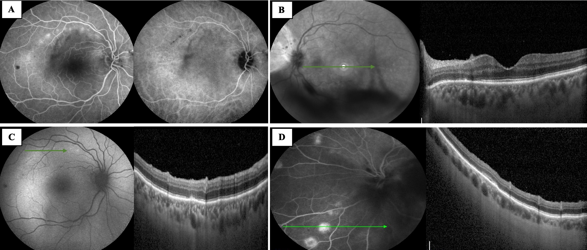
In this case, we present a young female with a continuous follow-up extending over a period of nine years where the lesions, probably due to HHT type 2, evolve over time. Her best-corrected visual acuity is unaffected at all time and there are no visual complaints despite the objective findings by fundus imaging, infrared images, OCT and OCTA verifying changes in the choriocapillaris and retinal pigment epithelial layers.
Whether the genetic variant influences on ocular changes is unknown. According to a study by Ines Gomez-Acebo e al. [4] ocular lesions are mainly associated with HHT 1 subtype. The patient in this case has the HHT 2 subtype, which does not correspond to the findings of Ines Gomez-Acebo e al. [4].
Earlier studies have found chorioretinal lesions in the eyes of elderly patients. To our knowledge chorioretinal changes have not been reported before in young patients. Sindhar et al. found no lesions in patients younger than the mean age (52 years), suggesting that the lesions may develop later in life [5].
Compared to conjunctival telangiectasias, chorioretinal findings are rare in patients with HHT. Although a study by Rinaldi et al. found 3 out of 8 patients with intraocular lesions described as widened and well-defined areas of choriocapillaris atrophy, and to a less extent involvement of the retinal pigment epithelium [6]. This could be the same type of lesion, seen in this case. In the study by Rinaldi et al. the average age of the patients with intraocular lesions is 60 years (range 57–62) and a visual acuity of (0.5—0.8). It has been suggested that an extension of the choriocapillary changes may lead to visual impairment [6].
Another case with similar changes reports retinal pigment modifications overlying choroidal ectatic vessels [7]. Here the authors suggest that the altered choroidal vessels could be the cause of changes in the retinal pigment epithelium, possibly owing to micro-excudation from the choriocapillaris layer.
Also, a case report by Mennel et al. [8] in 2005 reveals parafoveal telangiectasia in both eyes seen by fluorescein angiography in a 76- year old woman known with HHT. The authors conclude that a choroidal neovascularization (CNV) occurred secondly to the parafoveal telangiectasias in one eye. The CNV was treated with photodynamic therapy.
A study by Sindhar et al. [5], who examined eighteen patients having HHT with fluorescein angiography, found 83% of the patients with retinal alterations. The occurrence of ocular findings in these patients appears to be more frequent and as they suggest, the alterations might be over-looked with fundus photography.
A recent article by Abdolrahimzadeh et al. [9] discusses the different examination modalities of patients and that this may be a factor in the variation between the different frequencies of the retinal abnormalities found amongst studies. This aspect is also supported by Sindhar et al. [5] as they used both fluorescein angiography and OCTA in their study of HHT patients. They also found the highest occurrence of intraocular malformations (78%) and that is probably due to the fact of the different examination modalities [5, 9]. Ocular involvement in HHT patients is more common than assumed. Even though intraocular involvement is relatively rare. Conjunctival lesions seem to be more harmless, while the consequences of the intraocular lesions are unknown.
Our case is unique primarily because of the young age of the patient and because of the time spectrum. In this case, lesions develop over a long period in both eyes.
It is not possible to conclude from this case whether there is an indication for screening of this patient group. The disease is rare, the conjunctival affections are harmless and the consequences of the more uncommon retinal lesions are unknown.


留言 (0)