
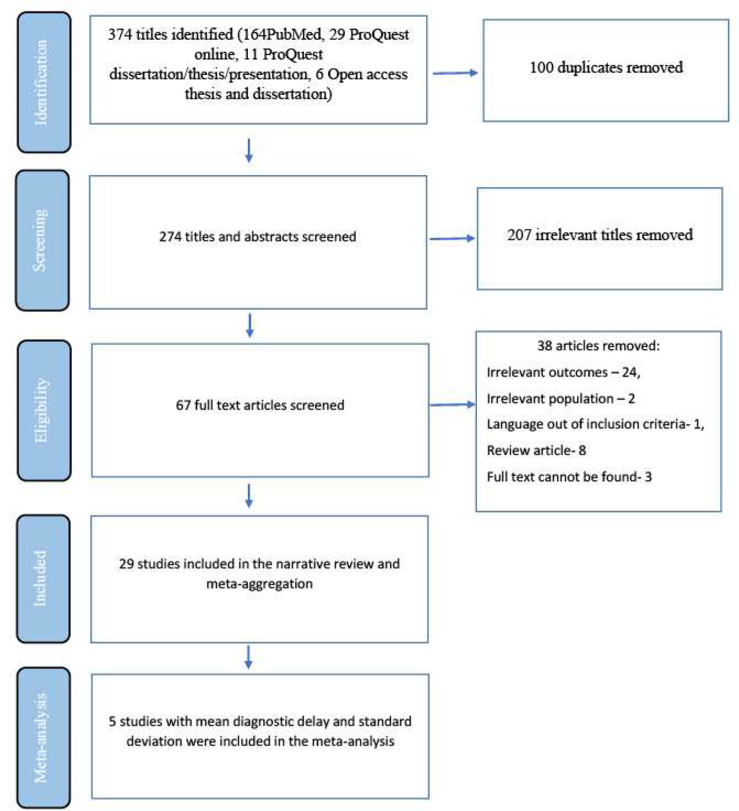
記住我


Poroshell Agilent 120 EC-C8 150 mm×2.1 mm, 4 μm (Agilent Technologies, Santa Clara, CA, USA) was chosen thanks to its capability to obtain higher, narrower and symmetrical peaks compared to the ones obtained with the other columns tested. In order to perform the analytes’ separation, the stationary phase was eluted with mobile phase A (water with 0.15% formic acid and 5 mM ammonium formate) and mobile phase B (acetonitrile:water 95:5 with 0.15% formic acid and 5 mM ammonium formate). The mobile phases were delivered in gradient mode as described in Table 1 in a total run of 14 min at a flow rate of 0.4 mL/min.
Table 1 Optimized gradient for LC-MS/MS analysis for the quantification of cysteamine in plasma.During the analysis, the samples were kept at 4 °C in the autosampler and the column oven was set at 30 °C. The injection volume was 3 µL. As shown in Fig. 1, the retention time of the analyte cysteamine and of the IS D6-cystine was 1.01 and 7.49 min respectively.
Fig. 1
Chromatogram showing the retention times of the analyte cysteamine (retention time = 1.01; blue line) and the IS D6-cystine (retention time = 7.49; red line).
MS conditionsThe m/z ratios of the precursor ions of cysteamine and of the IS used, D6-cystine, were already known from scientific literature; instead, the m/z ratios of product ions of each compound were set up through the product ion scan acquisition mode. Quantitative analysis was achieved with multiple reaction monitoring (MRM) scan mode in positive ionization. Among the three transitions selected for cysteamine, m/z = 61.000 was used to quantify and the others to confirm the analyte. Among the two transitions selected for D6-cystine, m/z = 131.300 was used to quantify and the other to qualify the IS. The compound dependent MS parameters were also optimized and were specified in Table 2 together with the m/z ratios
Table 2 Optimized MS parameters of the LC-MS/MS method related to the analyte or to the IS. The first transition of the product ion was chosen for the quantification and the other for the confirmation of the compound. DP declustering potential; EP entrance potential; CE collision energy; CXP collision cell exit potential. Selectivity and specificitySelectivity of the analytical method was assessed analyzing plasma samples from different healthy volunteers. No other compound, except for the analyte and IS, was detected in the run; therefore, the method proved to have specificity towards cysteamine and the IS used.
LinearityCalibration standards (2.5, 5, 10, 25, 50 µM) were analyzed in triplicate in three different days. The calibration curve was constructed plotting the areas of each concentration level corrected with the IS area versus the nominal values. QCs were also analyzed in each analytical run and the concentration were calculated comparing the ratio between the analyte peak area and the IS peak area with the calibration curve relation. Table 3 showed three calibration curves and the calculated concentration of the QCs analyzed with the same analytical runs.
Table 3 Peak areas and peak heights of analyte and IS referred to the calibrators and to the QCs with the corresponding calculated concentration. CAL calibrators. NA not available. Cps counts per second calculated concentration and accuracy SensibilityDilutions of the calibrator with the lowest concentration (2.5 µM) were carried out in order to identify the lowest concentration of analyte that the method is able to detect, corresponding to limit of detection (LOD), and able to quantify, corresponding to LLOQ. As reported in Table 4, testing the dilution 1:2 (1.25 µM), the concentration of cysteamine resulted to be detectable and quantifiable with an accuracy of 105%; this concentration can be considered the LLOQ. Instead, testing the dilution 1:10 (0.25 µM), the signal was detectable but was not quantified with a sufficient accuracy and precision; therefore, it was defined as the LOD. Chromatograms related to calibrator 1, LOQ and LOD respectively are shown in Fig. 2.
Table 4 Peak areas and peak heights of analyte and IS referred to the calibrator CAL1 (2.5 µM) and to its dilutions 1:2 (1.25 µM) and 1:10 (0.25 µM) with the corresponding calculated concentration and accuracy. ND not detectable. Cps counts per second. Fig. 2
Chromatograms obtained after injecting calibrator 1 (A), dilution 1:2 of calibrator 1 corresponding to LOQ (B) and dilution 1:10 of calibrator 1 corresponding to LOD (C)
Accuracy and precisionAccuracy and precision were evaluated both for calibrators and QCs; in particular QCs’ concentrations were calculated on the basis of calibration curve. Table 5 shows results of inter-day accuracy, and precision, represented by the coefficient of variation (CV%). The percentage of accuracy varied between 97.80 and 106.00% and CV% between 0.90 and 6.93%.
Table 5 Inter-day accuracy and precision of calibrators and QCs. Carry overCarry over was assessed by injecting blank samples after the calibrator with the highest analyte concentration and, as shown in Fig. 3, the signal derived was not greater than 20% and 5% of that of the analyte and of IS respectively.
Fig. 3
Chromatogram obtained after injecting a blank sample after the calibrator with the highest analyte concentration. The blue and red lines consist in respectively the signal along the chromatogram derived from the transition of cysteamine and of IS used to quantify
Stability of cysteamineThe stability of the 100 µM cysteamine in plasma was evaluated using the working solutions for the preparation of calibrators and QCs after 1, 2 and 3 months from its preparation. The solution was stored at -20 °C and was found to be stable up to 2 months at these conditions.
Application of the method to samplesCysteamine and intracellular cystine were measured in 4 patients suffering from nephropathic infantile cystinosis in order to test the applicability of the analytical method. As shown in Tables 6 and 7, the concentrations of plasmatic cysteamine and of intracellular cystine were in line with those expected for patients after 6 h since the oral administration of cysteamine bitartrate.
Our results confirmed previously reported data: cysteamine succeeds in lowering the concentrations of intracellular cystine below the recommended value of 1 nmol of hemicystine/mg of protein [9,10,11].
Table 6 Measurements of plasmatic concentrations of cysteamine in 4 patients suffering from nephropathic infantile cystinosis after 6 h since the administration of cysteamine bitartrate Table 7 Measurements of intracellular concentrations of cystine in 4 patients suffering from nephropathic infantile cystinosis after 6 h since the administration of cysteamine bitartratePearson correlation test did not evidence a linear relationship between the cystine and cysteamine concentrations (p = 0.142).
Furthermore, we tested also a healthy individual not affected by cystinosis and not undergoing treatment with cysteamine, as negative control. As shown in Fig. 4, no peak of the analyte cysteamine distinguishable from background noise was evidenced. The only peak present was that of the IS.
Fig. 4
Chromatogram obtained after injecting a sample derived from a healthy individual not affected by cystinosis and not undergoing cysteamine treatment. The blue and red lines consist respectively in the signal derived from the transition of cysteamine and of IS.
留言 (0)