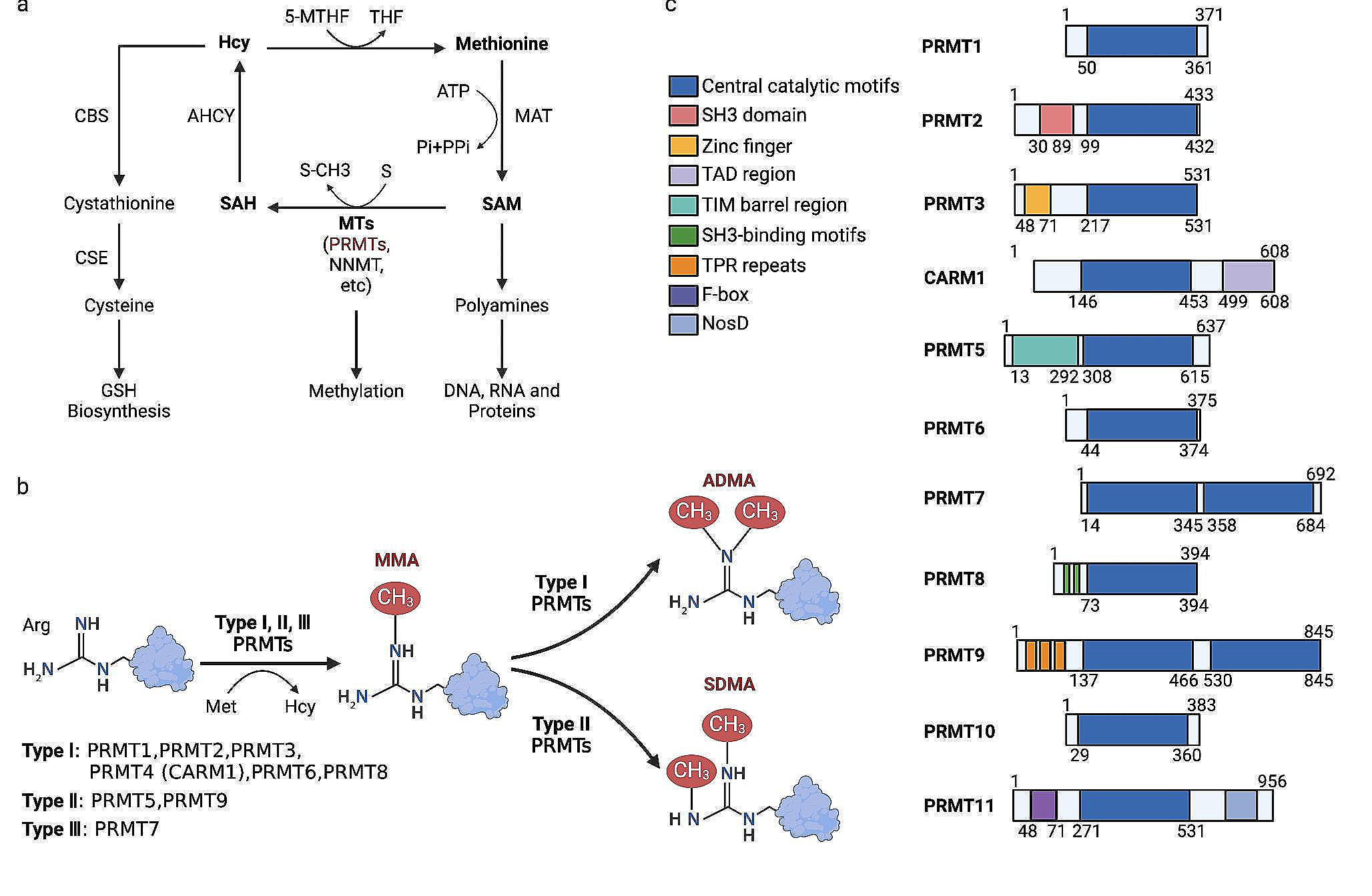
Cell culture and CoQ10 treatment
Human GBM U251 and human monocytic leukemia Thp-1 cells were obtained from ATCC. Human umbilical vein endothelial cells (HUVEC) were obtained from Lonza (C2517A). Cells were cultured at 37℃ and 5% CO2. U251 and Thp-1 were maintained in DMEM (#D5796; Sigma-Aldrich) and RPMI-1640 (#R8758; Sigma-Aldrich) respectively, with 10% fetal bovine serum (FBS) (#F4135; Sigma-Aldrich) and 1% antibiotic/antimycotic (#A5955; Sigma-Aldrich). HUVECs were grown in EBM-2 Basal Medium (CC-3156) and with EGM-2 SingleQuots Supplements (CC-4176). CoQ10 was generously provided by Kaneka Corporation. When indicated, cells were treated for 24 h with 5 µM CoQ10 or vehicle (ethanol; control).
Mice models
Xenograft and orthotopic implantation studies were performed with athymic nu/nu mice (Envigo). Mice were fed with autoclaved pelleted food and water ad libitum. Tumor generation, measurement, and processing were performed as previously described [15,16,17]. For xenograft, U251 cells mixed in Matrigel (1:1) (A1413201; Thermo Fisher) were injected subcutaneously into the flank of each mouse. Tumor volume was calculated every week using a digital caliper. After twelve weeks, animals were sacrificed, and the tumor samples were collected and processed (n = 7; 4 Vh & 3 CoQ10).
For orthotopic intracranial implantation, mice were anesthetized with an intraperitoneal injection of ketamine and xylazine. The animals were placed in a Kopf (Tujunga, CA) stereotaxic apparatus, and the skull trepanned at the injection spot into the striatum (0.5 mm in front of the Bregma, 2.1 mm lateral position, and 3 mm ventral position from the dura). U251 cells (3 × 10⁵ cells in a volume of 2 μL) were implanted intracranially using a micro syringe (10 μL NeurosModel 1701 RN, point style 4, SYR, Hamilton Co.). The animals were housed on a standard 12/12 h light/dark cycle at 21 °C with food and water ad libitum. Once the different models were established, the treatment with CoQ10 (100 mg/Kg mouse) or vehicle (saline solution) was performed intraperitoneal, every four days, starting on day seven. Tumor-bearing animals (n = 9; 4 Vh & 5 CoQ10) were sacrificed at week 9. Animal procedures followed European (Directive 2010/63/EU) and Spanish (RO 53/2013) legislation on the protection of animals used for scientific purposes. Experiments were previously approved by the Ethical Committee of Animal Research of the University of Castilla-La Mancha (PR-2012–6-03).
Immunofluorescence and immunohistochemical procedures
Immunohistochemistry and immunofluorescence were performed as standard protocols previously described [14, 18]. Images were captured through an LSM 710 Zeiss confocal microscope (Oberkochen, Germany) and an Eclipse TiU inverted microscope (Nikon, Tokyo, Japan). All data were processed and analyzed using ImageJ 1.53 software (National Institutes of Health [NIH], Bethesda, USA). Supplementary Materials and Methods provide a complete description of antibodies and methodology.
Hypoxia quantification
Hypoxia was measured using Hypoxyprobe (pimonidazole hydrochloride) as previously described [19]. Briefly, before the animals were sacrificed, they were injected with pimonidazole, a compound that forms adducts in hypoxic regions, which can then be examined by fluorescence microscopy by immunostaining. Once mice were sacrificed, tissues were processed, and the adducts generated by hypoxia were imaged by confocal microscopy or a Cytation 5 cell multimode reader and quantified with ImageJ.
Tumor volume estimation
The area of the primary tumor and infiltration was determined through vimentin immunostaining. Volumetric values were obtained using serial cuts, applying the Cavalieri estimator method [20]. The same approach was used to calculate the maximum volume of infiltration. A full description of the methodology is provided in Supplementary Materials and Methods.
Proteomics
Proteomic studies were performed as previously described [17]. The proteome was assessed in a RP-LC–MS/MS using an Easy-nLC II system coupled to a LCQ Fleet ion trap mass spectrometer (Thermo Scientific). Peptides were detected in survey scans from 400 to 1600 amu (1 μscan), followed by three data-dependent MS/MS scans (Top 3), using an isolation width of 2 mass-to-charge ratio units. Clustering and paired analysis for changes in protein levels were assessed with the free software MEV 4.9.
Immunocytochemistry
Cells were treated for 24 h with 5 µM CoQ10 or vehicle (ethanol). Then cells were fixed for 15 min in 4% paraformaldehyde (PFA), blocked, and stained with different primary antibodies (full description in Supplementary Materials). Primary antibody binding was detected using secondary antibodies conjugated with Alexa 488 (goat anti-mouse A32723; goat anti-rabbit A32731; rabbit anti-goat A-11078, Life Technologies) and Alexa 594 (goat anti-mouse A11005; goat anti-rabbit A11012; Life Technologies). Images were captured through an Eclipse TiU inverted microscope (Nikon, Tokyo, Japan). All data were processed and analyzed using ImageJ 1.53 software (NIH, Bethesda, USA).
Western blotting
Western blot was performed as previously described [10]. Briefly, cells were treated for 24 h with 5 µM CoQ10 or vehicle (ethanol). Then, cells were washed with PBS and lysed in RIPA buffer or SDS-DTT buffer. Lysates were obtained by centrifugation and quantified using BCA protein assay kit (71,285-M; Merck). Equal amounts of protein were separated using 10% or 12% acrylamide gel, and the proteins were transferred to a nitrocellulose membrane. Then, membranes were incubated with primary antibodies (see Supplementary Materials). After that, membranes were washed and incubated with secondary antibodies goat anti-rabbit (P0448; Dako), goat anti-mouse (P0447; Dako), or rabbit anti-goat (P0449; Dako). The relative density of the immunoreactive bands was analyzed using ImageJ 1.53 software (NIH, Bethesda, USA).
Functional metabolic assays
Oxygen consumption rate (OCR) and extracellular acidification rate (ECAR) were quantified using a Seahorse XFP analyzer (Seahorse Biosciences) as previously described [21, 22]. Briefly, cells were seeded in XFp miniplates and treated for 24 h with 5 µM CoQ10 or vehicle (ethanol). Cells were incubated with Seahorse XFp base medium for 60 min at 37ºC without CO2 before loading into the analyzer. To study mitochondrial respiration, OCR was measured under different conditions. Three baseline OCR values were obtained during the first 20 min, after which the different mitochondrial inhibitors were added (oligomycin, 1 μM; carbonyl cyanide-p-trifluoromethoxyphenylhydrazone [FCCP], 0.3 μM; antimycin A and rotenone, 1 μM). Finally, 1 µg/mL Hoechst 33,342 was added, and the total number of cells per well was quantified using a Cytation 5 (Biotek). Respiration parameters were calculated using Wave 2.6 software. A similar protocol was followed for glycolysis, although incubating the cells for 60 min in Seahorse XFp medium base without glucose. Three baseline ECAR measurements were taken for each well within the first 20 min, and glucose (10 mM), oligomycin (1 μM), 2-deoxyglucose (50 μM), and Hoechst 33,342 (1 µg/mL) were subsequently injected for the evaluation of the different parameters. Results were normalized against the total number of cells per well.
Boyden chamber/invasion assay
Cell invasion assay was performed using a Boyden chamber with polycarbonate Matrigel-coated membranes, as previously described [15]. Briefly, after treatment, cells were plated into the upper well of the chamber in FBS-free medium, while the lower chamber was filled with FBS-supplemented medium. After 24 h, migrated cells were fixed with 4% PFA; images were acquired using fluorescence microscopy (Nikon TiU) and then analyzed using ImageJ 1.53 software (NIH, Bethesda, USA).
Matrix degradation
Cells were treated for 24 h with 5 µM CoQ10 or vehicle (ethanol). Then, cells were transferred to coverslips coated with Oregon Green 488-labeled gelatin (G13186; Thermo Fisher), as previously described [23]. After 24 h, coverslips were fixed with 4% PFA and stained with Actin-red (R37112; Thermo Fisher) and DAPI solution. Images were acquired using a Nikon TiU microscope and analyzed using ImageJ 1.53 software (NIH, Bethesda, USA).
Quantification of MMPs Activity
EnzChek Gelatinase/Collagenase Assay Kit (E12055; Thermo Fisher) was used to determine MMP 2/9 proteolytic activity. For this purpose, the cells were treated with CoQ10 or vehicle (ethanol) for 24 h, after an incubation of 16 h in the dark at 37℃, and a reaction solution with DQ gelatin (E12054; Thermo Fisher) and enzyme (purified collagenase type IV from Clostridium histolyticum) was added. Further, determination by fluorimetry (495/515) was also carried out. Results show the ratio of collagenase/gelatinase activity against control cells.
Cell tube formation
U251 cells were treated for 24 h with 5 µM CoQ10 or vehicle (ethanol; control) in 24-well plates. Conditioned cells medium was transferred to HUVEC cells plated onto GFR Matrigel-precoated 96-well plates. Cells were incubated for 6 h with a conditioned medium, and then the number of cell tubes was quantified using phase-contrast microscopy.
Angiogenesis array
For the determination of soluble inflammatory and angiogenic factors, the Human Angiogenesis Array Q2 was used (QAH-ANG-2–1; RayBiotech). The assay was conducted under manufacturer conditions. A full description of the methodology is provided in Supplementary Materials and methods.
THP1- HUVEC adhesion
HUVEC cells were incubated with conditioned media of U251 cells treated with vehicle (control) or CoQ10. Thp-1 monocytes loaded with Calcein-AM (#C34852; Thermo Fisher) were added to HUVEC monolayers. After a 24-h incubation period, unattached monocytes were removed, and endothelium-adhered monocytes (green) were quantified using fluorescence microscopy (Nikon Ti-U) and analyzed with ImageJ.
THP-1 transmigration assay
Thp-1 cells were incubated with conditioned media of U251 cells treated with vehicle (control) or CoQ10. Then, a Boyden chamber with untreated polycarbonate membranes was used. The conditioned cells were added to the upper chamber and incubated for 24 h. Then, Thp-1 monocytes that crossed the membrane were fixed, marked with DAPI, quantified using fluorescence microscopy (Nikon TiU) and analyzed with ImageJ.
Differentiation of THP-1 to monocytes
Human Thp-1 monocytes were incubated with conditioned media from U251 cells treated with vehicle (ethanol) or CoQ10. After 7 days, the cells in suspension were removed, and those that had adhered and differentiated to macrophages were fixed, marked with DAPI, quantified by fluorescence microscopy (Nikon TiU), and analyzed with ImageJ.
Statistical analysis
Different statistical analysis was performed using GraphPad Prism 7 software (GraphPad). Data are expressed as mean ± SEM. When comparing two groups, P values were calculated using two-tailed Student’s t-tests or a one-way ANOVA (Kruskal–Wallis’ test). Differences were considered significant at p < 0.05 (*, P < 0.05; **, P < 0.01; ***, P < 0.001; ****, P < 0.0001). Additional methods are described in the Supplementary Materials and Methods.


留言 (0)