
記住我


Following major joint arthroplasty, the moderate-to-severe pain many patients experience is frequently treated with opioids with their associated side effects and risk of misuse, dependence, and diversion. Neuromodulation is an analgesic alternative with few associated limitations.1 Percutaneous leads inserted under ultrasound guidance and subsequently attached to an external pulse generator provide postoperative analgesia for outpatient surgery2 and possibly knee arthroplasty.3 However, this technique requires physician-level skills, advanced equipment, and up to an hour to administer, targets only 1 nerve or plexus distribution per lead, and is—at least at the time of this writing—often cost-prohibitive.1
An alternative is percutaneous “auricular” neuromodulation involving the stimulation of nerves in and around the ear (Figure 1). The mechanism of action is complex and multifactorial, and remains under investigation,4,5 but undoubtably involves modulation of serotonergic, noradrenergic, and endorphinergic pathways with associated release of serotonin, norepinephrine, and endogenous opioids such as beta-endorphins.6 Auricular vagal stimulation further chemically modulates nociceptive (pain) processing, anxiety, and depression.
 Figure 1.:
 A percutaneous auricular nerve stimulation system (NSS-2 Bridge, Masimo). Each of the 3 electrodes has a 2-millimeter-long integrated needle/lead (inset), and the ground electrode has four 2-millimeter-long integrated needles—also termed “leads” (inset). Used with permission from Brian M. Ilfeld, MD, MS.
The United States Food and Drug Administration (FDA) has cleared a percutaneous auricular neuromodulation device to reduce symptoms associated with opioid withdrawal for up to 5 days (near-field stimulator system 2 [NSS-2] Bridge, Masimo; Figure 2).7–9 Three small nonrandomized studies suggest that this device may also provide “analgesia” in hospitalized patients following abdominal and pelvic surgeries.10–12 The device has multiple beneficial features, such as being small, disposable, medication-free, nonsurgical, and battery-powered. It has few contraindications, lacks systemic side effects and associated serious adverse events, is relatively simple to apply, and requires no additional equipment or advanced training. Finally, it has no potential for misuse, dependence, or diversion, and is a fraction of the cost relative to ultrasound-guided percutaneous neuromodulation devices.
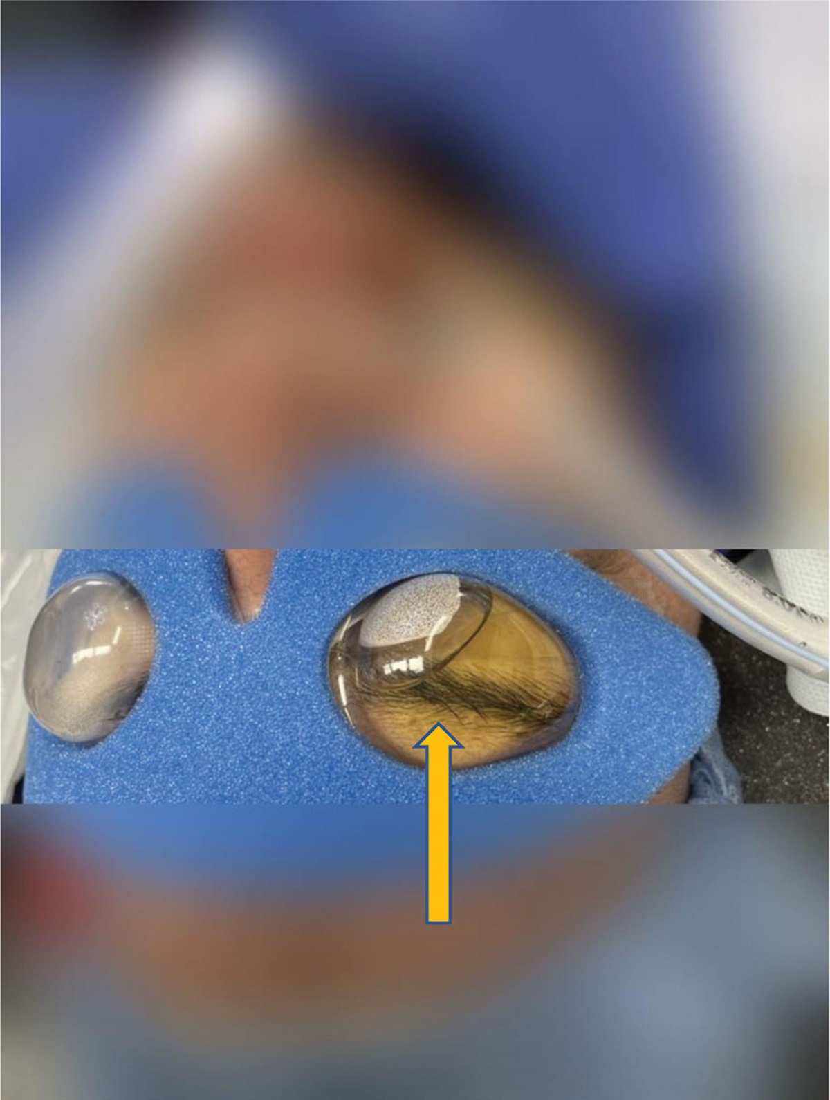
 Figure 2.:
 A percutaneous auricular nerve stimulation system (NSS-2 Bridge, Masimo). The pulse generator is adhered directly to the patient behind the ear over the mastoid process. Leads are placed: (1) at the most cephalad portion of the antihelix, (2) immediately cephaloanterior to the incisura and posterior to the superficial temporal arterial pulse, and (3) on the posterior ear opposite the antihelix at the level of the incisura. The ground electrode is inserted on the anterior side of the lobule (ear lobe). Used with permission from Baharin Abdullah, MD.
However, it remains unexamined whether percutaneous auricular nerve stimulation will provide analgesia following knee and hip arthroplasties and if patients will accept the device following discharge (including home removal). We now report 5 off-label cases to explore the possibility of treating pain following knee and hip arthroplasty with percutaneous auricular neuromodulation.
CONSENT FOR PUBLICATIONThe University of California, San Diego, Institutional Review Board, waives the review of case reports and short series. Written, informed consent documenting Health Insurance Portability and Accountability Act authorization and allowing for publication of nonidentifying medical information and in situ device imaging in the form of a case series were obtained from all patients.
CASE DESCRIPTIONFive patients undergoing unilateral, primary, total knee (n = 3) and hip (n = 2) arthroplasties were offered and consented for postoperative administration of percutaneous auricular neuromodulation. Preoperatively, patients having knee arthroplasties received an ultrasound-guided, single-injection adductor canal block with ropivacaine 0.5% and epinephrine (20 mL). Intraoperative anesthesia consisted of a bupivacaine subarachnoid block for all but 1 patient who preferred a general anesthetic. A mixture of bupivacaine 0.25% (50 mL), ketorolac (30 mg), and epinephrine (250 μg) was infiltrated throughout the surgical area intraoperatively for all cases. Following surgery, in a semirecumbent position, each patient received intravenous fentanyl 25 µg, and the application locations were wiped with an alcohol pad and benzoin over the mastoid process for the pulse generator placement and at the 4 points of electrode placement (Figure 2).
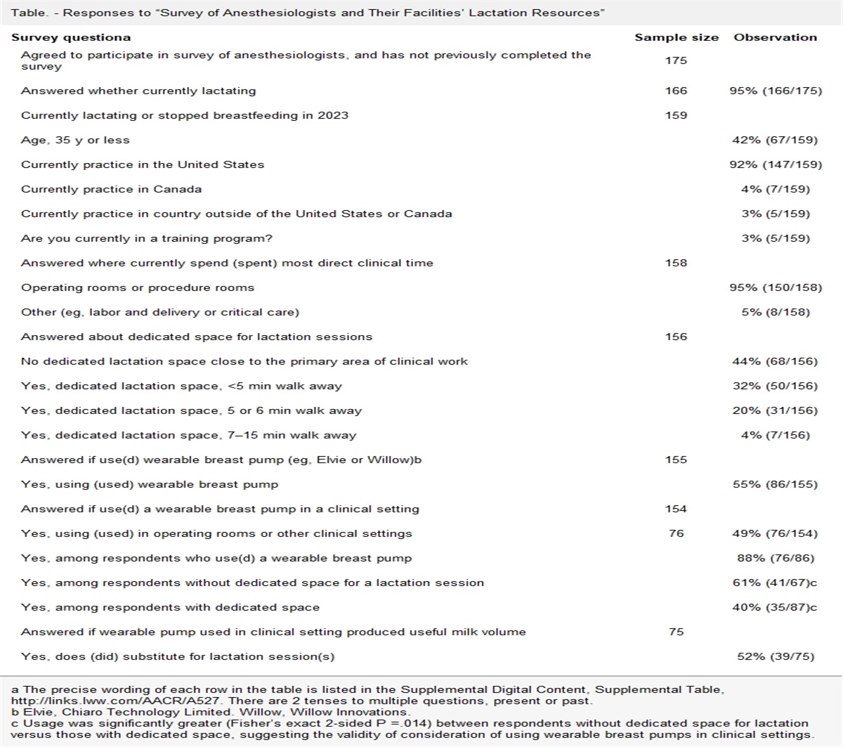
The pulse generator was applied posterior and slightly caudad to the preferred ear (contralateral to the side on which the patient slept) with a double-sided adhesive pad, which was further secured with a clear adhesive dressing. Each of 3 electrodes and a ground has a 2-millimeter-long integrated needle(s) and was affixed with a small, round adhesive bandage (Figure 2). Specific lead locations on the outer ear were guided with transillumination to optimize effects and avoid placement into neurovascular bundles, which can cause pain and bleeding. Neurovascular bundles were identified by placing an included pen light against the skin and viewing the opposite side of the ear. For the most cephalad anterior electrode and electrode on the posterior side of the ear, the needles were inserted 1 to 2 mm away from a neurovascular bundle and never immediately opposite to each other.
The first lead was placed at the most cephalad portion of the antihelix by simply pressing the electrode directly into the skin and affixing with an overlying dressing (Figure 2). The second electrode was inserted immediately cephaloanterior to the incisura and posterior to the superficial temporal arterial pulse. The third electrode was inserted on the posterior ear opposite to the antihelix at the level of the incisura. The ground electrode with four 2-millimeter-long integrated needles was inserted on the anterior side of the lobule (Figure 1, inset). All patients tolerated the procedure without wincing or complaint, and total duration for each application was approximately 5 minutes.
Postoperatively, patients received acetaminophen 975 mg 4 times daily, celecoxib 200 mg twice daily, and, if needed, the synthetic oral opioid oxycodone (5 mg tablets). Patients were instructed to keep the pulse generators and leads dry with the use of a shower cap when bathing.
The 4 women and 1 man had a mean (standard deviation) age of 71 (3) years, height of 159 (9) centimeters, weight of 72 (12) kilograms, and body mass index of 28.5 (3.4). During the device use through postoperative day 5, average daily pain at rest and while moving was a median of 0 to 2 as measured on the 0 to 10 numeric rating scale (Figure 3). Maximum daily pain was a median of 2 to 4, and median daily oxycodone requirements were 0 to 2.5 mg during this same time period (Figure 3). Most patients perceived various periauricular sensations during the first 24 hours, but rarely after postoperative day 1. The sensations were described as “warmth,” a soft “thumping,” or “pulsing,” which were never disturbing.
 Figure 3.:
 Pain and opioid consumption following total knee and hip arthroplasty with percutaneous auricular nerve stimulation for the first 5 postoperative days. Each circle represents 1 patient, and the median for each time point is denoted with a horizontal line.
The pulse generators automatically ceased functioning after 120 hours (5 days), and patients or their caretakers then detached the device by first removing the round bandage of the grounding electrode, which extracted the electrode from the patient along with the bandage. The remaining 3 electrodes were subsequently removed in the same manner followed by the pulse generator, after which the single-use, disposable device was discarded. Average pain scores and opioid consumption were similar or just slightly greater following device removal on postoperative day 5 (patients were followed through postoperative day 8), although worst pain scores did increase to a greater degree (Figure 3). No device-related localized irritation, systemic side effects, or complications were identified.
DISCUSSIONThese cases demonstrate that percutaneous auricular neuromodulation following major orthopedic surgery is feasible during both the inpatient and outpatient portions of the first postoperative week and may be an effective analgesic enabling decreased opioid consumption considering the relatively low pain scores and opioid use of the current patients to historic patterns at our institution. Moreover, the device used in this report is FDA-cleared to reduce symptoms associated with opioid withdrawal, which includes anxiety, insomnia, muscle aches, nausea, and vomiting, all of which are frequent following surgery.7–9
Other percutaneous auricular neuromodulation devices have been used to treat pain following various surgical procedures with various degrees of success, from decreased pain to no effect and even “increased” pain.13,14 Pulse duration, frequency, amplitude, duty cycle, and additional parameters such as the number and location of electrodes vary greatly among devices. These parameters determine the properties of the generated electric field. Therefore, differing devices can have considerably different physiologic effects. This inconvenient reality dramatically decreases generalizability of the results from any 1 clinical trial to other devices. The pulse generator of the current report uses an integrated 3-volt battery and has a load impedance range of 1k to 10k Ω with 3.2 volt maximum, and symmetrical, biphasic stimulation cycles occur at a frequency of 0.125 Hz with periodic rest.
Importantly, multiple studies demonstrate that neurologic effects of auricular stimulation outlast the stimulation itself,15 which is why we chose to follow these 5 patients for a total of 8 days. Indeed, the patients in the current report experienced little increased pain and opioid requirements following removal on postoperative day 5.
The auricular neuromodulation device described in this report has few contraindications listed on its label: (1) use of cardiac pacemakers, (2) hemophilia, and (3) psoriasis vulgaris. In addition, the skin where the leads are applied should be intact. The only reported complications have been minor skin bleeding (0.91%) and dermatitis from the adhesive bandages (0.91%).9 For the pivotal studies of the device to reduce the symptoms of opioid withdrawal (n = 1207), no analgesic was administered for electrode placement, and only 2 participants complained of “significant” pain (0.17%).9
These cases demonstrate that percutaneous auricular neuromodulation is feasible for knee and hip arthroplasties and may be an effective analgesic enabling decreased opioid consumption both during hospitalization and at home following discharge. Considering the ease of placement, few contraindications, applicability to any anatomic surgical location, low patient and provider burden, lack of systemic side effects and serious adverse events, as well as no misuse, dependence, or diversion potential, further study with a randomized, controlled trial appears warranted to document and quantify potential analgesic and opioid-sparing benefits.
DISCLOSURESName: John J. Finneran IV, MD.
Contribution: This author helped develop the intervention pathway, identify and contact appropriate patients, administer the intervention, write the initial manuscript, revise the manuscript, and approve the final draft.
Name: Engy T. Said, MD.
Contribution: This author helped develop the intervention pathway, identify and contact appropriate patients, administer the intervention, revise the manuscript, and approve the final draft.
Name: Scott T. Ball, MD.
Contribution: This author helped introduce patients to the intervention, follow patients, revise the manuscript, and approve the final draft.
Name: Krishna R. Cidambi, MD.
Contribution: This author helped introduce patients to the intervention, follow patients, revise the manuscript, and approve the final draft.
Name: Baharin Abdullah, MD.
Contribution: This author helped assist in patient management, revise the manuscript, and approve the final draft.
Name: Brian M. Ilfeld, MD, MS (Clinical Investigation).
Contribution: This author helped identify the intervention, identify and contact appropriate patients, follow patients, collect data, write the initial manuscript, and approve the final draft. The University of California San Diego has received funding and/or product from the following companies for other research studies of the authors: Epimed International (Farmers Branch, TX), SPR Therapeutics (Cleveland, OH), Infutronix (Natick, MA), and Avanos Medical (Irvine, CA).
This manuscript was handled by: BobbieJean Sweitzer, MD, FACP.
REFERENCES 1. Ilfeld BM, Finneran JJ. Cryoneurolysis and percutaneous peripheral nerve stimulation to treat acute pain. Anesthesiology. 2020;133:1127–1149. 2. Ilfeld BM, Plunkett A, Vijjeswarapu AM, et al.; PAINfRE Investigators. Percutaneous peripheral nerve stimulation (neuromodulation) for postoperative pain: a randomized, sham-controlled pilot study. Anesthesiology. 2021;135:95–110. 3. Ilfeld BM, Ball ST, Gabriel RA, et al. A Feasibility study of percutaneous peripheral nerve stimulation for the treatment of postoperative pain following total knee arthroplasty. Neuromodulation. 2019;22:653–660. 4. Kaniusas E, Kampusch S, Tittgemeyer M, et al. Current directions in the auricular vagus nerve stimulation I - a physiological perspective. Front Neurosci. 2019;13:854. 5. Babygirija R, Sood M, Kannampalli P, Sengupta JN, Miranda A. Percutaneous electrical nerve field stimulation modulates central pain pathways and attenuates post-inflammatory visceral and somatic hyperalgesia in rats. Neuroscience. 2017;356:11–21. 6. Lockard JS, Congdon WC, DuCharme LL. Feasibility and safety of vagal stimulation in monkey model. Epilepsia. 1990;31(suppl 2):S20–S26. 7. Miranda A, Taca A. Neuromodulation with percutaneous electrical nerve field stimulation is associated with reduction in signs and symptoms of opioid withdrawal: a multisite, retrospective assessment. Am J Drug Alcohol Abuse. 2018;44:56–63. 8. Qureshi IS, Datta-Chaudhuri T, Tracey KJ, Pavlov VA, Chen ACH. Auricular neural stimulation as a new non-invasive treatment for opioid detoxification. Bioelectron Med. 2020;6:7. 9. Roberts A, Sithole A, Sedghi M, Walker CA, Quinn TM. Minimal adverse effects profile following implantation of periauricular percutaneous electrical nerve field stimulators: a retrospective cohort study. Med Devices (Auckl). 2016;9:389–393. 10. Chelly JE, Monroe AL, Planinsic RM, Tevar A, Norton BE. Auricular field nerve stimulation using the NSS-2 BRIDGE((R)) device as an alternative to opioids following kidney donor surgery. J Complement Integr Med. Published online Nov 1, 2021. doi: 10.1515/jcim-2021-0208. 11. Ahmed BH, Courcoulas AP, Monroe AL, Gourash WF, Chelly JE. Auricular nerve stimulation using the NSS-2 BRIDGE device to reduce opioid requirement following laparoscopic Roux-en-Y gastric bypass. Surg Obes Relat Dis. 2021;17:2040–2046. 12. Lim G, LaSorda KR, Monroe AL, Chelly JE. Auricular percutaneous nerve field stimulator device as alternative therapy for cesarean delivery analgesia: proof of concept. Can J Anaesth. 2019;66:1522–1523. 13. Wigram JR, Lewith GT, Machin D, Church JJ: Electroacupuncture for postoperative pain. Physiotherapy Pract. 1986; 2:83–88 14. Sator-Katzenschlager SM, Michalek-Sauberer A. P-Stim auricular electroacupuncture stimulation device for pain relief. Expert Rev Med Devices. 2007;4:23–32. 15. Sator-Katzenschlager SM, Scharbert G, Kozek-Langenecker SA, et al. The short- and long-term benefit in chronic low back pain through adjuvant electrical versus manual auricular acupuncture. Anesth Analg. 2004;98:1359–1364.
留言 (0)