Airway assessment can be used to quantify the morphological features of an upper airway with complicated geometry, which is a key step prior to performing general anesthesia. Complications associated with improper airway management are the most common cause of morbidity and mortality [16]. As such, difficulties in establishing a clinical airway are an ongoing challenge for anesthesiologists [2]. Clinical practice guidelines recommend specific strategies to ensure patient safety during the management of difficult or unexpected airway. However, no gold standard method has been published [17]. Visibility-enhancing applications, such as fiberoptic bronchoscopy and video laryngoscopy, have greatly reduced the incidence of intubation challenges of difficult airway during the perioperative period. However, none of these techniques can fully eliminate the risk. Image-based 3D models developed in this study display the anatomical structure in detail, and thus provide a novel cutting-edge digital technology to detect potential risks of difficult airway.
Rapidly developing medical imaging techniques, such as ultrasound, computed tomography (CT), and MRI, that generate images of internal organs and tissues have significantly improved clinical diagnoses. Kelly et al., [18] created a resin model of the nasal cavity based on computerized x-ray images to study the distribution of the gas-flow field within the nasal cavity. Chun et al., [19] developed an MRI-based kinetic model for the upper airway of rats to investigate muscle characteristics, airway shape, and anatomical structure to be applied to patients with obstructive sleep apnea (OSA). Yu et al., [20] created a 3D finite element model of airway filling based on spiral CT images of healthy individuals and patients with obstructive sleep apnea–hypopnea syndrome (OSAHS) using a surface rendering method. Using this model, the original shape of the upper airway was accurately preserved, and the airflow of the entire respiratory cavity was numerically simulated using finite element analysis. Zhou et al., [21]reconstructed a 3D model of the upper airway of patients with small-jaw deformity and OSAHS and further measured the minimum cross-sectional area of the upper airway-related sagittal, cross-sectional, and coronal planes. Upper airway stenosis of patients with OSAHS was mainly detected in the sagittal plane, with the most marked stenosis in the pharyngeal segment. Liu et al., [22]constructed an upper airway 3D finite element model based on spiral CT data from 10 patients with OSA and performed fluid dynamic simulation to assist clinical diagnosis and treatment.
However, despite these advances, there are currently only a few published studies addressing finite element analysis of the upper airway. Fan et al., [23] used 3D CT to observe changes in the intrinsic oral volume based on the tongue position in patients with a difficult airway and found an increased volume ratio between the front of the tongue before and after sticking out the tongue. Most of the current research is based on CT scans, which are insufficient for identifying airway soft tissue and other structures. Therefore, in this study we used MRI imaging to create an upper airway filling 3D model in which soft tissues can be clearly identified.
The morphological data measured in the developed model agree with published studies [17]. The model precisely depicted detailed morphological structure and further investigated the anatomical specificity of the difficult airway group. Volumetric values of the upper airway changed with variation in body positions. Volume increased in both normal and difficult airway as patients moved from the supine to maximum elevation position. Difficult airway showed a relatively low increase in volume rate. These findings agree with a published study addressing different intubation oropharyngeal models [24].
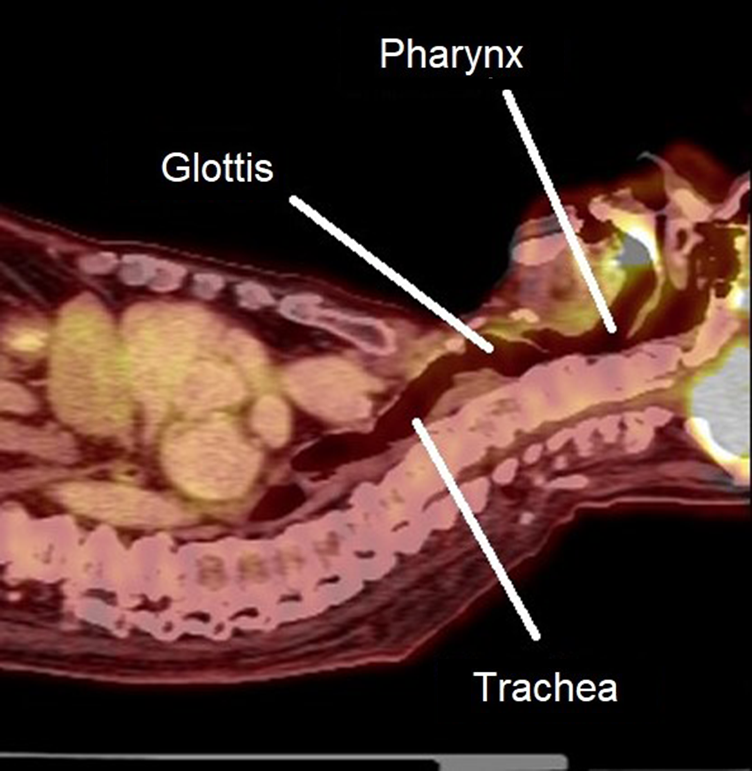
The special anatomical features of the upper airway technically support our results. The muscular structures in the oropharynx (e.g., soft palate, tongue, PA) play a key role in supporting the upper airway due to the absence of bony structures. Therefore, the airway opening is controlled by not only bony structures, such as the mandible and teeth, but also relevant soft tissues. Anatomical positions of those tissues are movable with variations in body position. The increased volume of the upper airway favored the placement of the laryngoscope to expose the glottis and smooth insertion of the endotracheal tube during endotracheal intubation.
Morphological data of the sagittal plane play a key role in assessing the potential occurrence of a difficult airway. Morphological datasets on three projection planes proposed in this study quantified the irregular geometry of an upper airway. Technically, four sets of geometric data—maximum longitudinal radial distance, maximum transverse radial distance, surface area, and corner angles—were associated with volumetric changes. Those data showed marked changes on the sagittal planes with respect to variation in body position, while no obvious changes were present in the coronal and horizontal planes. It should be noted that an upper airway filling 3D model can display an irregular geometric shape. We simplified the geometric expression for the 3D model to four sets of data in our analysis. Similar methods have been used in a knee meniscus model [14, 15]. The above-mentioned results demonstrated that the morphological variation of the upper airway in the sagittal plane was an appropriate indicator of a difficult airway when a patient moves from the supine position to maximum elevation. Image-based morphological assessments can initially screen patients with a high risk of a difficult airway and can further provide comprehensive evaluation using indicators such as the Mallampati score, Cormack-Lehane classification, and Wilson’s score, as well as physical features like mouth opening less than 30 mm, TMD less than 60 mm, and restrictions of neck flexion and extension.
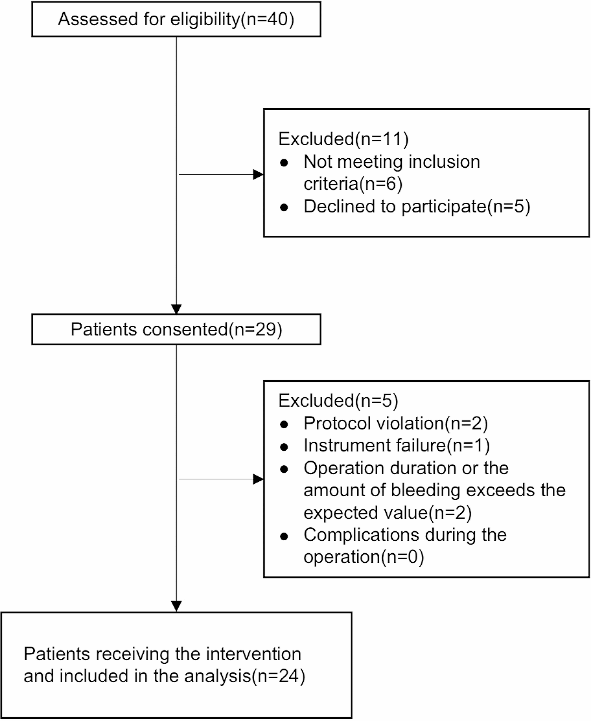
Several limitations of this study should be mentioned. First, the technique in our study indicated free volume in the airway, but this volume also varied when the soft tissue was "crushed" by the intubation tool. Secondly, statistical significance of dimensional changes may be difficult to correlate with clinical correlates of intubation or ventilation difficulties because there is no reliable and accurate method to evaluate and predict difficult airway at present. Thirdly, this study only included a single group of patients, and thus these results may not apply to patients with trauma, head and neck pathologies, or prior difficulty. Moreover, this was a retrospective study with a small patient sample size. Large sample multi-center and randomized controlled trials should will be conducted in future studies to better inform actual clinical application. Finally, the use of this digital assessment technique in daily practice is limited, which was mainly used to evaluate the impact of position-specific morphological changes in difficult airway difficult airway when difficult intubation is highly suspected or in cases of compressive tumor airway surgery.


留言 (0)