
記住我


This study was a hospital-based, case–control study conducted at the Third Affiliated Hospital of Kunming Medical University. Patients with cervical cancer (International Classification of Diseases version 10, ICD-10 C53) and patients with benign gynecologic diseases (ICD-10 D25-28, N70-73, N80-85), as controls, were both newly diagnosed and treated from January 2015 to December 2017. A total of 4648 patients with cervical cancer and 1002 benign disease controls were identified consecutively during the study period. The exclusion criteria for cervical cancer cases were as follows: (1) without a blood test for HBV and HCV infection before treatment; (2) with HIV infection; (3) previous or synchronous malignant tumors at another organ site; (4) a specific pathological type other than squamous cell carcinoma, adenocarcinoma and adenosquamous carcinoma, such as cervical neuroendocrine carcinoma, carcinosarcoma, adenosarcoma, lymphoma, or melanoma; (5) without a test result for HPV infection at initial diagnosis; (6) non-Chinese residency. The controls comprised patients who were pathologically confirmed to have benign gynecologic diseases that were considered to be unrelated to HBV infection, mainly including uterine leiomyoma, endometriosis, mature teratoma, ovarian cystadenoma and cysts. The exclusion criteria for controls were the same as cases, except for the cancer diagnosis. Finally, there were 3748 qualified participants among the cervical cancer cases and 838 qualified participants among the benign disease controls. The list of pathologic diagnoses of benign disease is shown in Additional file 1: Table S1. To balance the baseline characteristics between the cases and controls, cases and controls were frequency matched at a ratio of 1:1 by age, ethnicity, and place of birth. 838 cases were selected for the controls to build a 1:1 case–control study (Fig. 1). Relevant information, such as cigarette smoking, alcohol drinking, family history of cancer, number of full-term pregnancies, and BMI was collected from detailed medical records reviews for both cases and controls.
Fig. 1
Patient flow chart. HBV, hepatitis B virus; HCV, hepatitis C virus; HPV, human papillomavirus
The study was conducted in accordance with the Declaration of Helsinki and was approved by the Ethics Committee of Yunnan Cancer Hospital (NO. KYLX2022033). Individual consent for this retrospective analysis was waived.
Serological assaysVenous blood was collected from both cases and controls before treatment. Serum was separated and examined for the presence of HCV antibodies (anti-HCV), HBV serological markers (including hepatitis B surface antigen (HBsAg), hepatitis B surface antigen (anti-HBs), and hepatitis B e antigen (HBeAg)), antibodies to the hepatitis B e antigen (anti-HBe), antibodies to the hepatitis B core antigen (anti-HBc), and antibodies to HIV, using enzyme-linked immunosorbent assays (WANTAI BioPharm, Beijing, China). Quality control for the measurements was performed in accordance with the protocols provided by the manufacturer.
Clinical significance of hepatitis virus infection-related antigens and antibodiesThe serological markers of HBV included HBsAg, anti-HBs, HBeAg, anti-HBe, and anti-HBc. HBsAg is a hallmark of infection, including chronic HBV infection and inactive HBsAg carrier state. Anti-HBs positivity represents acquired immunity to HBV infection, and might result from both current or prior HBV infection and vaccination. HBeAg and anti-HBe were used to show viral replication and infectivity. Anti-HBc appears 1–2 weeks after the appearance of HBsAg and is a serum marker for current or prior HBV infection [32]. HBV infection status was divided into three categories according to the status of HBsAg and anti-HBc. Individuals that were both HBsAg-negative and anti-HBc-negative were defined as never exposed to HBV; those that were HBsAg-positive and anti-HBc-positive were defined as chronic carriers of HBV; those that were HBsAg-negative and anti-HBc-positive were defined as having past exposure to HBV [17]. Individuals who were positive for anti-HCV antibodies were defined as having chronic HCV infection [33].
HPV-DNA testFor patients from both the case and control groups, before a biopsy and pelvic examination, the surface of their cervix or vaginal canal were gently rubbed several times for DNA collection using a brush. Either a Digene Hybrid Capture 2 (HC2) High-Risk HPV DNA Test (Qiagen corporation, Hilden, Germany) or a quantitative real-time polymerase chain reaction (qPCR)-based test (BioPerfectus, Jiangsu, China) was used to evaluate the presence of HPV DNA. The HC2 test detects the presence of 13 high-risk HPV types (16/18/31/33/35/39/45/51/52/56/58/59/68). The qPCR test detected 18 high-risk HPV types (16/18/31/33/35/39/45/51/52/56/58/59/68/53/66/73/26/82).
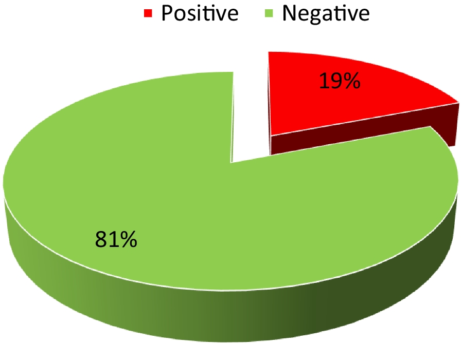
Immunohistochemical staining for HBsAg and HBcAg in cervical cancer tissuesThe formalin-fixed, paraffin-embedded tissues of 50 seropositive HBsAg patients were cut into 4 mm sections. The tissue sections were deparaffinized and incubated to block the endogenous peroxides in 0.3% H2O2 for 10 min. Antigen retrieval was performed by treating heating sections in 10 mmol/L citric acid buffer in an autoclave for 10 min. Sections were then incubated overnight at 4 °C with antibodies against HBsAg (ZM-0122; ZSGB-BIO, Beijing, China) and antibodies against HBcAg (ZA-0121; ZSGB-BIO). Horseradish peroxidase-conjugated secondary antibodies (Kit-5010; MXB Biotechnologies, Fuzhou, China) were used as detection reagents. The reaction products were visualized using 3,3′-diaminobenzidine as the chromogen and were finally counterstained using hematoxylin. The immunohistochemical detection of each marker was visualized under a Leica CMS GmbH Image Viewer (Leica, Wetzlar, Germany). Immunohistochemically stained slides were viewed independently by two pathologists. Any positive reaction for HBsAg and HBcAg, irrespective of percentage of reactive cells, was recorded as positive.
Statistical analysisThe statistical analyses were performed using SPSS 23.0 statistical software (IBM Corp., Armonk, NY, USA). A chi-squared test was used to compare the differences in baseline characteristics between the cases and controls. Binary logistic regression analysis was used to analyze the associations between cervical cancer and the characteristics of HCV infection, HBV antibodies and antigens, HPV infection, family history of cancer, smoking, alcohol drinking, BMI [underweight (< 18.5), normal (18.5–25), overweight (25–30), obese (> 30)], number of full-term pregnancies (0, 1, 2, 3, > 4)). Variables significant in the univariate analyses and baseline characteristics including age, ethnicity, and place of birth, were included for multivariate analyses. Multivariate logistic regression analysis was used to estimate the adjusted odds ratio (AORs) and 95% confidence interval (CI). The HBV serological markers and the three HBV infection states might have a collinear relationship; therefore, we included the HBV serological markers and the integrated HBV infection states separately in to the multivariate analyses. Tests for interaction were calculated using a multiplicative logistic regression model. A two-tailed P-value of < 0.05 was set as the criterion for statistical significance.
留言 (0)