Mucormycosis species being ubiquitous and an uncommon commensal in human paranasal sinuses and oral cavity turns pathogenic only if the host immunity is compromised. This can occur due to various reasons including prolonged steroidal therapy, immunocompromised patients, uncontrolled diabetes, malignancies such as lymphomas and leukaemias, renal failure and in patients infected with COVID-19 infection [4]. In the present study, 70% of participants tested Reverse Transcriptase-Polymerase Chain Reaction (RT-PCR) positive, 10% were symptomatic but tested negative, and 12.5% had no COVID-19 association. Immune dysregulation caused by the SARS-CoV-2 virus and the use of broad-spectrum antibiotics and corticosteroids particularly in patients with poorly controlled diabetes with ketoacidosis were proposed to be the cause of infection [4].
Factors attributable to COVID-19-related mucormycosis include hyperglycemia, sera response to infection, altered adipose tissue sensitivity and β-cell destruction, altered mucosal clearance and local immunity, and use of immune-mediated therapies (Corticosteroids, Tocilizumab, etc.). Additionally, mucosal erosion secondary to aggressive use of steam inhalation or the use of high flow oxygen has also been considered as a factor promoting fungus colonisation.
Uncontrolled diabetic patients and patients under corticosteroid usage normally present with the reduced capability of the host in combating fungal infections. It can lead to impaired ciliary motility of the nasal mucosa thereby leading to ineffective phagocytosis of the invading organisms, instead providing them with an excellent substrate for proliferation. The presence of ketone reductase in the fungi helps them thrive through critical situations when there is ketoacidosis and metabolic acidosis [5].
Infection is caused by asexual spore formation. In an immunocompromised host, when airborne spores settle on the oral or nasal mucosa, germination will follow and hyphae will develop as polymorphonuclear leukocytes are less effective in removing the invading hyphae. Hyphae then begin to invade arteries, wherein they propagate within the vessel walls and lumens causing thrombosis, ischaemia, and infarction with dry gangrene of the affected tissues. Later haematogenous spread to other organs can occur leading to the spread of the infection [5]. Due to their relatively bigger dimensions as compared to other species, they are easily retained in the paranasal sinuses.
The time lapse between the onset of symptoms and diagnostic procedures was proved to be associated with mortality [6]. Chamilos et al. demonstrated that delayed antifungal therapy increases mortality in patients with haematologic malignancy that have invasive mucormycosis infection. They emphasised the importance of not just the timely administration of antifungal agents, but also aggressive diagnostic strategies [7]. According to Jeong et al., timely debridement of necrotic tissue can reduce the extent of infecting moulds and improve the penetration of antifungal agents to the site of infection [6]. In the absence of reliable diagnostic tools except for direct examination and culture of infected tissues, molecular approaches are currently just in the phase of trial and economic feasibility is difficult in developing countries like India with an overburdened health management system.
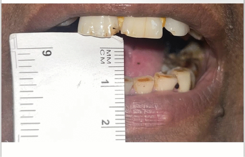
Successful management of this deadly disease is early diagnosis and early treatment which includes a combination of medical and surgical debridement. Knowing the importance of early diagnosis, which is the most challenging aspect of mucormycosis management, we were left with the option of microscopic examination of tissue specimens in patients who were clinically suspected to have a fungal infection. Commonly patients with rhinomaxillary infection presented with mobile tooth (70%), numbness in the infraorbital region (40%), palatal ulceration (25%), blurred vision (7.5%), nasal discharge (20%), intraoral pus draining sinus tract or fistula (65%), exposed bone [alveolar (20%), palatal (5%), both (7.5%) and absent (67.5%)] with common history of COVID-19 infection. Most of the patients were hospitalised under oxygen and were usually given steroids to combat the deadly viral infection. Computed tomography and magnetic resonance imaging revealed thickening of the maxillary sinus (right—5%, left—10%), any two sinuses (47.5%), three sinuses (32.5%) and all paranasal sinuses in 5% of the cases. Subtle erosive changes of the maxillary bone (80%) with alveolar bone involvement and without alveolar erosion were seen in 35% and 45% of the cases, respectively. Changes extending to zygomatic bone were seen in 15% of the cases.
Infection usually starts in the nasal cavity and spreads to the paranasal sinuses. Bhandari et al., in 2021 stated that the humid environment of the nose and paranasal sinuses favour the growth and invasion of fungi. Early implantation of fungi is common in the maxillary sinus with a mass of fungal growth called a fungal ball and with no evidence of bone erosion. The most common site involved in mucor is middle turbinate, followed by middle meatus and septum. In undiagnosed or untreated cases, the infiltration of the bone is common [8].
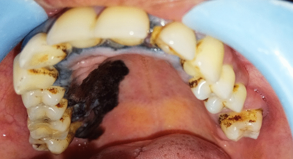
When the infection extends through the nasal turbinates, the orbit frequently becomes involved, and orbital cellulitis, extraocular muscle paresis, proptosis and chemosis are commonly found. Extension of the infection posteriorly into the brain results in the formation of abscesses and necrosis of the frontal lobes. When the disease invades inferiorly into the mouth, a black, necrotic eschar is often found on the palate, this finding is highly suggestive of the presence of invasive mucormycosis [9].
In the present setup, various tissue specimens were sent for microscopic examination (using KOH stain) and fungal culture. Swabs were sent from nasal mucosa, sinus tract or tissue specimen from necrotic bone or suspected tissue from paranasal sinus with the aid of endoscopy. Few of these showed fungal elements giving way for hospitalisation and definitive treatment, while some turned negative despite clinically evident disease leading to delayed medical management. Multimodal therapy is key to reducing the disease burden which involves both surgical debridement and systemic antifungal therapy. Without antifungal therapy, the disease burden and recurrence increase even after performing surgical debridement.
Harrill et al., stated that the median time from the symptom onset to diagnosis was 7 months [1]. In the present study, the mean number of days for delayed diagnosis among various study groups was 56.33 ± 37.53, 32.86 ± 19.53 and 22.00 ± 12.94 days, respectively, which was statistically significant.
Because of the nonspecific clinical signs, a high index of suspicion should be made in the existence of risk factors. Nasal scrapings and fine-needle aspiration cytology were performed to give the diagnostic results which showed fungal hyphae. Swabs were taken, and cultures from sinuses were negative in most of the cases (25%). Thus in the rhinomaxillary group which presents with common disease presentations, we found 52.5% of participants to show fungal hyphae from the oral tissue (necrotic bone or sinus lining via Caldwell Luc), 12.5% from nasal tissue/ pus as well as from both tissue specimens with extensive involvement.
Mucorales appear in the tissue as irregularly shaped, broad hyphae with right-angle branching. The hyphae are easily visualised in routine haematoxylin–eosin-stained sections as well as in tissue stained by the periodic acid–Schiff reaction or by Grocott-Gomori methenamine-silver nitrate stains. Hyphae are usually found in the midst of an acute neutrophilic infiltrate along with the hyphal invasion of the blood vessels [9].
In a report to check the diagnostic values of KOH examination, histological examination, culture for onychomycosis and periodic acid–Schiff stain (PAS) were highly specific but poorly sensitive. KOH was highly sensitive but poorly specific and Grocott’s methenamine-silver stain (GMS) was both highly sensitive and specific [10].
Various other techniques for diagnosis include immunohistochemistry, polymerase chain reaction (PCR)-based methods and matrix-assisted laser desorption or ionisation time-of-flight mass spectrometry (MALDI-TOF MS). A study done by combining a semiquantitative method using high-resolution melt analysis and real-time quantitative PCR (RQ PCR) assays showed sensitivity and specificity of 100% and 93%, respectively, and a high negative predictive value (99%) in detecting mucormycetes. Despite their higher sensitivity and specificity, these diagnostic aids are not available in clinical setups in India for it to be used commonly giving way to conventional techniques. Thus, KOH mount setup is feasible for the Indian scenario; however, the sites should be carefully selected to collect suitable samples showing hyphae infiltration in the necrotic tissue helping to predicate the diagnosis at an early stage rendering them to medical management with systemic antifungals.
The limitation of the study is its retrospective temporality with limited sample size. The present study concurs that in the rhinomaxillary mucormycosis group, the fungal spores have two different patterns of invasion evident with oro-nasal signs and symptoms. To avoid the chance of delayed diagnosis or false-negative results, it is best to collect samples from both nasal tissues as well as the most representative site in the dentoalveolar segment depending on the extensiveness of the disease.


留言 (0)