The interaction between anti-thyroid antibodies (ATAs) and reproductive disorders has been attracted a great deal of attention over the past two decades. Although a significant progress has been achieved in understanding the pathophysiology underlying thyroid autoimmunity and reproductive disorders, the exact mechanism of pathogenesis is not fully elucidated. This review will explore the possible mechanism of ATAs and their correlation with implantation and pregnancy complications.
Reproductive disorders including recurrent pregnancy loss (RPL) or recurrent spontaneous abortion (RSA) and recurrent implantation failure (RIF) are multifactor disorders. RPL, also is referred to RSA and recurrent miscarriage, and is defined as two or more consecutive pregnancy losses prior to the 20th week of gestation (Medicine, 2012; RPL et al., 2018). RIF is also described as failure in achieving a pregnancy, after at least four good-quality embryos transfer or in vitro fertilization (IVF) attempts in a woman (Bashiri et al., 2018, Coughlan et al., 2014). In this regard, the role of several factors have been confirmed in the etiology of these disorders, including chromosomal, anatomical and immunological abnormalities, as well as endocrine disorders and infections (Dosiou & Giudice, 2005).
Autoimmune thyroid disorders (AITD), is caused due to immunologic abnormalities, and includes inflammatory thyroid diseases, such as Graves’ disease (GD) and Hashimoto’s thyroiditis (HT) as the most common AITDs (McLeod & Cooper, 2012). Considering the immunostimulatory effect of estrogen and immunoinhibitory role of progesterone and androgens, women are more susceptible to AITD (Moulton, 2018). AITD is related to excessive inflammation due to the over production of autoantibodies, which are also responsible for reproduction failure and pregnancy adverse outcome (Poppe and Glinoer, 2003, Prummel and Wiersinga, 2004).
Thyroid hormones play a pivotal role in the development of all type of cells in human body, including female reproductive organs (Cho, 2015). Triiodothyronine (T3) in combination with follicle-stimulating hormone (FSH) affects the luteal and granulosa cells proliferation and also directly affects the invasion of extravillous trophoblasts by regulating matrix metalloproteinases (MMPs) expression (Oki et al., 2004; C. Zhang et al., 2013). Maturation process of oocyte and implantation process is also affected by thyroid hormones such as thyroid-stimulating hormone (TSH). Hence, all reproductive processes including folliculogenesis, implantation and etc. are affected and regulated by thyroid hormones, hypothyroidism inevitably is associated with infertility and reproductive disorders (Lata et al., 2013). It has been suggested that there is a potential hypothyroidism in ATA positive women. In addition, thyroid autoantibodies in patients with AITD and even in euthyroid women, may lead to pregnancy adverse outcome. The presence of these wide range autoantibodies is also correlated with the higher risk of spontaneous abortion and pregnancy loss (Fausett & Branch, 2000) and can be considered an indicator of activated immune system (Lata et al., 2013). An uncontrolled immune system in the process of implantation and pregnancy, would hamper the maternal tolerance toward embryo or fetus, as a semi allograft, and induces a pro-inflammatory situation, which increases the risk of miscarriage (Challis et al., 2009). Taken together, the documents indication that abnormal levels of thyroid hormones, could determine ovarian dysfunction and infertility.
Thyroid peroxidase antibodies (TPOAbs), thyroid globulin or thyroglobulin antibodies (TgAbs) and TSH receptor antibodies (TRAbs) are the anti-thyroid antibodies (ATAs), which are directly correlated with AITD (Kahaly et al., 2018, Ross et al., 2016). TRAbs are divided into TSHR-stimulating antibodies (TSAbs), TSHR-blocking antibodies (TBAbs), and neutral TRAbs (N-TRAbs) according to their function. TRAbs are able to cross the placenta, therefore they can disturb the receptors in both mother and fetus and lead to delayed thyroid gland development (Bucci et al., 2017, Fröhlich and Wahl, 2017). Additionally, TPOAbs bind to the thyroid peroxidase (TPO) enzyme and disturb its function. Therefore, TPOAbs are responsible for hypothyroidism by acting as a competitive inhibitor for TPO action (Bhattacharyya et al., 2015). TPOAbs are also able to induce complement activation and immunologic responses, destroy thyrocytes and induce oxidative stress (Kaczur et al., 1997, Ruggeri et al., 2016). It has been confirmed that these antibodies are capable of passing through the placental barrier (Balucan, Morshed, & Davies, 2013). On the other side, TgAbs are capable of binding to the different antigenic determinants of thyroglobulin in thyroid gland colloid and disturb the production and secretion of these hormones (S. Matalon, Blank, Ornoy, & Shoenfeld, 2001). This group of antibodies can also pass through the placenta (Fröhlich & Wahl, 2017). General characteristics of these antibodies are summarized in Table 1.
These antibodies mainly belong to the immunoglobulin G1 (IgG1), and with a lower content to IgG2, IgG3, IgG4 and IgA (Fröhlich and Wahl, 2017, McLachlan and Rapoport, 2004). In the case of HT, the autoreactive CD4+ T lymphocytes which are infiltrated the thyroid gland, are responsible for inducing the production of these antibodies by B cells. Attachment of these antibodies to the basal membrane of thyroid follicles would activate the complement system, leading to thyrocyte apoptosis and necrosis caused by cytotoxic CD8+ T cells-secreted perforin (Fröhlich & Wahl, 2017). Thyroid autoantibodies could undesirably influence reproductive status of woman and so have been proposed to have a role in adverse pregnancy outcomes.
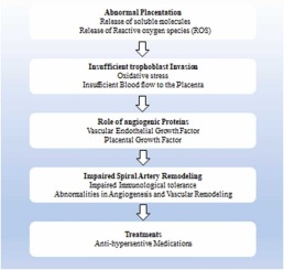
According to the literature, it has been confirmed that there is a strong connection between adverse pregnancy outcome and maternal thyroid disorders. Some of gestational hypothyroidism-related complications are low fetal birth weight or fetal death, anemia and hypertension, preterm labor, preeclampsia, intrauterine growth restriction and stillbirth (Phoojaroenchanachai et al., 2001, Yalamanchi and Cooper, 2015). Higher level of TPOAbs have also been reported in RPL patients compared to normal women (5.4-20% versus 14-33%, respectively) (Poppe, Velkeniers, & Glinoer, 2008). Indeed, increased level of ATAs has been reported in infertile women compared to the normal fertile women, and the presence of these antibodies may increase the risk of pregnancy wastage (S. Matalon et al., 2001; Poppe et al., 2002). On the other hand, women with TPOAbs prior to pregnancy, are in 2 fold of greater risk to develop hypothyroidism when they get pregnant (Lin, Zhang, & Long, 2014). Similar trend has also been observed in women with high titer of TPOAbs and TgAbs in serum, who experienced abortion after in vitro fertilization (IVF)/intracytoplasmic sperm injection (ICSI) (Busnelli et al., 2016, Bussen et al., 2000). Additionally, thyroid gland in women with higher ATAs titer, does not properly response to the human chorionic gonadotropin (hCG) and the risk of premature delivery is increased in this condition. The ability of ATAs for transplacental passage is also another risk factor, which may also induce hypothyroidism in fetus (Korevaar et al., 2017). Additionally, the presence of ATAs indicates a vast underlying immune abnormality and autoimmunity, which may compromise the fetal development (Thangaratinam et al., 2011).
Thyroid dysfunction have been related to adverse fertility and pregnancy outcomes. This review aimed to explore the probable ATAs action mechanisms in hypothyroid state, and pregnancy complication pathophysiology, while special attention is paid to the generalized underlying immunologic abnormalities in AITD patients.


留言 (0)