
記住我


The role of biopsy without any doubt has been proven time and again for confirming the diagnosis of various spinal pathologies. Indications for spinal biopsy include spinal infections [tuberculosis (TB), bacterial, etc.], postoperative discitis, osteoporotic vertebral compression fractures (pseudoarthrosis fluid), suspected metastasis, multiple myeloma, and primary tumors. Spinal TB used to be diagnosed in endemic regions such as India based on clinicoradiologic judgment. Tissue diagnosis was not considered necessary as this was the most common spinal infection by far. However, with the emergence of drug-resistant TB and atypical infections, tissue diagnosis through biopsy and culture have become mandatory.1 TB reported based on magnetic resonance imaging (MRI) can turn out to be other non-TB infection or more sinister lesions such as round cell tumors and other malignancies. Moreover, even if we get the radiologic diagnosis right, it does not give any information about the drug sensitivity on the bug—a very important prerequisite to treat TB in today’s era of drug resistance alone. In the older populations biopsy can be performed with minimal morbidity and high diagnostic yield as an outpatient procedure.2
Biopsy procedures can be done using several image-guided techniques such as with computed tomography scan or fluoroscopy. The objective of this technical report is to describe our experience using fluoroscopy and 2 syringes for percutaneous transpedicular biopsy of spinal lesions.
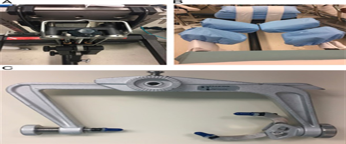
TECHNIQUEFluoroscopy-guided percutaneous transpedicular biopsy is a safe procedure with high adequacy and accuracy and low complication rate for thoracic and lumbar vertebral body lesions. The procedure is performed in prone position on a radiolucent table under local anesthesia (LA) with anesthetist around. The target pedicle is marked in both true anteroposterior (AP) and lateral view. LA is given by injecting plain bupivacaine along the proposed needle tract (Fig. 1A).
 FIGURE 1:
 A, Surface marking on fluoroscopy before giving local infiltration along the tract of biopsy. B, Lateral fluoroscopy image showing the step when the trocar is withdrawn from the bone biopsy needle.
A stab incision is given on the skin, a tract is created through deep fascia and muscle by a T-handle Bone marrow biopsy needle (BONEAID 8 G×10 cm) manufactured by Advanced Life Sciences Pvt Limited, India. The entry point into the pedicle is created by gently tapping under fluoroscopy guidance; it lies on the superolateral margin of the pedicle in AP view. The correct position of entry point is also confirmed in lateral view and cephalocaudal inclination is adjusted to reach the target area for biopsy of vertebral body as per MRI image. Once entry point is ensured, the biopsy needle along with its trocar is gently tapped into the pedicle under fluoroscopy guidance. Once it reaches the posterior border of vertebral body in lateral view, an AP image is obtained to make sure that the tip of the needle is at the center of pedicle and has not reached the medial edge of pedicle. Now the pointed trocar is withdrawn (Fig. 1B) and the serrated cannula is advanced into the vertebral body towards the target area (granulation tissue/pus pocket) and desired position is checked under fluoroscopy.
Fifty milliliter Luer lock syringe is attached to corresponding locking hub on cannula of the biopsy needle. Luer lock syringes avoid disengagement from biopsy needles during the entire procedure. After stabilizing the cannula, content of lesion is to be aspirated. In the conventional technique, the plunger of 50 mL syringe tends to recoil back, as it is difficult to maintain suction pressure with 1 hand and simultaneously hold the cannula-syringe assembly in position with another hand for 1 person. This step of aspiration usually requires an assistant. To overcome this, we propose to place the plunger of a 10 mL syringe in between the end of barrel and plunger of 50 mL syringe as shown in the intraoperative image (Fig. 2). This way it helps to maintain suction pressure and avoid recoiling at the same time effortlessly. It acts as a stopper while maintaining optimum suction pressure. In addition, the target area is not overshot or missed during the hustle of aspiration. This simple and cost-effective modification in standard daycare procedure makes it easy for 1 surgeon to do it in a controlled manner.
 FIGURE 2:
 Dual syringe and cannula assembly during the biopsy procedure.
Gentle cephalocaudal and mediolateral rocking movements of the cannula-syringe assembly while withdrawing, dislodges the bone from the vertebral body into the lumen of biopsy needle. Cannula along with its attached dual syringe assembly is withdrawn. Fifty milliliter syringe is unlocked from the Luer lock system. Ten milliliter syringe plunger is removed and contents of 50 mL syringe are collected in sterile containers. Specimen trapped into the cannula is removed with a blunt stylet. Biopsy material is sent for histopathology, Gram and Ziehl Nielsen stain, Gene Xpert MTB/RIF, manufactured by Cepheid. Gene Xpert MTB/RIF is a nucleic acid amplification test for simultaneous rapid TB diagnosis and rapid antibiotic sensitivity testing. The sample is also sent for microbiology (TB/pyogenic/fungal) culture and drug sensitivity. The patient is permitted to go home after 2 hours of observation to ensure that the pain is controlled and no evidence of evolving hematoma. Patients are instructed to return to the hospital if they develop any radicular symptoms.
EXPECTED OUTCOMESWith the advent of better imaging modalities such as fluoroscopy, computed tomography scan, and MRI, percutaneous transpedicular vertebral biopsy has now become a popular minimally invasive technique for biopsy of vertebral lesions. The first pedicular approach to the vertebral body was performed in 1928 by Duncan et al, whereas the first percutaneous biopsy was reported in 1935 by Robertson and Ball.3 Fine needle aspiration cytology should be avoided in spine as the yield is consistently lower than core biopsy. To optimize the outcomes, a biopsy from granulation tissue is ideal, but if no granulation tissue is available, bone biopsy can be done with good results. Yield wise; granulation tissue >> pus > bone are preferred for biopsy.4 It is vital to carefully study the imaging to plan the site of biopsy. On the T2-weighted image of MRI (Fig. 3A), pus is brighter than granulation tissue. On contrast study, pus shows peripheral enhancement and granulation tissue shows complete enhancement. On T1-weighted image it is easy to appreciate erosions in bony lesions (Fig. 3B).
 FIGURE 3:
 A, T2 sagittal image showing abscess around L5 vertebra. B, T1 axial image showing right sided erosions in L5 vertebral body.
COMPLICATIONSLA enables monitoring of nerve roots during the biopsy. The estimated complication rate of procedure is 0.2%.5 A retrospective review of around 80 patients in last 2 years showed that there were no nerve root injuries, pneumothorax, or sinus tract formation during follow-up in the first 3 months. Other possible complications such as hematoma due to vascular injury, pseudoaneurysm, transient paresis, and medial pedicle breach were also not encountered as entire procedure is conducted in a controlled environment.
Thus, the dual syringe technique described above can be a very handy tool and a worthy technical tip to optimize the final results of the important and commonly performed percutaneous transpedicular biopsy procedure for spinal lesions.
REFERENCES 1. Shah K, Nene A. Tuberculosis of the spine: the current clinical landscape. Astrocyte. 2017;4:94–99. 2. Dave BR, Nanda A, Anandjiwala JV. Transpedicular percutaneous biopsy of the vertebral lesions: a series of 71 cases. Spinal Cord. 2009;47:384–389. 3. Robertson RC, Ball RP. Destructive spinal lesions: diagnosis by needle biopsy. J Bone Joint Surg. 1935;17:749–758. 4. Kim C-J, Kang S-J, Choe PG, et al. Which tissues are best for microbiological diagnosis in patients with pyogenic vertebral osteomyelitis undergoing needle biopsy? Clin Microbiol Infect. 2015;21:931–935. 5. Murphy WA, Destouet JD, Gilula LA. Percutaneous skeletal biopsy: a procedure for radiologists-results, review, and recommendations. Radiology. 1981;139:545–549.
留言 (0)