
記住我


Patients with displaced pertrochanteric proximal femur fractures require operative reduction and stabilization to regain mobility and decrease pain. Cephalomedullary nails have become the implant of choice for many orthopedic surgeons treating unstable pertrochanteric fractures.1–3 Obtaining an acceptable reduction of the fracture and appropriate placement of the implant in the proximal femur are 2 variables that effect outcome.4
A fracture or traction table is one method currently used to assist the surgeon with regaining and maintaining alignment during surgery of proximal femur fractures.5 Patients may have associated conditions in the operative leg that make placement of the foot in a traction boot and manipulating with traction and rotation questionable. Examples of this include patients with an amputation of the lower portion of the operative leg, a fracture in the ipsilateral leg below the knee, or soft tissue compromise of the lower leg. Morbidly obese patients can be a challenge to position on the fracture table.
Femoral shaft fractures have been treated with intramedullary nailing on a radiolucent table using manual traction or the large distractor for many years with good published results.6–9 I encountered patients with pertrochanteric femur fractures I felt were poor candidates for utilizing the fracture table. The described technique for cephalomedullary nailing using the Synthes large distractor on a radiolucent table was performed in these cases and subsequently used on all pertrochanteric fractures treated with an intramedullary nail. This manuscript describes the technique used and results.
METHODSIRB approval for Study Number 150216-4 was obtained from the Mount Carmel Institutional Review Board on February 23, 2015. All surgeries were performed at 1 hospital with a surgical team consisting of a circulating nurse, radiology technician, and either 2 surgical technicians or 1 surgical technician and 1 orthopedic physician’s assistant.
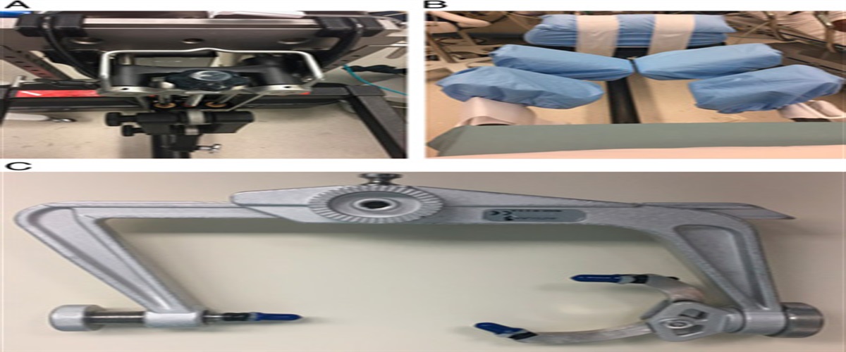
Patients were positioned supine on a radiolucent table with a bump under the ipsilateral buttock. The surgical prep was performed starting at the hip to extend proximally to at least the iliac crest and then circumferentially around the thigh and leg to the foot. The foot was placed in an impervious stockinette and split drapes were used to isolate the affected leg (Fig. 1). An obturator oblique fluoroscopy image was used to identify the supra-acetabular column of bone and the cortex is opened with the appropriate drill bit. A 5 or 6 mm threaded half pin was placed in the ilium above the acetabulum by hand allowing the pin to proceed between the tables of the ilium but not penetrate posteriorly10,11 (Fig. 2). A 5 mm threaded half pin was placed in the metaphysis of the distal femur lateral or medial to the mid-line to allow eventual intramedullary nail placement without the pin impeding the nail (Fig. 3). Provisional reduction was obtained with manual traction on the leg and the Synthes large distractor was applied to provide traction throughout the procedure.
 FIGURE 1:
 Patient prepped and draped on radiolucent table.
 FIGURE 2:
 Fluoroscopy obturator oblique image of supra-acetabular pin.
 FIGURE 3:
 Fluoroscopy image of pin in distal femur.
The assistant positioned the leg to allow the appropriate proximal starting point to be achieved. I have found with this technique I can adduct the operative leg more freely and not cause a significant varus deformity at the fracture site. Before adopting this technique when I used the fracture table to reduce pertrochanteric fractures in patients with obese or muscular thighs I felt the perineal post often caused varus at the fracture site. In fractures with flexion or abduction deformity of the proximal fragment after traction was applied, a ball tipped reduction instrument was placed percutaneously under fluoroscopy in the anterior or lateral thigh to reduce the deformity. This was performed before any reaming or implant preparation. Standard percutaneous technique was used to obtain the appropriate starting point for the implant in the medial aspect of the greater trochanter. (Fig. 4) A long cephalomedullary or short cephalomedullary implant was chosen based on the fracture pattern. When a long implant is desired, the reaming guide rod was placed and confirmed to be central to the distal half pin. Fluoroscopy was used to confirm the reamer and therefore the nail could be inserted past the distal half pin. The implant was locked proximally and distally before removal of the distractor (Figs. 5A–D).
 FIGURE 4:
 Proximal reamer with large bone distractor in place.
 FIGURE 5:
 A, Anterioposterior fluoroscopy image proximal femur pin placement for proximal fixation. B, Lateral fluoroscopy image proximal femur pin placement for proximal fixation. C, Anterioposterior fluoroscopy image distal femur with rod past the distal pin of distractor. D, Lateral image distal femur before distal locking.
The fractures were classified according to the Orthopaedic Trauma Association system and the surgical time was obtained from the operating room records completed by the circulating nurse. I attempted to follow all pertrochanteric fracture patients until there was clinical and radiographic fracture healing. An attempt was made to locate and follow patients who were lost to follow up before these criteria were met. Radiographs were performed in my outpatient clinic during follow up and the tip to apex distance and neck-shaft angle were recorded.
RESULTSThe technique was successful in allowing cephalomedullary nailing in all cases it was attempted. The study group was typical of an elderly fracture population, predominately female with 30 female and 6 male patients and age range from 48 to 95 years and an average age of 81 years. Surgery was performed on the day of admission in 12 cases, 1 day following admission in 18 cases, and 2 days after admission in 6 cases. There were 19 right sided fractures and 17 left sided fractures. A long Smith and Nephew Intertan intramedullary implant (Memphis, TN) locked distally in the metaphyseal region was used in 33 cases, a short 180 mm Smith and Nephew Intertan implant locked distally in the femoral shaft in 2 cases, and a short 160 mm Synthes TFN (Paoli, PA) locked distally in the femoral shaft in 1 case. Table 1 has the Orthopaedic Trauma Association classification of the fractures in this study.12
TABLE 1 - OTA Fracture Classification12 1.1 1.2 2.1 2.2 2.3 3.1 3.2 3.3 Total 1 2 8 11 5 2 2 5 36Three patients died during the index hospitalization at 6, 19, and 23 days following surgery from medical complications. The family of 1 patient who did not follow up after surgery was contacted and reported the patient died 6 months after surgery, and one patient resided out of the area and no follow up could be obtained. Thirty-one patients had been followed in the office with clinical and radiographic signs of healing in 30, and nonunion with proximal hardware failure in 1 patient. No additional surgery related to the affected hip or femur was performed in the 30 patients who were determined to have clinical and radiologic signs of healing at final follow up. The patient with a nonunion and failure of proximal fixation was scheduled for revision to total hip arthroplasty. No deep surgical site infection requiring surgery occurred during the follow-up period. There were no noted complications related to the pins used with the large distractor. No patients in follow up reported symptoms of lateral femoral cutaneous nerve injury and no patients returned to our hospital or reported a periprosthetic femur fracture during the follow-up period.
The total time in the operating room averaged 89 minutes with a range of 72 to 111 minutes. Total surgical time defined as time from incision until wound closure averaged 53 minutes with a range of 40 to 69 minutes. The average tip to apex distance was 14 mm and the average femoral neck-shaft angle of the injured hip was 129 degrees at final follow up.
DISCUSSIONThis pilot study was performed to report the results of treating pertrochanteric fractures with a cephalomedullary implant using the Synthes large distractor to provide traction on a radiolucent table. I treated pertrochanteric femur fractures that were considered to have the potential for significant collapse with shortening of the proximal femur and medialization of the femoral shaft with a cephalomedullary implant.5 Patients with an ipsilateral below knee amputation can be a challenge to place on a fracture table. At my hospital this would require a traction pin distal to the fracture and traction bow attachment for the fracture table which was seldomly used and difficult to locate. Patients with ipsilateral fractures below the knee may not be safe to have traction applied though the leg with the foot in the traction boot. Significant contractures of the foot and ankle are difficult to secure in the traction boot and the surgeon may not want to place a foot with poor soft tissues or wounds in the traction boot. By using the large distractor to provide traction, there was no need to place the ipsilateral foot in a traction boot or place traction or rotational force across the ipsilateral lower leg and foot.
At my hospital hip fracture cases were often performed later in the day and on the weekend when the circulating nurse was frequently not well trained or comfortable adjusting the fracture table. Once I was scrubbed into surgery the circulating nurse was responsible for adjusting the traction and changing the position of the operative leg when using the fracture table. By utilizing the large distractor either the scrubbed assistant or I can adjust the traction and position of the leg throughout the procedure.
The technique was used in 36 patients to reduce and stabilize pertrochanteric fractures with a cephalomedullay nail. I was able to reduce the fracture and allow acceptable cephalomedullary implant placement with an average Tip-Apex distance of 14 mm and a maximum distance of 20 mm.4 The average neck-shaft angle of the injured proximal femur at final follow up was 129 degrees. I believe this demonstrates that acceptable reduction and implant placement can be obtained with this technique. This technique has been used continuously since this pilot study was completed with similar results over the next 4 years.
The pertrochanteric fractures in this elderly population healed following the original procedure in 30 of the 31 patients followed until fracture union. One patient who with diabetes and chronic renal failure had failure of proximal fixation 3 months following fracture and the recommendation was made to revise with total hip replacement. No patients developed a deep infection or periprosthetic fracture during follow up. The mortality rate of 11% in this study group is similar to reported series of pertrochanteric fractures.5 Although no complications were noted relating to the placement of the pins used in the large distractor, in this retrospective review it was possible that they were not adequately reported in clinic notes.
This retrospective pilot study has many limitations that affect the conclusions which can be made from our data. The operative times are dependent upon appropriate input by the circulating nurse. The postoperative radiographs were taken per protocol at the author’s office by multiple radiologic technicians without formal standardization. Patients were followed until the author determined the fracture to be healed clinically and radiographically, but no long-term follow up is available. There is no group of patients treated with the use of the fracture table to provide comparison data. The strength of this study is the fact that it reports a consecutive series of patients were treated with this technique by 1 surgeon with commonly available operative room equipment and assistants.
In conclusion I believe pertrochanteric femur fractures can be appropriately reduced and stabilized with a cephalomedullary nail using the Synthes large distractor on a radiolucent table. This technique can be an option for treating patients who may not be good candidates for use of the fracture table.
REFERENCES 1. Bridle SH, Patel AD, Bircher M, et al. Fixation of intertrochanteric fractures of the femur: a randomized prospective comparison of the gamma nail and the dynamic hip screw. J Bone Joint Surg Br. 1991;73:330–334. 2. Radford PJ, Needoff M, Webb JK. A prospective randomised comparison of the dynamic hip screw and the gamma locking nail. J Bone Joint Surg Br. 1993;75:789–793. 3. Bohl DD, Basques BA, Golinvaux NS, et al. Extramedullary compared to intramedullary implants for intertrochanteric hip fractures. J Bone Joint Surg. 2014;96:1871–1877. 4. Baumgaertner MR, Curtin SL, Lindskog DM, et al. The value of the tip-apex distance in predicting failure of fixation of pertrochanteric fractures of the hip. J Bone Joint Surg. 1995;77:1058–1064. 5. Lindskog DM, Baumgaertner MR. Unstable intertrochanteric hip fractures in the elderly. J Am Acad Orthop Surg. 2004;12:179–190. 6. Baumgaertel F, Dahlen C, Stiletto R, et al. Technique of using the AO-femoral distractor for femoral intramedullary nailing. J Orthop Trauma. 1994;8:315–321. 7. Mcferran MA, Johnson KD. Intramedullary nailing of acute femoral shaft fractures without a fracture table: technique of using a femoral distractor. J Orthop Trauma. 1992;6:271–278. 8. Wolinsky PR, McCarty EC, Shry Y, et al. Length of operative procedures; reamed femoral nailing performed with and without a fracture table. J Orthop Trauma. 1998;12:485–495. 9. Stephen DJ, Kreder HJ, Schemitsch EH, et al. Femoral intramedullary nailing: comparison of fracture table and manual traction. A prospective, randomized study. J Bone Joint Surg. 2002;84:1514–1521. 10. Haidukewych GJ, Kumar S, Prpa B. Placement of half-pins for supra-acetabular external fixation: an anatomic study. Clin Orthop. 2003;411:269–273. 11. Langford JR, Burgess AR, Liporace FA, et al. Pelvic fractures: part II. Contemporary indications and techniques for definitive surgical management. J Am Acad Orthop Surg. 2013;21:458–468. 12. Marsh JL, Slongo TF, Agel J, et al. Fracture and dislocation classification compendium. J Orthop Trauma. 2007;21:S31–S32.
留言 (0)