
記住我


A man sitting in the back seat of a moving car was accidentally shot by a bullet fired from the opposite side of a highway. During the crime scene investigation, the metal wire fence placed between the two carriageways was examined, and a small, yet noticeable deflection was documented and sampled (Fig. 1a). There was a hole on the left rear window of the car with radial cracks in the glass (Fig. 1b). The victim was found in a sitting position with two GSWs to the neck, one on his left side and one on the right side, still wearing a necklace chain with two breaks at the level of the GSWs (Fig. 2a). The bullet, once retrieved during site examination, was found to be partially deformed at the ogive with a damaged jacket (Fig. 3). The event’s dynamics was initially reconstructed from the testimonies collected by bystanders who saw a man shooting from the opposite side of the highway, where a cartridge case was found.
Fig. 1
Case 1 intermediate targets. a First intermediate target: the wire fence dividing the two carriageways. Interaction with the bullet caused a “watermelon slice”–like deformation of the metal and subsequent ricochet from the original trajectory. b Second intermediate target: glass of the rear window. Note the shape of the hole which accurately represents the lateral projection of the bullet
Fig. 2
Case 1 third intermediate target. a The necklace worn by the victim with two breaks at the level of the entrance (*) and exit (arrow) wounds. b Repositioning of the necklace during the autopsy with the margins at the level of the entrance wound. c Repositioning of the necklace during the autopsy with the margins at the level of the exit wound
Fig. 3
Cal. 9-mm bullet retrieved during crime scene investigation, characterized by a deformation produced by the contact with the metal wire fence (upper arrow) and the glass (lower arrow)
Based on the conducted investigations, it was postulated that the bullet had impacted the metal fence with subsequent deviation from its original trajectory (first intermediate target), the rear window with the fragmentation of the glass (second intermediate target), and, lastly, the necklace worn by the victim (third intermediate target) before reaching his neck and producing a perforating GSW to the neck. For the sake of this analysis, before providing a description of the injuries caused by the deviated and deformed bullet, each of the intermediate targets will be examined.
First intermediate target—metal wire fenceDuring the crime scene investigation, the judicial police recognized that the wire fence dividing the two lanes had acted as an obstacle to the projectile's motion. A deformation was detected on the central portion of one of the cylindrical elements, orthogonally everted with respect to the plane of the wire and curved towards the side where the car was located (Fig. 1a).
According to the reconstructions, the bullet drew this portion of the intermediate target causing a glove-like semi-invagination, and then deviated to the left of the wire as suggested by the morphology of the impression, which was compatible with the cylindrical-ogival shape of the deforming agent and limited to the contact area (Fig. 3).
Second intermediate target—glass of the rear windowBy analyzing the shape of the hole in the glass of the rear window of the car, it was postulated that an impact with the lateral surface of the bullet had occurred. The morphology was attributed both to the perturbation of the projectile’s trajectory after contact with the wire fence, and to the convexity of the glass itself (Fig. 1b).
Third intermediate target—necklaceTwo breaks with deformed margins were found on the necklace: the one on the left side of the neck had produced some metallic fragments which deposited on the subcutaneous tissue of the cervical region, while the one on the right had branching-out metallic fibers, hence confirming the correct positioning with respect to the entrance and exit wounds (Fig. 2b–c). The bullet had caused an initial bending of the necklace, followed by tearing of the folded area, defeating the elastic capacity of the necklace.
Field emission gun–scanning electron microscopy (FEG-SEM) and scanning electron microscopy–energy dispersive spectroscopy (SEM–EDS) analysis were conducted on the necklace’s fragments and allowed the investigators to document additional blood traces at the level of the right break.
Final target—the victimA D-shaped 1.3 × 0.4 cm entrance wound was found in the left latero-cervical region of the neck, and was surrounded by a slightly depressed, triangularly shaped abrasion collar and a brown-to-black oval contusion ring (Fig. 4a). On the internal side of the cutaneous layers, golden metallic fibers were visible.
Fig. 4
Case 1 GSWs. a D-shaped entrance wound produced by the lateral projection of the bullet coupled to the cutaneous fold of the neck region. b Jugular vein with a D-shaped hole. c D-shaped hole in the trachea, which caused bleeding and bronchoaspiration of blood. d Shored exit wound produced by the presence of the necklace. All the pictures have been oriented as follows: TOP–BOTTOM, LEFT–RIGHT. Note that some of the structures have been partially displaced during the autoptic examination to exhibit and document the wounds
On the right side of the neck, 1 cm above the clavicle, a 1 × 0.4 cm oval exit wound was detected, which was surrounded by a small abrasion collar-contusion ring complex (Fig. 4b).
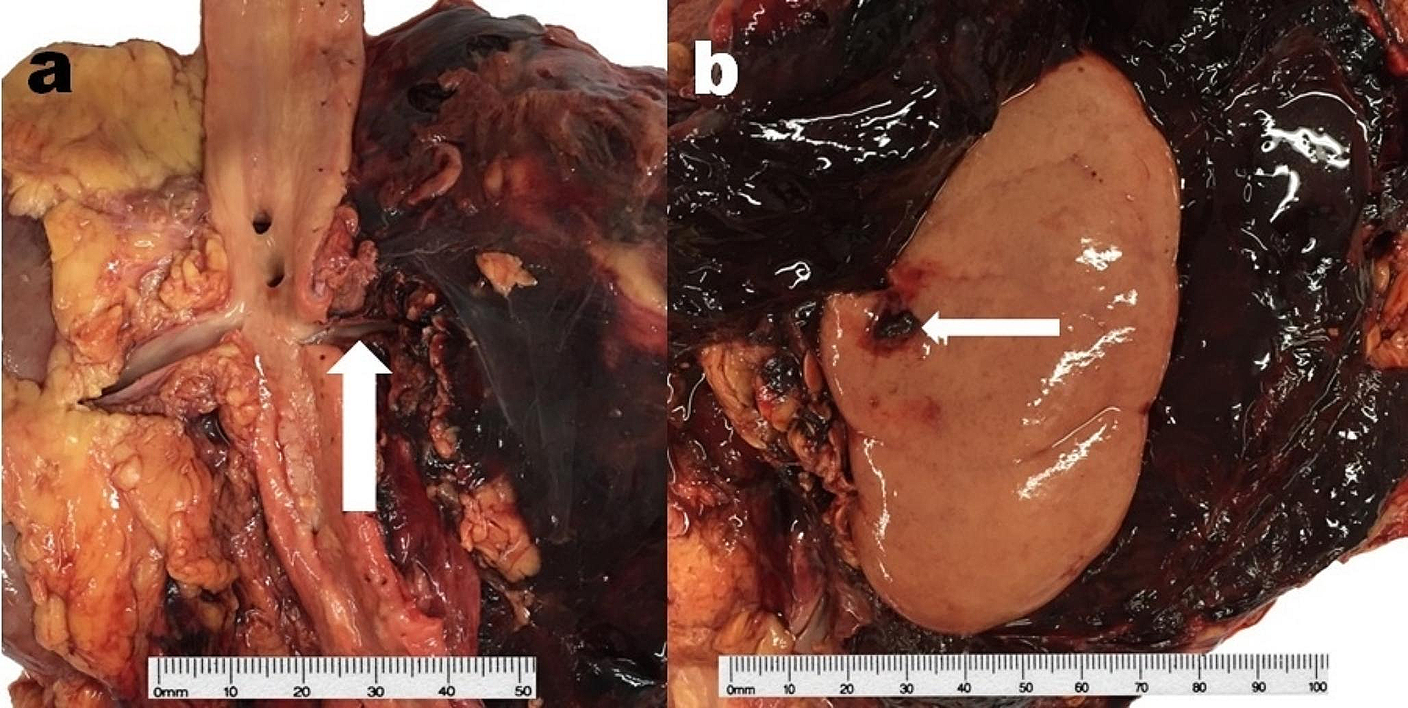
Anterior and lateral neck dissection revealed a D-shaped hole in the middle third of the left jugular vein, perfectly reproducing the bullet’s lateral surface, where the tip was cranially oriented (Fig. 4c). The ventral wall of the left common carotid artery was lacerated with irregular margins (Fig. 5). Another D-shaped hole was detected at the level of the trachea, just beneath the cricoid cartilage (Fig. 4d). Both the lungs were pink with multiple red-to-purple polygonal punctuations on the pleural surface, which were compatible with bronchoaspiration.
Fig. 5
Lacerated carotid wall compared to the venous wall
Based on the autoptic findings, it was possible to accurately reconstruct the intrasomatic bullet course: left cervical skin, left infrahyoid muscles, left jugular vein, left common carotid artery, trachea, right intrahyoid muscles, right cervical skin.
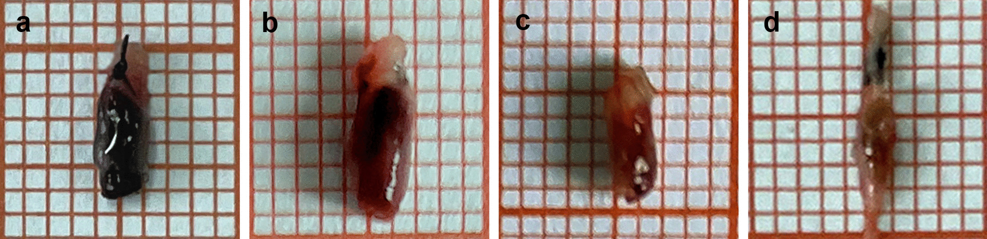
Bullet’s characteristicsThe 7.40 g, cal. 9 Parabellum bullet was analyzed (Fig. 3):
(a)The tip of the bullet was deformed and flattened by the impact with the glass of the rear window;
(b)The latero-ogival region of the base of the bullet was curved, as a result of the impact with the wire fence, which was also demonstrated by SEM–EDS analysis;
(c)The analysis on the bullet’s surface also detected the presence of silver and cadmium residues, which were the main components of the necklace;
(d)Additionally, silica fragments from the rear window were present on the projectile’s ogive; therefore, contact with the intermediate targets was confirmed.
Case 2The incident details and crime scene investigationA family of three was assaulted by two robbers while walking on a sidewalk. A single bullet was fired from a revolver and struck both the man and his 11-month-old daughter. The emergency response system was activated, and cardiopulmonary resuscitation was performed, without any successful restoration of consciousness nor perfusion of the victims.
When the medical examiner arrived, the man was leaning on his right side on the sidewalk, while the infant had been moved into the ambulance by the rescuers for the resuscitation maneuvers. Several blood traces were found and documented during the site inspection. The right half of the infant’s face was covered in blood, with diffuse blood traces on her clothes. A D-shaped GSW was detected at the level of the sternum of the man’s chest, with no evidence of an exit wound. No cartridge case was retrieved during the crime scene investigation.
Intermediate target—the infant’s headUpon removal of the blood from the infant’s forehead, a 0.6 cm diameter round entrance wound with a prominent V-shaped projection surrounded by an abrasion collar with a contusion ring was revealed on the superior portion of the glabella (Fig. 6a). On the left occipital region, a round exit wound, 0.5 cm in diameter, was detected (Fig. 6b).
Fig. 6
Case 2 intermediate target. a Entrance wound on the forehead of the infant, with a prominent V-shaped projection. b Irregular exit wound on the occipital region
In the occipital region, the internal surface of the scalp was diffusely infiltrated by blood, while the frontal bone had a 0.8-diameter hole. The two wounds were connected by an intrasomatic bullet path, with a ventro-dorsal, right-to-left, top-to-bottom bullet course. The left frontal lobe, the left temporal lobe, and the left cerebellar lobe were collapsed with diffuse parenchymal and subarachnoid hemorrhage accompanied by cerebral edema. The skull was affected by a comminuted cranial base fracture with fragment dislocation and fracture lines extending up to the temporal and occipital bones.
Final target—the victimA chest and abdomen X-ray was conducted prior to the autopsy and highlighted the presence of a semiconical radiopaque object with an ogival tip localized on the right side of the twelfth thoracic vertebra.
On the left side of the midsternal line, 10.2 cm below the left jugular notch, an oval entrance wound was detected. This wound was 1.7 × 1.2 cm in dimension and was surrounded by an abrasion collar with a contusion ring (Fig. 7a).
Fig. 7
Case 2 GSWs. a D-shaped entrance wound on the sternal surface of the thorax. b Removed sternum with an irregularly shaped hole and bony fragments. c D-shaped hole on the pericardial sac. Anterior d and posterior e cardiac walls with D-shaped lesions, which appear longer and thinner than the pericardial ones. All the pictures have been oriented as follows: TOP–BOTTOM, LEFT–RIGHT. Note that some of the structures have been partially displaced during the autoptic examination to exhibit and document the wounds
A 1 × 0.5 cm irregularly shaped wound was documented on the body of the sternum at the level of the IV intercostal space. Fragments from the outer layer of the sternal bone were detected together with three black metallic fragments embedded in the trabecular bony tissue.
Upon removal of the sternum, osseous fragments protruding towards the internal thoracic cavity were detected around a 1.2 × 0.6 cm wound (Fig. 7b). In the intraosseous bullet path, an additional black metallic fragment was retrieved. On the parietal surface of the pericardium, a D-shaped GSW was documented and massive hemopericardium was present (Fig. 7c).
Two similar wounds were detected both on the anterior and the posterior walls of the right ventricle (Fig. 7d–e).
On the diaphragmatic surface of the pericardium, an irregular wound was documented, with signs of the left hepatic lobe involvement. A small round lesion was detected on the lesser omentum and a cal. 9-mm bullet was found in the peritoneal cavity, at the level of the posterior surface of the right hepatic lobe.
Therefore, the intrasomatic bullet path was reconstructed: left midsternal line, body of the sternum, pericardial sac, anterior surface of the right ventricle, posterior surface of the right ventricle, diaphragmatic surface of the pericardial sac, left hemidiaphragm, left hepatic lobe, lesser omentum, epiploic retrocavity (XII thoracic vertebral body). The bullet course had an antero-posterior, left-to-right, up-to-down direction.
Bullet’s characteristicsThe cal. 9-mm bullet retrieved during the autopsy showed no signs of deformation nor fragmentation, with an intact jacket.
留言 (0)