Primary cell culture
Human lung microvascular endothelial cells (HLMEC; Sigma) were grown to confluence in endothelial basic medium-2 (EBM-2; Lonza) supplemented with 5% FBS, human recombinant epidermal growth factor, human recombinant insulin-like growth factor-1, human basic fibroblast growth factor, vascular endothelial growth factor, hydrocortisone, ascorbic acid, heparin, gentamicin, and amphotericin B. Endothelial cells (passages 5–10) were used for following experiments.
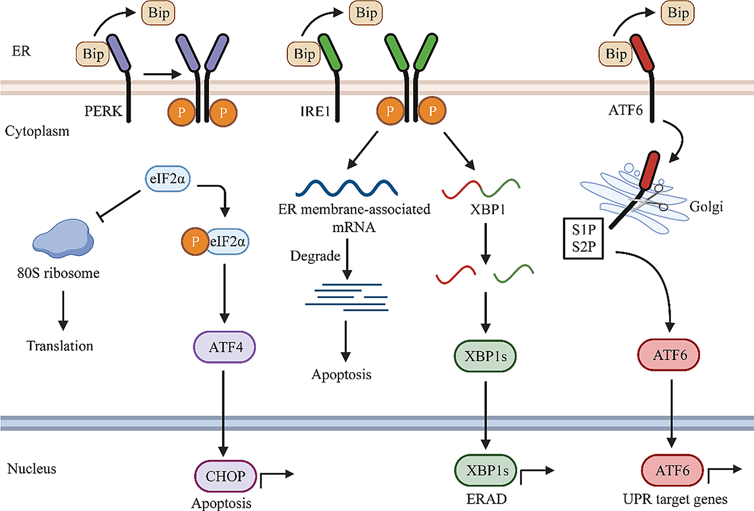
Hypoxia/reoxygenation (H/R)
H/R was used as an in-vitro model of hemorrhagic shock and was conducted as we described previously (Wu et al. 2021). For normoxia, cells were cultured in EBM-2. For H/R, cells were cultured in EBM2 and exposed to 94% N2, 1% oxygen, and 5% CO2 for 18 h followed by normoxia for indicated periods of time in the figure legends.
Western blotting
Endothelial cells were lysed in NuPAGE LDS samples buffer (Thermo Scientific) for Western blot analysis using antibodies including anti-c-Jun (#9165, Cell Signaling Technology). Blots were also probed with anti-GAPDH antibody (PA1-987, Thermo Scientific) for the reference of sample loading.
RNA extraction and quantitative real-time PCR
Total RNA was extracted from cells using Trizol reagent and was reversely transcribed using Qiagen miScript RT kit. Real-time PCR was performed using Qiagen miScript SYBR Green PCR kit and miR-19b-3p miScript Primer (Qiagen). Pri-miR-19b (hsa-mir-92a-1 primer assay) and mature miR-19b primer was obtained from Taqman. RNU6 miScript Primer (Qiagen) was used as an endogenous control. Relative RNA amount was calculated using the 2−ΔΔCt method.
siRNA and miRNA oligo inhibitor transfection
HLMECs were seeded in 6-well plates and grown for 24 h in antibiotic-free EBM-2 containing 5% FBS and supplements. Cells were then transiently transfected by incubation with 100 nM miR-19b oligo inhibitor, c-Jun siRNA, or scrambled RNA (scRNA) and 2.5 ul/ml Lipofectamine 2000 (Thermo Fisher Scientific) in antibiotic-free Opti-MEM for 24 h. The medium was then changed to the growth medium, and the cells were cultured for another 48 h prior to assays. Silencing of the respective proteins was validated by quantitative real-time PCR or Western blot analysis.
Overexpression of c-Jun
HEK293T cells were transfected with pMIEG3-c-Jun overexpression vector or empty vector (pMIEG3) obtained from Addgen (Watertown, MA) using Lipofectamine 2000 following the manufacturer’s recommended procedures. After 3 day-transfection, the expression of c-Jun-GFP was examined under inverted fluorescent microscopy and images obtained. The cells were further exposed to hypoxia for 18 h or exposed to normoxia only for pri-miR-19b induction.
Luciferase reporter assay
The full human miR-19b promoter (miR-17-92 gene cluster promoter, positions 5786 to 8494 in accession# NG_032702) was inserted into lentivirus pEZX-LvPG04 dual-reporter vector (GeneCopoeia, MD). This vector uses Gluc (Gaussia Luciferase) as the promoter reporter and SEAP (secreted Alkaline Phosphatase) as the internal control for signal normalization. Mutated miR-19b promoter vector was produced by GeneCopoeia by deleting the two c-Jun binding sites at positions 6237–6243 (TGACTCT) and 7495–7501 (TGTGTCA) in accession# NG_032702 as predicted by PROMO (http://alggen.lsi.upc.es/cgi-bin/promo_v3/promo/promoinit.cgi?dirDB=TF8.3) using 3% dissimilarity in comparison to the c-Jun consensus sequence (TGAC/GTCA). HEK293T cells were transfected with the vectors containing wild-type miR-19b promoter, mutated miR-19b promoter, or empty vectors in 96-well plates. Some HEK293T cells were also co-transfected with pMIEG3-c-Jun overexpression vector (Addgen, MA) to overexpress c-Jun or empty vector (pMIEG3) for negative control. Some HEK293T cells were exposed to hypoxia for 18 h then normoxia for 3 h to induce c-Jun overexpression. The culture medium of the transfected cells was harvested for assays of promoter activity using Secrete-Pair™ Gaussia Luciferase Dual Luminescence Assay Kit (GeneCopoeia). In the assay, the activities of Gluc and SEAP were detected and Gluc activity was normalized to SEAP activity.
Syndecan-1 immunofluorescence staining
HLMECs were grown directly on 8-well chambers (Corning). Cells were then transiently transfected by incubation with 100 nM c-Jun siRNA or scrambled RNA (scRNA) and 2.5 μl/ml Lipofectamine 2000 in antibiotic-free Opti-MEM for 24 h. The medium was then changed to the growth medium, and the cells were cultured for another 48 h prior to assays. Silencing of the respective proteins was validated by Western blot analysis. After being exposed to hypoxia (94% N2, 1% oxygen, and 5% CO2) for 18 h, cells were fixed in 4% paraformaldehyde for 15 min and blocked with 2% bovine serum albumin (BSA) in PBS for 1 h at room temperature. Cells were then incubated with anti-syndecan1 antibody (1:100; Cell Signaling) in 1% BSA at 4 °C overnight and followed by incubating with Alexa fluor 488 conjugated anti-mouse IgG (1:200; Invitrogen) in 1% BSA for 2 h at room temperature. The fluorescence intensity was quantified using Quantity One and reported as relative fluorescence units.
Endothelial barrier integrity
HLMECs were seeded on culture inserts (3 μm pore size, Costar) in 24-well companion plates and grown to confluence in EBM-2 containing 5% FBS and supplements. In some experiments, monolayers were transfected with 100 nM miR-19b oligo inhibitor, c-Jun siRNA, or scRNA before seeding the inserts. Subsequently, FITC-labeled dextran (40 kDa, Sigma) was added to the upper chamber at a concentration of 100 μg/ml, and phosphate buffered saline (PBS) to the lower chamber (to prevent the formation of an oncotic pressure gradient) for 1 h. Medium was collected from the lower chamber, and the fluorescence was measured using a fluorimeter (485 nm excitation, 530 nm emission). The fold change in FITC-dextran fluorescence intensity over controls was used as a measure of monolayer permeability.
Chromatin immunoprecipitation assay
After exposure to hypoxia for 18 h then normoxia for 3 h, cells were fixed with 1% formaldehyde to crosslink chromatin. Chromatin immunoprecipitation (ChIP) analysis was performed using Piece Magnetic ChIP kit (cat# 26157) and anti-c-Jun antibody (cs#39165, Cell Signaling Technology). The bound-DNA was isolated and purified for quantitative PCR. As described above, the miR-19b promoter contains two c-Jun binding sites at positions 6237–6243 (TGACTCT) and 7495–7501 (TGTGTCA). The primers to detect DNA around position 6237–6243 were 5′-CCTTGTGCGACATGTGCTG -3′ and 5′-GATGGCATGCCGTTAATTTT -3′ (174 bp) and around position 7495–7501 were 5′-GCCACGTGGATGTGAAGATT -3′ and 5′- AAGTGGTGGCTCTTCCAATG -3′ (165 bp). Isotype IgG was used as a negative control. DNA isolated from the whole cell lysates served as input DNA control.
Human study
Available plasma samples from a recently completed prospective observational clinical study in trauma patients (Zeineddin et al. 2022) were used to measure c-Jun. The present study was approved by the Institutional Review Board of the University of Maryland Baltimore. Informed consent was obtained from all patients and included the consent to investigate biologic markers of endothelial dysfunction in the present study. Samples were de-identified and stored prior to bulk analysis. All experimental procedures were conducted in compliance with the University of Maryland Baltimore and the National Institutes of Health guidelines. This study included severely injured patients in hemorrhage shock, defined as a systolic blood pressure < 90 mm Hg and requiring blood component therapy upon arrival. Plasma was collected at admission in the trauma bay and stored in – 80 °C until time of experiment. For healthy donor controls, aliquots were obtained from 13 random donor units of fresh frozen plasmas obtained from Tennessee Blood Services (Memphis, TN). Plasma c-Jun was measured by ELISA (catalog# NBP2-75279, Novus Biologicals).
Statistical analysis
Data are expressed as mean ± SE. Values from different groups were analyzed by T test or one-way analysis of variance (ANOVA) with Bonferroni multiple comparison tests with significance set at level at p < 0.05.


留言 (0)