Strains, plasmids, and materials
The pET28a(+) vector (GenScript Biotechnology Co., Ltd, China), pretreated with NcoI and XhoI, was used to clone the alkaline pectin lyase (BacPelA) gene (NCBI accession no. KR819891.1) from Bacillus clausii synthesized by GenScript Bioengineering Co., Ltd, China. Escherichia coli BL21 (DE3) (laboratory storaged) was the gene expression analysis host strain. TB culture medium was purchased from Beijing Coolebo Technology Co., Ltd, China. Apple and citrus pectin and polygalacturonic acid (PGA) were purchased from Sigma-Aldrich (St. Louis, MO, USA). All restriction enzymes were obtained from Thermo Fisher Scientific (China) Co., Ltd. Medium preparation reagents, agar and yeast extract powder, and peptone were obtained from Beijing Aoboxing Biotechnology Co., Ltd, China. Kanamycin, ampicillin, and isopropyl-β-d-thiogalactoside (IPTG) were purchased from Qingdao Sangon Biotechnology Co. Ltd, China. The chemicals used to prepare the buffers and other reagents were of reagent grade.
Gene cloning and expression plasmid construction of PGLA
The NCBI BLAST program was used to search for the nucleotide sequence of BacPelA (Zhou et al. 2017a). The SignalP5.0 server (http://www.cbs.dtu.dk/services/SignalP/) was used to predict the signal peptide, which was removed. The BacPelA encoded DNA fragment comprised 304 amino acids (AA), with a total of 912 base pairs. The fragment’s two ends were modified with NcoI and XhoI, subjected to codon optimization to obtain the gene PGLA (accession no. OP355468), and were then ligated to the pretreated pET28a(+) vector, creating the ligation product pET28a(+)-PGLA (Additional file 1: Fig. S1). This recombinant plasmid was PGLA and then was transformed into E.coli BL21(DE3) competent cells, spread on Luria-Bertani (LB) agar (yeast dip powder, peptone, and NaCl; 5, 10, and 10 g/L, respectively) plates containing 50.0 µg/mL kanamycin, and incubated overnight at 37 °C. The cultured colonies were collected using a sterile pipette tip and transferred to tubes containing 1 mL of LB solution. These were shaken and incubated at 37 °C for 14 h. Thereafter, 1 mL of 50% sterilized glycerol was added to each tube, and the components were mixed and stored at – 80 °C.
Modification of gene sequence of mutated alkaline pectin lyase with high enzyme activity
Sukhumsiirchart et al. (2009) studied pectin lyase Pel SWU (accession no. AB428424) from Bacillus sp. RN1, which has superior heat resistance compared to other similar enzymes. The NCBI program provided the nucleotide sequence of Pel SWU. The SignalP5.0 server predicted the signal peptides, which were subsequently removed. The 5′- and 3′-ends of the fragment were modified using NcoI and XhoI, respectively, and then ligated into the pretreated pET22b vector (GenScript Biotechnology Co., Ltd, China), creating the ligation product pET22b-Pel.
The AA sequences of PGLA and Pel were compared using ESPript 3.0 (https://espript.ibcp.fr/ESPript/ESPript/index.php). Four DNA fragments had a low sequence similarity at the N-terminus; this part was replaced. All the oligonucleotides used in the fragment replacement procedure are listed in Additional file 1: Table S1. The first replaced 5′ fragment was short. Thus, we used the PGLA1-F and PGLA1-R primer pairs to clone the entire recombinant plasmid (pET28a(+)-PGLA-rep1), via inverse PCR, with the pectin lyase pET28a(+)-PGLA DNA as the template. The second, third, and fourth N-terminal DNA fragments were then replaced by seamless cloning.
The replacement gene vector fragment was obtained by PCR amplification of the pectin lyase pET28a(+)-PGLA DNA template, with the primer pairs PGLA2-F, PGLA34-F, PGLA23-R, and PGLA4-R. The PCR amplification conditions are as follows: one reaction cycle at 95 °C for 3 min; pre-denaturation at 95 °C for 15 s, denaturation at 60 °C for 15 s, and extension at 72 °C for 3.2 min; 30 final extension cycles at 72 °C for 5 min; and storage at 4 °C.
We obtained the replacement gene fragment via PCR amplification using the primer pairs rep23-F, rep4-F, rep2-R, rep3-R, and rep4-R and the DNA template of pectin lyase pET22b-Pel. PCR amplification conditions: one cycle of reaction at 95 °C for 3 min; 30 cycles of pre-denaturation at 95 °C for 15 s, denaturation at 60 °C for 15 s, and extension at 72 °C for 20 s; a final extension at 72 °C for 5 min; and storage at 4 °C.
The above three pairs of DNA fragments were subjected to sodium dodecyl sulfate-polyacrylamide gel electrophoresis (SDS-PAGE). Thereafter, gel recovery was carried out with a product purification kit (Nanjing Novizan Biotechnology Co., Ltd, China), and the concentration of DNA fragments was measured with an MD2000 ultra-micro spectrophotometer (Shanghai Meixi Instrument Co., Ltd, China). The replacement gene fragment and vector fragment were ligated using the seamless cloning kit C112 (Nanjing Novizan Biotechnology Co., Ltd, China) to obtain the recombinant plasmids pET28a(+)-PGLA-rep2, pET28a(+)-PGLA-rep3 and pET28a(+)-PGLA-rep4. The recombinant plasmids were transformed into E. coli BL21(DE3) competent cells, spread on LB agar plates and incubated overnight at 37 °C. The cultured colonies were collected using a sterile pipette tip and transferred to a tube containing 1 mL of LB solution. The tubes were shaken and incubated at 37 °C for 14 h. Thereafter, 1 mL of 50% sterilized glycerol was added to each tube, mixed, and stored at – 80 °C.
Culture conditions for expression of alkaline pectin lyase in E. coli
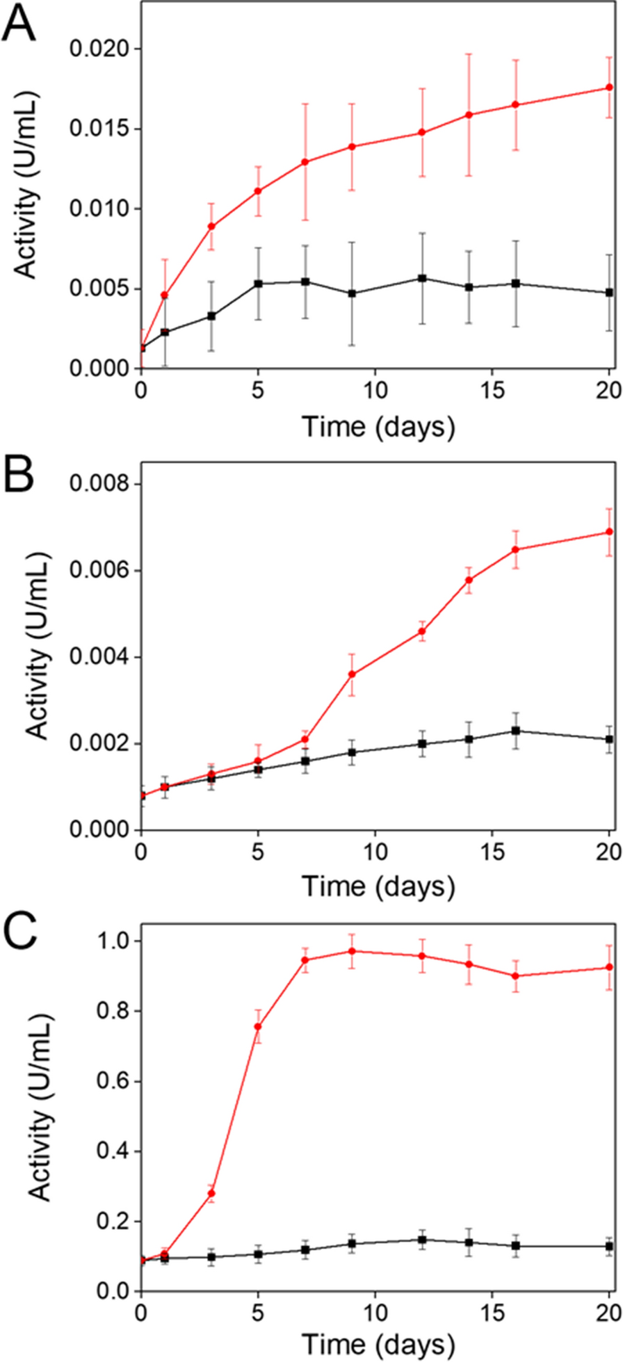
Approximately 100 µL of frozen (– 80 °C) glycerol bacteria was added to 50 mL of LB medium containing 50 µg/mL kanamycin and cultured overnight at 37 °C to collect exponential phase cells. 300 uL seed medium containing exponential growth cells was inoculated into 50 mL TB medium containing 50 ug/mL kanamycin. The content of these flasks was cultured at 37 °C and 200 r/min. When the optical density at 600 nm (OD600) reached 0.6–0.8, isopropyl-β-d-thiogalactoside (IPTG) was added at a final concentration of 0.5 mM. Consequently, the protein was expressed in shaking flasks (200 r/min) for 24 h at 25 °C.
Determination method and operation steps of enzyme activity
The activity of alkaline pectin lyase was assayed by measuring the increase in unsaturated bonds at 235 nm (A235 method). The fermentation broth (30 mL) was centrifuged to remove the supernatant, resuspended the pellet in 8 mL of phosphate-buffered saline (pH 7.4), and sonicated for 20 min. After centrifugation, the supernatant was collected to obtain the enzyme solution and stored at 4 °C. The enzyme activity assay was performed as described by Zhou et al. (2017a) with some modifications. We added 190 mL of glycine-NaOH (Gly-NaOH) buffer (pH 11.0), containing 0.2% pectin substrate and 100 µL of appropriately diluted enzyme solution, to a 25 mL colorimetric tube. The contents were mixed and allowed to react at 70 °C for 10 min. The reaction was terminated by adding 3 mL of 30.0 mM H3PO4. We used an V-5600 (PC) UV–Vis spectrophotometer (Shanghai Youke Instrument Co., Ltd, China) to measure the absorbance of the unsaturated product at 235 nm. One enzymatic activity unit is defined as the amount of enzyme required to cleave PGA to produce an equivalent of 1 µmol unsaturated oligogalacturonic acid per min. The molar absorption coefficient of unsaturated polygalacturonic acid at 235 nm was 4600 L/mol/cm. All enzyme activity measurements were performed in triplicate.
Effects of pH, temperature and metal ions on enzyme activity and stability
To determine the optimum pH, we measured the absorbance of the enzyme solution at pH 8.5–11.5 with 50 mM Gly-NaOH buffer containing 0.2% apple pectin at 55 °C for 10 min. The optimal temperature was determined by measuring the absorbance of the enzyme solution (pH 11.0) at 55–85 °C (intervals of 5 °C) for 10 min. The pH stability was determined by the residual enzyme activity after 7 h incubation in Na2HPO4-citrate buffer (pH 4.0–7.0), 50 mM Tris-HCl buffer (pH 8.0), and Gly-NaOH buffer (pH 9.0–12.0) at 50 °C. The stability of pectin lyase was measured every hour for 5 h based on the residual enzyme activity after incubating in Gly-NaOH buffer (pH 11.0 and 12.0) at 25 °C. The thermal stability of pectin lyase was measured every hour for 5 h by testing the residual enzyme activity after incubating the enzyme solution at 60 and 70 °C. All experiments were repeated three times.
We incubated the enzyme, at room temperature, for 60 min, in a solution containing 1 mM of one of the following metal ions: K+, Ca2+, Na+, Mg2+, Cu2+, Mn2+, Fe2+, Zn2+, Fe3+, and Ni+. We then measured the relative enzyme activity under standard reaction conditions (pH 11.0, 70 °C) to determine the effect of metal ions on enzyme activity. We performed a control assay in the absence of metal ions to determine purified enzyme activity.
Substrate specificity, molecular dynamics simulation, and kinetic parameter calculation
We determined substrate specificity by measuring the residual enzymatic activity of purified PGLA-rep4 on different substrates, including apple and citrus pectin and PGA, at a concentration of 0.2% under standard conditions (pH 11.0, 70 °C). Using the Swiss-model website (https://swissmodel.expasy.org/), we performed a sequence alignment on the five pectin lyases before and after the fragment was replaced. Thereafter, the Modeller 10.2 software (University of California San Francisco, San Francisco, CA, USA; http://salilab.org/modeller) was used to perform three-dimensional (3D) modeling. Molecular dynamics (MD) simulations of PGLA, PGLA-rep1, PGLA-rep2, PGLA-rep3, and PGLA-rep4 were performed using Gromacs 4.5 package (Royal Institute of Technology, Stockholm, and Uppsala University, Uppsala, Sweden; http://www.gromacs.org/), with the GROMOS 96 forcefield, and a simple point charge (SPC) water model. The protein was placed in a square box, with the edge of the box no closer than 1.5 nm to the protein, and 15,000 water molecules were added to the solvate proteins. After adding two Na+, the net charge of the system was zero, reaching an equilibrium state. Subsequently, to minimize the energy of the system, 1500 steps of steep descent and 2000 conjugate gradients were performed. Molecular dynamics simulations were performed at a constant temperature and pressure for 20 ns, with each step comprising 0.02 ps. The B-factor value of the amino acid residue was generated after MD simulation of the three-dimensional structure of the protein, that is, the atomic displacement parameter. The Km and Vmax enzyme values were calculated using nonlinear regression. All data were expressed as the average of three experiments.


留言 (0)