Cell cultures
The human GBM cell lines U87(RRID: CVCL_0022), LN229 (RRID: CVCL_0393), U251 (RRID: CVCL_0021), and A172 (RRID: CVCL_0131) and human embryonic kidney cell line (293FT, RRID: CVCL_6911) were purchased from ATCC. The cells were cultured in DMEM supplemented with 10% FBS, 1% penicillin, and streptomycin. All cells within passages 8 to 15 were used and passed the detection of mycoplasma contamination by Myco-LumiTMMycoplasma Kit (Beyotime, China).
Lentivirus preparation and infection
The shRNA and negative control of VHL were constructed in the pLKO.1 vector, and the overexpression and negative control of VHL were constructed in the PLVX-Flag vector (IGEbio,China). The shRNA and negative control S100A16 were constructed in the Tet-pLKO vector (IGEbio,China), and DOX (Sigma, USA) at 1 μg/mL induced shRNA expression. The sequences of the knockdown genes were as follows:
The S100A16 shRNA targeting sequences are listed below:
S100A16 shRNA-1: 5′-CAG TCA TTG TCC TGG TGG AAA TTT CCA CCA GGA CAA TGA CTG-3′;
S100A16 shRNA-2: 5′-CGA TGA GTA CTG GAC CTT GAT ATC AAG TCC AGT ACT CAT CG-3′;
S100A16 shRNA-3: 5′-CAG CCT GGT CAA GAA CAA GAT ATC TTG TTC TTG ACC AG -3′;
1 μg/mL Puromycin (Beyotime, China) was used to screen stable strains of cells.
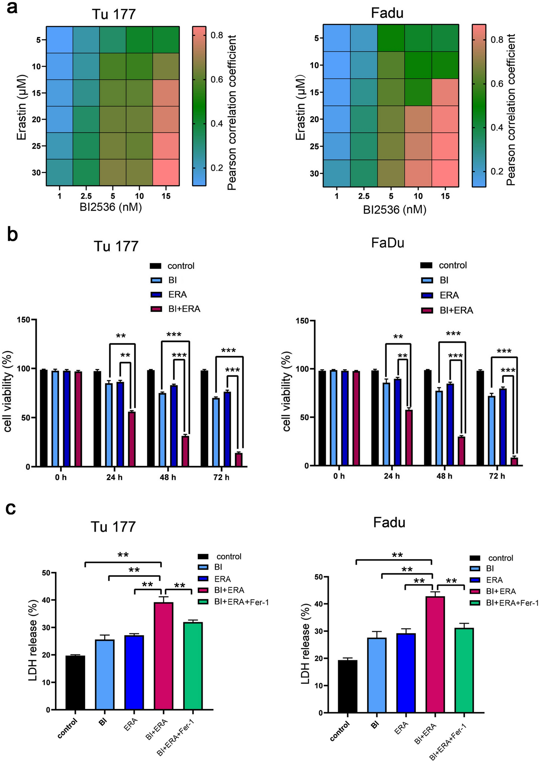
Cell viability and proliferation assay
GBM cells (2 × 10³/well) were cultured overnight in 96-well plates; GBM cells were treated with GNE987, JQ1, ARV825, dBET1 (MedChemExpress, USA) or DMSO (Sigma, USA) for 3 days,5 days or 7 days, and the absorbance of a 96-well plate was measured using a microplate reader with CCK8 (Dojindo, Japan). Cell viability and proliferation rates were calculated using GraphPad Prism8.4.0.
Clone formation assay
GBM cells (1 × 10³/well) were seeded into 6-well plates, and DMSO or GNE987 was incubated for 2 weeks. GBM cells were washed with PBS and fixed with methanol and stained with Giemsa (Solarbio, China). Pictures were taken and clones were counted.
Cell cycle analysis
GBM cells (20 × 104/well) were seeded into 6-well plates, and DMSO or GNE987 was incubated for 3 days. Cold 70% ethanol was applied overnight to GBM cells, followed by one wash with PBS the next day. Light-free incubation using the cell cycle analysis kit (Cat#C1052, Beyotime, China) for 30 min. Flow cytometry (Beckman Gallios, USA) was used to test the cells.
Cell apoptosis assay
GBM cells (20 × 104/well) were seeded into 6-well plates, and DMSO or GNE987 was added and incubated for 3 days. And GBM cells were stained using the FITC-Annexin V Apoptosis Kit (Cat#556547, BD, USA). Flow cytometry (Beckman Gallios, USA) was used to test the cells and analyzed to determine the proportion of apoptotic cells.
EdU staining analysis
GBM cells (5 × 10⁴/well) were seeded into 24-well plates and treated with GNE987 or DMSO for 3 days, then stained by the EdU staining kit (BeyoClick™ EdU-488 Cell Proliferation Assay kit, Beyotime, China) according to a previous protocol [14]. Cells were treated with EdU working solution for 2 h, 4% paraformaldehyde for about 10 min, 3% BSA for 1 h, and then A 30 min click reaction at room temperature away from light, and DAPI for 5 min.
In vivo xenografts
For the GBM subcutaneous transplanted tumor model, 5 × 10⁶ U87 cells were inoculated in the left- back of nude mice (Shanghai ling chang biotech, 4 weeks of age, female, n = 6/group), and tumor-bearing mice were randomly divided into two groups, the drug treatment group, and the vehicle group. The day the tumor was received was defined as day 0, from the 3rd day after the tumor, and the drug treatment group was given an intraperitoneal injection of GNE987 (0.25 mg/kg) every 2 days according to a previous protocol [7]; the vehicle group was administered the same dose of 5% Kolliphor®HS15 as GNE987. The mice were weighed and measured every two to three days to determine their body weight and tumor volume. The survival endpoint was defined as when the tumor in the vehicle group exceeded 1 cm3. The Animal Ethics Committee of Soochow University approved this research (CAM-SU-AP#: JP-2018-1).
Immunohistochemistry (IHC)
IHC was performed using the IHC Kit (Cat#KIT-9720, MXB Biotechnology, China). Sections were subjected to dewaxing, hydration, antigen retrieval, Ki67 antibody (Cat. ab15580, Abcam, UK), DAB chromogenic reaction (Cat#DAB-2031, MXB Biotechnology, China), hematoxylin staining (Beyotime, China), and dehydration. Brown-yellow indicated positive expression, and violet-blue indicated the nucleus.
RT-qPCR
Cells were harvested using TRIzol reagent (Invitrogen, USA), total RNA was extracted using Chloroform reagent, isopropanol reagent, and cDNA was synthesized according to a previous protocol [7]. RT-qPCR was performed using LightCycler 480 Real Time System (Roche, Germany). The relative mRNA expression was calculated using the 2–ΔΔCT method. Glyceraldehyde-3-Phosphate dehydrogenase (GAPDH) was used for interior management. The primer sequences used were as follows:
GAPDH: 5′-ATC ATC CCT GCC TCT ACT GG-3′ (forward) and 5′-CCC TCC GAC GCC TGC TTC AC-3′ (reverse).
cyclinB: 5′ TCG CCT GAG CCT ATT TTG GT-3′ (forward) and 5′-GCA TCT TACT TGG GCA CAC AA-3′ (reverse).
cdc2: 5′ AGT CTG GTC TTT CTT TGG CTG TCA G-3′ (forward) and 5′-AAA CAC CTA CAA CCA CCA CTC TGC-3′ (reverse).
WNT5A: 5′-TAC GAG AGT GCT CGC ATC CTC A-3′ (forward) and 5′-TGT CTT CAG GCT ACA TGA GCC G-3′ (reverse).
ZMYND8: 5′-AAG CGC CAG ATT CGT AGC AGG T-3′ (forward) and 5′-TCC TCC GAA TCG CTG TGC TCT A-3′ (reverse).
BCL2L1: 5′-GCC ACT TAC CTG AAT GAC CAC C-3′ (forward) and 5′-AAC CAG CGG TTG AAG CGT TCC T-3′ (reverse).
CAV1: 5′-CCA AGG AGA TCG ACC TGG TCA A-3′ (forward) and 5′-GCC GTC AAA ACT GTG TGT CCC T-3′ (reverse).
TBX2: 5′-AGC AGT GGA TGG CTA AGC CTG T-3′ (forward) and 5′-GGA TGT CGT TGG CTC GCA CTA T-3′ (reverse).
STEAP3: 5′-TGC AAA CTC GCT CAA CTG GAG G-3′ (forward) and 5′-AGG CAG GTA GAA CTT GTA GCG G-3′ (reverse).
POU2F2: 5′-TCC TGG AGA AGT GGC TCA ACG A-3′ (forward) and 5′-ATG CTG GTC CTC TTC TTG CGT C-3′ (reverse).
EPHA2: 5′-ACT GCC AGT GTC AGC ATC AAC C-3′ (forward) and 5′-GTG ACC TCG TAC TTC CAC ACT C-3′ (reverse).
KCNJ15: 5′-TGT GCT TGG TGA TTC AGG TAG CC-3′ (forward) and 5′-GAC AGT GGC TTG GTT GAG GAG A-3′ (reverse).
EPSTI1: 5′-ACT GAA ACG GCA GCA GCA AGA G-3′ (forward) and 5′-TCC AAC AGC CTC CAG ATT GCT C-3′ (reverse).
PALMD: 5′-GAG GAA GAC AAA CTA AAG CAC CAG-3′ (forward) and 5′-CTC TTC CTG TTC TTT TCC GCT GC-3′ (reverse).
S100A16: 5′-GCT CCA GAA AGA GCT GAA CCA C-3′ (forward) and 5′-ATG CCG CCT ATC AAG GTC CAG T-3′ (reverse).
Western blot
GBM cells were treated with GNE987 or DMSO for 48 h. Then, proteins were extracted with RIPA supplemented with protease and phosphatase inhibitors, Western blot was performed using appropriate primary and secondary antibodies. The specific antibodies included the following: BRD4 (Cat#13440 s, CST, USA), BRD2 (Cat#5848 s, CST, USA), β-TUBULIN (Cat#2146, CST, USA), FLAG (Cat#14793S, CST, USA), VHL (Cat#68547S, CST, USA) and C-Myc (Cat#9402; CST, USA), BRD3 (Cat#11859-1-AP, Proteintech, USA), and GAPDH (Cat# MA3374, Millipore, USA).
RNA sequencing analysis
For RNA-seq (Novogene Ltd., China), U87 cells were treated with GNE987 and DMSO for 48 h, and then harvested using TRIzol reagent (Invitrogen, USA). Differentially expressed genes were identified by the Bioconductor DESeq2.
Chromatin immunoprecipitation sequencing (ChIP-seq) data processing
ChIP was performed according to a previous protocol [7]. First, 3 × 10⁷ U87 cells were cross-linked with 1% paraformaldehyde at room temperature for 10 min, neutralized with 0.125 M at room temperature for 5 min, centrifuged to collect the cells, and lysed with cell lysis buffer on ice for 5 min; cells were lysed by repeatedly aspirating the solution with an insulin needle. The solution was centrifuged, pellets were resuspended in shearing buffer, and genomic DNA was cleaved into fragments of approximately 500 bp using a sonicator (M220, Covaris, USA). After centrifugation, the supernatant was collected and the H3K27Ac antibody (Cat. ab4729, Abcam, UK) was added to it overnight at 4 °C; the next day, Dynabeads Protein G beads (Thermo Fisher Scientific, USA) were added to the supernatant at 4 °C for 4 h for immunoprecipitation. The magnetic beads were washed with TE buffer (Sigma, USA); EB buffer was added, and shaken for 15 min to separate the magnetic beads, antibodies, proteins, and DNA; the supernatant was taken, and 5 M NaCl was added to it, and the mixture was heated at 65 °C overnight. The antibodies, proteins, and DNA were separated using a PCR purification kit (QIAGEN, Germany). ChIP-seq was provided by BGI Ltd (China).
Statistical analysis of data
Data and graphs were processed using GraphPad Prism (version 8.4.0, USA). Comparison between the two groups was performed by Student’s t test. ANOVA was used for comparison between multiple groups. In the graphs, mean ± standard deviation (SD) is represented; P value < 0.05 denoted statistical significance (*P < 0.05, **P < 0.01, ***P < 0.001).

留言 (0)