
記住我


Q fever is a worldwide zoonosis caused by Coxiella burnetti. Farm animals are the main reservoirs to human infection,1 being the most common mode of transmission inhalation of contaminated aerosols.2
Acute Q fever is asymptomatic in up to 60% of cases, whereas most of the remaining subjects will develop a mild illness.1,2-4 The top clinical symptoms of primary infection in children are respiratory symptoms, malaise, and gastrointestinal signs.4 Children with primary infection are more often asymptomatic than adults,1,3-5 and less than 1% develop a chronic infection, especially osteoarticular disease.2 The optimal antimicrobial therapy for this infection remains unclear.3,5
CLINICAL CASEA 2-year-old healthy Caucasian girl was diagnosed with suppurative arthritis. A bone scintigraphy showed an increased uptake in the right distal femoral metaphysis, and a Brodie abscess was confirmed by magnetic resonance imaging (MRI), which was drained (negative culture and 16SrRNA gene PCR). She was treated with intravenous (IV) and oral (PO) cefuroxime. A Mantoux and different serology tests, including Bartonella and Brucella, were negative.
A month later, she developed a recurrence of the abscess. Thus, the patient was switched to vancomycin and cefotaxime and transferred to our center. On arrival, antibiotics were switched to IV cefuroxime, with a favorable outcome without surgery, and she was discharged on PO cefuroxime.
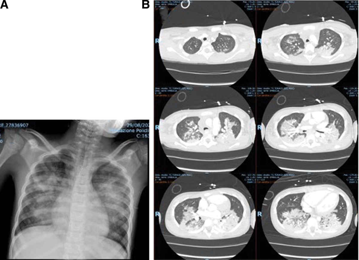
Two months later, the patient relapsed again with a dorsomedial side of her right foot swelling. Ultrasound and MRI showed a soft tissue abscess, osteomyelitis of the right first metatarsal, and persistence of the previous abscess with bone involvement in the ipsilateral femoral distal metaphysis (Fig. 1A,B). She was hardly symptomatic, without fever, or limping. A bone scintigraphy only showed a slightly increased activity on the right talocalcaneal joint, and a surgical drainage was performed. A presumptive diagnosis of chronic multifocal recurrent osteomyelitis (CRMO) was suspected, and antibiotics were stopped.
 FIGURE 1.:
 (A) Knee MRI. Axial T1 contrast-enhanced image. (B) Knee MRI. Coronal PDW contrast-enhanced fat suppressed image. (C) Left foot. (D) PET-CT showing 2 foci. MRI = magnetic resonance imaging; PDW = proton density-weighted; PET-CT = positron emission tomography–computed tomography.
Since the child had a dog and had been exposed to goats, C. burnetii serology (IFI) was ordered, showing a positive phase I IgG (1/1600) and IgA (1/50), with negative IgM; phase II IgG was also positive (1/100), with negative phase II IgM and IgA. Since the 16SrRNA PCR from surgical samples was negative and considering serology results, a specific C. burnetii PCR was performed, with positive result. An echocardiogram was normal, as it was an extensive immunologic study including immunoglobulins and complement levels, neutrophil function tests, lymphocyte and natural killer cell counts, and T-cell response to PHA stimulation.
PO rifampin and ciprofloxacin combination was initiated but the child continued having relapses. One month later, the abscess of the right foot fistulized and inflammation on the medial malleolus of her left ankle appeared (Fig. 1C). She was treated with IV doxycycline and rifampin for 10 days, with some improvement, and continued with the previous regimen, but 1 month later, she again underwent surgical drainage of both sites. MRI reported an involvement of the first metatarsal and the first cuneiform of the right foot, and the calcaneus of the left foot. She was discharged on PO levofloxacin and rifampin.
Because she continued developing abscesses despite antimicrobial and surgery therapies, adjuvant immunomodulatory treatment with subcutaneous (SC) INF-γ was added. A defect of the interleukin-12/INF-γ pathways was excluded. A few weeks later, and because of the persistence of intermittent swelling, azithromycin, and trimethoprim-sulfamethoxazole (TMP-SMX) were also added, which were maintained for more than 6 months. After these new treatments, she experienced a clear improvement of the swelling, requiring only 1 additional surgery. An antibiotic tapering was initiated, starting with azithromycin, levofloxacin being the last antibiotic stopped. Overall, she received 48 months of antibiotics with fluoroquinolones and rifampin as the backbone therapy. Moreover, she completed 51 months of SC INF-γ, with a maximum dose of 100 mcg/m2, 3 times a week. INF-γ was stopped 7 months after antibiotics.
She has been followed at our clinic, with multiple laboratory tests and radiological studies (MRI, scintigraphy’s, PET-TAC [Fig. 1D]) performed, as well as cardiology assessments. Besides the extensive local osteoarticular involvement, no other pathological findings or adverse events were observed. She is currently 11 years of age and has been off treatment for more than 5 years, remaining asymptomatic. Maximum C. burnetti phase I IgG titer was 1/3200, maintaining 1/400 in the last visit. The rest of the C. burnetii antibodies are currently negative.
DISCUSSIONChronic Q fever has rarely been described in children, possibly because it has fewer and milder symptoms than in adults, but it might be underdiagnosed.1-4 Children generally are diagnosed at young age (2–5 years) and are predominantly male.1 This age may reflect a later acquisition of natural immunity against C. burnetti or a lack of maturation of the innate immune system.3 Most frequent clinical presentations include osteoarticular infection (often recurrent and multifocal) in previously healthy children,2-4 and endocarditis in children with underlying cardiac disease.1,2,4,5
We identified 28 pediatric cases of Q fever with osteoarticular infections (OAI) in the literature; 71% male.2-6 The most common involved sites were lower limbs, pelvis, vertebrae and chest wall, being unexplained the predilection for the latter location.3 Furthermore, 17 (61%) had multifocal involvement, and two-thirds an initial diagnosis of CRMO, as our patient. Most children experienced symptoms long before diagnosis: from 2 weeks to 5 years (Table 1 shows a review of the literature with the characteristics of children with this disease).
TABLE 1. - Cases of Osteoarticular Infection DUE TO C. burnetti Reported in the Literature in Children up to 17 Years of Age Patient (Year of Publication) Age (Years)/Sex, Referencexx Previous diseases/Risk Factors Duration of Symptoms Before Diagnosis Bone and Joint involvement; Soft Tissue Involvement (^) Diagnosis Phase I IgG: at Diagnosis/Maximum Treatment* Complications/Sequelae 1 (1995) 7/male2 No/exposure to goats 8 mo Lumbar (3) vert, R hip Serology, culture 1:512/1:512 2 yr DOX + 6 mo RIF CRMO for 8 months/No 2 (1995) 9/male2 No/cat at home NR Calcaneus Serology 1:5120/1:5120 6 mo LIN + bone graft None 3 (1995) 2/female2 No/exposure to goats 5 mo before diagnosis NR L fibula, radius and humerus; R carpus Serology, culture, PCR 1:1600/1:3200 6 mo RIF →OFX+THI→ 4 yr TMP-SMX 3 surgical debridement CRMO for 3 years/No 4 (2004) 4/male3,2 No/living in a dairy farm 5 yr Thoracic (10) vert, R navicular, both feet^, chest wall^ Serology, PCR 1:2560/1:2560 CLM + TMP-SMX→ DOX + RIF→ 4 mo CIP→3 yr DOX+HCQ 6 surgical debridement. CRMO for 5 years/No 5 (2004) 7/male3,2 No/living in a cattle farm. Father Q fever 9 mo L tibia, R radius and shoulder, Lumbar (5)-sacral (1) vert Serology 1:10240/1:10240 4 mo TMP-SMX+DOX + RIF→ 18 mo CIP→ 18 mo DOX+ HCQ 4 surgical debridement CRMO for 13 months/No 6 (2004) 3/male3,2 No/living in a horse-cattle property 4 mo L radius and tibia, R femur, calcaneus and ankle Serology, PCR 1:10240/1:10240 7 mo RIF+ CIP→ 18 mo DOX+HCQ→ 18 mo DOX 4 surgical debridement. CRMO/No 7 (2011) 3/female2 No/rural area. Contact with horses 12 mo L humerus, R femur, tarsus, knee and wrist, thoracic (9-11) vert, chest wall^ Serology, PCR 1:32000/1:32000 40 mo RIF+CIP + 17 mo INF-γSurgery Spinal abscesses. CRMO for 30 months/No 8 (2014) 4/female2 No/rural area. Exposure to cats 4 mo Ulna/Elbow Serology, PCR 1:512/1:1024 6 mo TMP-SMX + 12 mo RIF+CIP+AZT 3 surgical debridement No/Minor limitation of elbow extension 9 (2015) 6/male2 No/living in a farm. Father worked in an abbatoir 4 mo L femur Serology, PCR 1:512/1:512 1 surgical debridement No antibiotic administered None 10 (2015) 6/female2 No/rural area. Exposure to farm animals 3 mo R femur (also growth plate) Serology, PCR 1:6400/1:6400 18 mo RIF+ CIP None 11 (2015) 5/female2 No/rural area. Exposure to cats/cattle 2 weeks Thoracic (2) vert, sternum Serology, PCR 1:25600/1:25600 18 mo RIF+ CIP None 12 (2016) 2/female3,2 No/unknown potential exposure risk NR R ilium, sternum, chest wall^ Serology, PCR 1:3200/NR 12 mo DOX+HCQ 3 surgical debridement CRMO/No 13 (2016) 3/male3,2 No/Unknown potential exposure risk NR L calcaneus, femur, tibia and knee Serology, PCR >1:1280/NR 5 mo CIP+RIF→36 mo AZT+CIP+RIF 6 surgical debridement CRMO/No 14 (2016) 4/male3,2 No/Rural area 3 mo Both femur, R tibia, talus and ankle, L cuneiform, knee and foot^ Serology, PCR 1:3200/NR 7 mo D+HCQ→ 4 mo AZT+DOX+HCQ→ 20 mo DOX+HCQ. 9 mo.INF-γ 11 surgical debridement CRMO/Valgus deformity of L femur, corrected with osteotomy at 8 years of age 15 (2016) 2/male3,2 No/Unknown potential exposure risk NR Both femur, iliac crest and navicular, L cuneiform, R acetabulum, calcaneus, tibia, metatarsal and wrist Serology, PCR 1:3200/NR 2 mo AZT→ 22 mo& DOX+HCQ 2 surgical debridement CRMO/NR 16 (2016) 4/male3,2 No/Unknown potential exposure risk NR L femur and knee Serology, PCR 1:3200/NR 12 mo AZT 2 surgical debridement CRMO/No 17 (2016) 6/female3,2 No/Unknown potential exposure risk NR L acetabulum, hip and obturator^ Serology, PCR >1:1280/NR 18 mo DOX+HCQ 1 surgical debridement CRMO/No 18 (2016) 3/male3,2 No/Unknown potential exposure risk NR L cuneiform, and radius, R 7th rib, cuboid and femur, chest wall^ Serology, PCR 1:800/NR 16 mo& DOX+HCQ 4 surgical debridement CRMO/No 19 (2016) 2/male3,2 No/Unknown potential exposure risk NR L femur and knee, sternum, chest wall^ Serology, PCR 1:3200/NR 4 surgical debridement CRMO/No 20 (2016) 4/male3,2 No/Unknown potential exposure risk NR L femur and knee Serology 1:3200/NR 2 surgical debridement CRMO/No 21 (2018) 3/male5,2 No/Exposure to dogs, cats and cattle. NR Talus Serology, PCR 1:600/1:6400 18 mo DOX+RIF→ DOX+HCQ& NR/No 22 (2018) 13/male5,2 BMT/NR NR Hip PCR 1:100/1:100 1.5 mo DOX NR/No 23 (2018) 3/male5,2 No/NR NR Ankle Serology, PCR 1:6400/1:12800 RIF+CIP NR/No 24 (2018) 5/male5,2 No/NR NR Tibia Serology, PCR 1:800/1:3200 24 mo DOX+RIF → DOX+RIF+HCQ& CRMO/No 25 (2018) 3/male5,2 No/Exposure to sheep NR Tibia Serology 1:800/1:3200 5 mo RIF+CIP→ 12 mo DOX+RIF NR/No 26 (2018) 6/female5,2 No/Rural area NR CRMO (NR) Serology 1:3200/1:12800 12 mo DOX+RIF+HCQ → DOX+RIF CRMO/No 27 (2018) 14/male5,2 No/NR NR Hip Serology 1:1600/1:3200 3 mo DOX+RIF+HCQ → DOX+HCQ NR/No 28 (2018) 7/male5,2 No/Exposure to cats NR NR Serology 1:6400/1:25600 4 mo DOX+RIF→ 4 mo DOX+HCQ Suspected reactive arthritis /NR 29 Our case 2/female No/Feeding animal at the zoo Goat at school 3 mo R femur, first cuneiform, 1st MTT and talo-calcaneal, L calcaneus ankle and foot^ Serology, specific C. burnetti 16SrRNA PCR and ulterior Bartonella PCR 1:1600/1:3200 RIF+CIP→ DOX+RIF→ LEV+RIF→ CIP+RIF+AZI+TMP-SMX→ tapering until stopping treatment (5 yr total) 51 mo INF-γMultiple surgical debridement CRMO for 12 months/NoAZT: azithromycin. CIP: ciprofloxacin. CLM: clarithromycin. CRMO: chronic recurrent multifocal osteomyelitis. DOX: doxycycline. HCQ: hydroxychloroquine. HSCT: Hemopoietic stem cell transplantation. INF-γ: interferon gamma. L: left. LEV: levofloxacin. LIN: lincomycine. MTT metatarsal. Mo: months. NR: non-reported. OFX: ofloxacin. PCR: polymerase chain reaction. RIF: rifampin. R: right. THI: thiophenicol. TMP-SMX: trimethoprim/sulfamethoxazole (cotrimoxazole). Vert: vertebrae. Yr: years.
→ Change of treatment. &Continuing therapy. ^Soft tissue involvement. *Duration of treatment not always available.
XXReferences 2, 3 and 5 are revisions including the cases of the table, but reference for specific cases may be consulted in these revisions.
Patients 1, 2 and 3. Cottalorda J, Jouve JL, Bollini G, et al. Osteoarticular infection due to Coxiella burnetii in children. J Pediatr Orthop B. 1995;4(2):219-21. doi: 10.1097/01202412-199504020-00018. PMID: 7670995.
Patients 4, 5 and 6. Nourse C, Allworth A, Jones A, et al. Three cases of Q fever osteomyelitis in children and a review of the literature. Clin Infect Dis. 2004 Oct 1;39(7): e61-6. doi: 10.1086/424014. Epub 2004 Sep 13. PMID: 15472834.
Patient 7. Neth OW, Falcon D, Peromingo E, et al. Successful management of chronic multifocal Q fever Osteomyelitis with adjuvant interferon-gamma therapy. Pediatr Infect Dis J. 2011 Sep;30(9):810-2. doi: 10.1097/INF.0b013e31821487f5. PMID: 21372749.
Patient 8. Britton PN, Macartney K, Arbuckle S, et al. A Rare Case of Q Fever Osteomyelitis in a Child From Regional Australia. J Pediatric Infect Dis Soc. 2015 Sep;4(3): e28-31. doi: 10.1093/jpids/piu095. Epub 2014 Oct 17. PMID: 26407439.
Patient 9. Khatami A, Sparks RT, Marais BJ. A Case of Pediatric Q Fever Osteomyelitis Managed Without Antibiotics. Pediatrics. 2015 Dec;136(6): e1629-31. doi: 10.1542/peds.2015-0024. Epub 2015 Nov 16. PMID: 26574586.
Patient 10 and 11. Costa B, Morais A, Santos AS, et al. Q Fever Chronic Osteomyelitis in Two Children. Pediatr Infect Dis J. 2015 Nov;34(11):1269-71. doi: 10.1097/INF.0000000000000861. PMID: 26226441.
Patients 12 a 20. Francis JR, Robson J, Wong D, et al. Chronic Recurrent Multifocal Q Fever Osteomyelitis in Children: An Emerging Clinical Challenge. Pediatr Infect Dis J. 2016 Sep;35(9):972-6. doi: 10.1097/INF.0000000000001211. PMID: 27294309.
Patients 21 a 28. Sachs N, Atiya-Nasagi Y, Beth-Din A, et al. Chronic Q Fever Infections in Israeli Children: A 25-year Nationwide Study. Pediatr Infect Dis J. 2018 Mar;37(3):212-217. doi: 10.1097/INF.0000000000001790. PMID: 28938256.
Differential diagnosis may include CRMO and multifocal osteomyelitis secondary to other bacteria, such as Bartonella, Brucella, or Mycobacterium.1,6 In contrast to CRMO, soft tissue is typically involved in Coxiella granulomatous multifocal osteomyelitis, as was the case in our patient. In addition to that, chronic Q fever can solely occur in soft tissue in the absence of bone involvement.7 She did not show significant systemic symptoms, with only mild elevation of inflammatory parameters, both features seen in children with Q fever OAI.2,3,6
Children at greater risk for acquisition of acute Q fever infection are those in contact with farm animals or pets.1,3-8 Usually, chronic Q fever in adults evolves after an episode of acute Q fever in patients with risk factors, such as valvulopathy1,2,4,5 or immunosuppression.4,5 However, in children often there are no risk factors described for development of chronic infection (Table 1), as it was the case with our patient.
Chronic Q fever is diagnosed based on a phase I IgG titer >1/800.1 In our review (Table 1), most of the children (85%) debut with IgG titers ≥1/800. Our patient initially had titers of 1/1600, with a peak of 1/3200, but they were quite variable along the follow-up, stabilizing at 1/400 more than 5 years off symptoms. In fact, Raoult et al proposed that a positive serology should be confirmed by PCR or cell culture of the lesion. All children described except 2 had a serology consistent with chronic Q fever infection, with 6 cases not confirmed by culture/PCR. We initially thought that the serology was a false positive, especially with the negative 16SrRNA gene PCR result. A specific C. burnetii PCR confirmed the bacterium in surgical samples.
Limited data are available regarding the optimum treatment for C. burnetti OAI. Children with this infection have received different antibiotics, including rifampin, trimethoprim-sulfamethoxazole (TMP-SMX), ciprofloxacin, doxycycline, and hydrochloroquine, in some combination1,2,3,4,5 (see Table 1). But the optimal therapy and parameters to evaluate the response to therapy of Q fever OAI in children are still not known. In addition, there is no clear evidence that antimicrobial treatment makes a significant difference to outcome.6 Initially, our patient received cefuroxime, which includes coverage for S. pneumoniae taking into account her age (2 years of age), and according to the Spanish Guidelines, which recommend this antibiotic for children between 3 months to 5 years with uncomplicated OAI. Then, after the first recurrence, the antibiotic spectrum was broadened to vancomycin and cefotaxime to cover a possible nosocomial infection. However, when the child arrived to our hospital, the treatment was switched to cefuroxime because of the lack of additional complications. Finally, when a diagnosis of Q fever OAI was confirmed, she was switched to a combination of up to 4 antibiotics for a prolonged period of time, despite which she developed several relapses with multiple foci. Thus, these children should have prolonged follow-up because of possible late relapses.1,3,5,6
Khatami et al8 postulated that after surgical drainage, the host immune responses may be able to control the infection. A surgical debridement was performed at least once in 17 (61%) children with Q fever OAI (Table 1). Despite 4 surgical drainages and dual antibiotic therapy, our child continued with relapses. Therefore, a combination of 4 antibiotics was initiated since this approach had been done before in children (Table 1).
Andoh et al9 demonstrated the importance of T cells for the clearance of C. burnetti, with INF-γ and TNF-α playing a major role. Therefore, treatment with INF-γ has been used in children with some success. Nevertheless, IFN-γ therapy was not effective in 1 case3 (Table 1: patient 14), whereas in another case it was successful after having failed to the antibiotic therapy (Table 1: patient 7). Therefore, we started INF-γ in our patient along with the additional aforementioned antibiotics, which makes it difficult to interpret which therapy was more beneficial for her favorable outcome, or if any synergism was obtained. Our patient achieved full clinical response several months after INF-γ was added, and it was maintained until the symptomatology disappeared.
Decreased in C. burnetti antibody levels despite a favorable outcome has been described to be very slow. Raoult et al10 proposed a phase I IgA and IgG titers of <1:200 to be indicative of cure, but this rarely occurs within 2 to 3 years after initiation of treatment. The decline of antibody titers in our case varied greatly, reaching 1:400 without further changes despite a complete clinical cure.
The course and outcome of Q fever osteomyelitis in children are variable, with relapsing courses from 5 months to 10 years, and time between recurrences up to 3 years, despite treatment.3 Therefore, although some authors claimed that C. burnetti OAI in children may have a benign course regardless therapy,6 it can be a very destructive disease in certain cases, with multiple relapses and sequelae. Nevertheless, only 2 children published in the literature were reported to develop sequelae (Table 1). Our patient remains asymptomatic after more than 5 years off therapy. Whether the antibiotic treatment may help avoid sequelae or the immunomodulatory therapy had any role on the outcome of this infection is not yet known.
CONCLUSIONIn conclusion, Q fever OAI is a rare and likely underreported disease in children. The diagnosis should be considered in cases of OAI with negative culture, chronic relapsing, or multifocal osteomyelitis, particularly if previous exposure to farm animals. Both surgical debridement and prolonged antibiotic treatment may be important for its cure, and INF-γ have shown some effectiveness, but optimal therapy remains unclear. Long-term follow-up seems important to evaluate relapses and sequelae.
ACKNOWLEDGMENTSWe thank the patient’s family and the multidisciplinary team that contributed to the diagnosis and follow-up of the patient.
REFERENCES 1. Maltezou HC, Raoult D. Q fever in children. Lancet Infect Dis. 2002;2:686–691. 2. Robinson JL. Coxiella burnetti infection in children. Curr Infect Dis Rep. 2020;22:12. https://link.springer.com/content/pdf/10.1007/s11908-020-00721-2.pdf 3. Francis JR, Robson J, Wong D, et al. Chronic recurrent multifocal Q fever osteomyelitis in children: an emerging clinical challenge. Pediatr Infect Dis J. 2016;35:972–976. 4. Eldin C, Mélenotte C, Mediannikov O, et al. From Q fever to coxiella burnetii infection: a Paradigm Change. Clin Microbiol Rev. 2017;30:115–190. 5. Sachs N, Atiya-Nasagi Y, Beth-Din A, et al. Chronic Q fever infections in Israeli children: a 25-year nationwide study. Pediatr Infect Dis J. 2018;37:212–217. 6. Britton PN, Macartney K, Arbuckle S, et al. A rare case of Q fever osteomyelitis in a child from regional Australia. J Pediatric Infect Dis Soc. 2015;4:e28–e31. 7. Cohn A, Prebble J, Robson J, et al. Q fever as a cause of recurrent soft-tissue nodules and abscesses in a child. Pediatr Infect Dis J. 2012;31:525–527. 8. Khatami A, Sparks RT, Marais BJ. A case of pediatric Q fever osteomyelitis managed without antibiotics. Pediatrics. 2015;136:e1629–e1631. 9. Andoh M, Zhang G, Russell-Lodrigue KE, et al. T cells are essential for bacterial clearance, and gamma interferon, tumor necrosis factor alpha, and B cells are crucial for disease development in Coxiella burnetii infection in mice. Infect Immun. 2007;75:3245–3255. 10. Raoult D. Chronic Q fever: expert opinion versus literature analysis and consensus. J Infect. 2012;65:102–108.
留言 (0)